SURGICAL
MANAGEMENT OF URETEROPELVIC JUNCTION OBSTRUCTION IN ADULTS
(
Download pdf )
SANKAR KAUSIK, JOSEPH W. SEGURA
Department of Urology, Mayo Medical School, Mayo Clinic, Rochester, Minnesota, USA
ABSTRACT
Ureteropelvic
junction (UPJ) obstruction is a well-recognized entity that may present
at any time – in fetal life, infancy, childhood, or early or late
adulthood. As the most common site of obstruction in the upper urinary
tract, the UPJ is an area with which urologists should be well familiar.
There has been an improved understanding of the pathophysiology of primary
congenital UPJ obstruction that has been reflected in the evolution of
surgical options, from open surgical repair to minimally invasive surgery.
Although the primary scope of this review
is the surgical management of this condition, we will briefly review the
pathogenesis, clinical presentation, and diagnosis of UPJ obstruction.
Key words:
kidney; kidney pelvis; ureteral obstruction; surgery; percutaneous
Int Braz J Urol. 2003; 29: 3-10
PATHOPHYSIOLOGY
Ureteropelvic junction (UPJ) obstruction may be defined as a functional or anatomic obstruction to urine flow from the renal pelvis to the proximal ureter that results in symptoms or renal damage. UPJ obstruction does not represent a single anatomic entity, but rather a group of obstructive processes that result from multiple etiologic factors. Congenital UPJ obstruction most often is a result of an intrinsic process, specifically the presence of an aperistaltic segment of the ureter (1). Histologically the lumens of stenotic UPJs are lined with the usual transitional cell epithelium, but are surrounded by an abnormal longitudinal muscle bundle or fibrous tissue. As a result, patients have functional failure of effective peristalsis and inadequate luminal distension to accommodate urinary bolus. Although extrinsic compression by kinks, bands, polar vessels, and a high insertion of the ureter may be obvious, the primary lesion is intrinsic.
CLINICAL PRESENTATION AND DIAGNOSIS
Congenital
UPJ obstruction can present at any time, from intrauterine life to old
age. With the increased use of prenatal ultrasound, a number of infants
are found to have hydronephrosis. UPJ obstruction is one of the most common
causes of prenatal hydronephrosis.
In adults, the majority present with flank
pain that can be associated with nausea. Others may present with hematuria,
urinary tract infections, stone disease, or vague gastrointestinal complaints.
Radiographic evidence plays a key role in the diagnosis of UPJ obstruction.
In our opinion, the best radiographic study is a diuretic intravenous
pyelogram (2). Other useful studies include radionuclide renal scans,
computed tomography, ultrasonography, and retrograde pyelography. The
diagnosis of UPJ obstruction is based on the combination of clinical manifestations,
radiographic evidence of obstruction and impairment of renal function.
SURGICAL MANAGEMENT
The
primary indications for treatment of UPJ obstruction include relief of
pain and relief of physiologically significant obstruction. In addition,
recurrent stone formation or infection may indicate the need for surgical
reconstruction of the UPJ. The ultimate goal is to provide a drainage
system with unobstructed urinary flow (3).
Although there are a variety of surgical
approaches to correction of UPJ obstruction, they can be classified into
3 categories: 1)- Open surgical procedures – pyeloplasty; 2)- Endoscopic
(antegrade or retrograde) procedures; 3)- Laparoscopic procedures. While
considering these various options, it is important to weigh the potential
risks and benefits of these approaches, the success rates, and to keep
in mind that long-term results are pending in some cases.
Open Surgical Repair – General Considerations
Although
a number of incisions for performance of a pyeloplasty have been described,
the most popular anatomic approach to the UPJ is the extraperitoneal flank
approach (3). When this incision is utilized through the bed of the twelfth
rib, it typically provides excellent exposure of the UPJ. An anterior
extraperitoneal approach is useful in horseshoe kidneys, or where there
is anterior malrotation of the kidney. In addition, it can be utilized
in thin patients, or for those who have had prior flank operations. The
posterior lumbotomy approach can be considered in cases with a significant
extraperitoneal component of the UPJ; however it has never gained popularity
in the United States.
As a general rule with the open surgical
procedures, loss of functional parenchyma and ligation of renal vessels
should be avoided. The cases which involve the aberrant anterior crossing
vessels are actually lower pole segmental arteries, and these should be
preserved by placing them posterior to the reconstructed UPJ (1).
The routine intraoperative placement of
ureteral stents and nephrostomy tubes remain somewhat controversial, and
varies among the pediatric and adult practices. In our institution we
primarily use ureteral stents to decrease the amount of extravasation,
and thus limit the risk of secondary fibrosis. External drainage of the
operative field is crucial as it prevents urinoma formation, secondary
fibrosis, and scarring.
Dismembered Pyeloplasty
This
procedure was popularized and modified by Anderson & Hynes (4), and
can be easily applied or modified to reconstruct the vast majority of
UPJ obstructions. It is this versatility that makes it the most popular
of all open procedures. When compared to the flap procedures, only a dismembered
pyeloplasty allows the excision of the anatomically strictured area. In
addition, its utilization is not dependent on whether the ureteral insertion
is high or normal. One of the few scenarios where the dismembered pyeloplasty
does not provide a good result is when there is a lengthy proximal ureteral
stricture associated with a poorly accessible intrarenal pelvis.
After exposing the proximal ureter and renal
pelvis to identify the UPJ obstruction, care must be taken to handle the
periureteral tissue as atraumatically as possible. This is important in
preserving the delicate ureteral vasculature. Fine marking sutures should
be placed on the medial and lateral aspects of the renal pelvis, just
superior to the UPJ obstruction, and on the lateral aspect of the ureter,
inferior to the area that is to be transected. This will maintain proper
orientation. The UPJ area is then excised and the lateral aspect of the
ureter is spatulated. The superior aspect of the renal pelvis is closed
to its most dependent aspect where the ureteral anastomosis is performed.
The apex of the spatulated ureter is then anastomosed to the most inferior
aspect of the renal pelvis, while the medial portion of the ureter is
sutured to the superior aspect of the newly constructed UPJ. The anastomosis
should be performed with absorbable sutures placed full thickness through
the ureteral wall and renal pelvis, in an interrupted or running fashion
(Figure-1).

Culp-DeWeerd Spiral Flap
Although
only occasionally used because of the ease and success of the dismembered
pyeloplasty, this spiral flap has its utility (5). The primary role of
this procedure is when there is a proximal ureteral stricture associated
with a UPJ obstruction. To be effective, the spiral flap should be performed
in the presence of a large extrarenal pelvis, as the size of the flap
is limited only by the renal pelvis. UPJ obstruction associated with high
insertion of the ureter can be difficult to repair with this technique.
Figure-2 illustrates the procedure.

Foley Y-V Plasty
This
procedure has also been supplanted by the dismembered pyeloplasty. It
was originally developed to reconstruct the obstructed system associated
with a high ureteral insertion into the renal pelvis. It is not well suited
when a proximal ureteral stricture is present, where lower pole vessel
transposition is indicated, or when the reduction of the renal pelvis
is desirable. Figure-3 illustrates the procedure.

Scardino-Prince Vertical Flap
This
flap as described by Scardino & Prince is largely of historic interest
only (6). Its application was limited to obstruction of an already dependent
UPJ that was situated on the medial aspect of an extrarenal pelvis (Figure-4).
Although the vertical flap can be used to manage proximal ureteral strictures,
it cannot provide the length and versatility of the spiral flap.

Ureterocalycostomy
Ureterocalycostomy
is an important procedure in certain clinical situations. It is most commonly
employed as a salvage procedure after a failed pyeloplasty, particularly
in situations where a repeat pyeloplasty will likely fail secondary to
fibrosis of the renal pelvis. Ureterocalycostomy may also be used as the
primary reconstructive procedure for UPJ obstructions associated with
rotational or fusion anomalies, such as a horseshoe kidney. Indeed, ureterocalycostomy
in the presence of a horseshoe kidney allows for dependent drainage of
the unit without the need to sacrifice the isthmus. In addition, it can
be utilized when a small intrarenal pelvis is present.
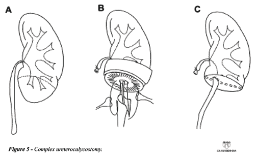
Ureterocalycostomy is performed by mobilizing
the kidney to allow easy access to the lower pole. The critical portion
is to resect sufficient lower pole parenchyma to prevent subsequent renal
cortical fibrosis. The inferior calyx is mobilized and then anastomosed
to the ureter that has been spatulated laterally. This is demonstrated
in Figure-5.

Endoscopic Management of UPJ Obstruction
Although open dismembered pyeloplasty remains the gold standard for repair of UPJ obstruction, with success rates between 90-95%, with the advent of endourologic equipment and techniques there are several minimally invasive techniques that are applicable to managing UPJ obstruction. Endopyelotomy has its roots dating back to the technique of intubated ureterotomy, which was popularized by Davis (7,8). Now it has become increasingly well accepted for optimal management of primary UPJ obstruction, with success rates that approach open pyeloplasty with significantly lower morbidity. The 2 approaches to endopyelotomy are the antegrade and the retrograde techniques, which will be reviewed further.
Antegrade Endopyelotomy
As
urologists gained experience with percutaneous management of stones in
the early 1980s, it became apparent that the same techniques could be
applied for the management of UPJ obstruction. Antegrade endopyelotomy,
as it is performed in the Unites States, was largely pioneered and refined
by Motola et al. (9). Although the initial success rates were not as good
as those of the open surgery, with increasing experience and advances
in equipment that has now changed, at our institution, antegrade endopyelotomy
has become a first-choice procedure for the management of UPJ obstruction
(10).
The initial step is to gain percutaneous
access through a lateral or upper pole calyx, with the patient in prone
position. The key step is the successful placement of a guidewire across
the UPJ and into the bladder. If this is not possible in an antegrade
fashion, it should be performed in a retrograde manner, and then exchanged.
A second safety wire is also useful, so as not to lose access to the UPJ.
We generally use a Lunderquist-Ring 0.038 torque wire as out working wire.
Since the endopyelotomy is performed with this wire, care should be taken
to prevent its kinking or bending. The tract is then dilated and a nephroscope
is placed. After careful inspection of the collecting system, the cutting
instrument is introduced over the wire (Figure-6). Although the endopyelotomy
incision was previously made in a posterolateral location, because of
the detailed anatomic findings described by Sampaio & Favorito (11)
we now make our incision in the lateral position. If the UPJ does not
appear wide enough to accommodate the knife, it should be balloon-dilated.
The instrument of choice at our institution is a cold-cut knife, and the
incision is made through the full thickness of the renal pelvis and ureteral
wall (Figure-7). This is confirmed by the presence of the wispy retroperitoneal
fat identified after the incision. The incision should extend down the
ureter at least 1 cm beyond the area of UPJ obstruction, and should be
continued laterally up into the renal pelvis an additional 1 or 2 cm.
Care should be taken to avoid any aberrant vessels; these can be identified
by their pulsation. If significant bleeding is encountered at the time
of the incision, allowing clot formation in the renal pelvis with subsequent
vascular tamponade, typically controls the bleeding. If arterial bleeding
were to persist despite this, urgent angiography with embolization will
be required.


After the incision, a stent should be used
and we usually use the 8F double-J stent. In addition, we place a 22F
nephrostomy tube in the renal pelvis. The nephrostomy tube is typically
left indwelling for 48 hours, and a trial of clamping is completed prior
to its removal. The majority of patients is discharged on post-operative
day 2 or 3, and is able to return to normal activity within a week. Cystoscopic
removal of the double-J stents is done at 6 weeks, and an intravenous
pyelogram is obtained a few weeks later. All patients are then followed
at periodic intervals for signs or symptoms of late failure. We consider
the percutaneous endopyelotomy successful if the patient is asymptomatic
and the intravenous pyelogram shows improved function or drainage.
Retrograde Endopyelotomy
Retrograde
management of UPJ obstruction can vary from simple balloon dilation (high
failure rate) to the use of Acucise™ cutting balloon device to ureteroscopic
endopyelotomy. A recent Internet survey of over a 1,000 practicing American
urologists revealed that Acucise™ endopyelotomy was the most frequently
selected therapy for adults with UPJ obstruction (12). The technique of
performing endopyelotomy with the Acucise™ cutting balloon involves
placement of the Acucise™ catheter over a guidewire across the region
of the UPJ. The region of the stricture is noted by the characteristic
“waist” seen when inflating the balloon with contrast. The cutting-wire
is then positioned laterally and, as the balloon is reinflated, the stricture
is simultaneously incised. Extravasation of contrast should be observed,
and then an 8F double-J stent should be placed. Follow-up is similar to
that of antegrade endopyelotomy.
Ureteroscopic endopyelotomy has gained popularity
in the treatment of UPJ obstruction (13). Again a wire is passed into
the UPJ obstruction, and a rigid or flexible ureteroscope is used to inspect
the UPJ and determine the location and length of the narrowed area. An
endoscopic incision may then be performed with electrocautery or holmium:YAG
laser. The incision is made from just inside the renal pelvis to normal
ureter by withdrawing the ureteroscope and the electrode as a unit. The
incision is then balloon-dilated to 24F. A ureteral stent should be placed
for 6-8 weeks.
Laparoscopic Pyeloplasty
The first laparoscopic pyeloplasty was performed in 1993 as an alternative to the standard flank pyeloplasty. Details of this procedure have been published recently (14) and are beyond the scope of this review. The principles of laparoscopic pyeloplasty are similar to those of open repair, and dismembered pyeloplasty is the most common approach.
FINAL CONSIDERATIONS
There are many factors to consider in deciding the optimal surgical procedure to a given patient. Anatomic considerations, past surgical procedures, patient expectations, and the surgeon’s experience all contribute to the success of the procedure. The gold standard with success rates of 90-95% is still the open dismembered pyeloplasty. However, with the trend toward decreasing morbidity and hospitalization, endoscopic management and laparoscopy have come to the forefront. At our institution, the preferred initial management is antegrade endopyelotomy. We have reported an overall success rate of 88% and this is similar to other published success rates (9,10,15). Although retrograde endopyelotomy is even less invasive, there may be a trade off in terms of success. There have been some comparable results with the retrograde approach; however the follow-up has been inferior. Lastly, laparoscopic pyeloplasty, although technically challenging, also provides early durable results.
REFERENCES
- Kletscher BA, Segura JW: Surgical management of UPJ obstruction in adults. AUA Update Series 1996; vol XV: lesson 18.
- Blute ML, Malek RS: Contemporary concepts in diagnosis and management of UPJ obstruction in adults. AUA Update Series 1990; vol IX: lesson 5.
- Novick AC, Streem SB: Surgery of the Kidney. In: Walsh PC, Retik AB, Stamey TA, Vaughan Jr ED (eds.), Campbell’s Urology. Philadelphia, WB Saunders, 7th ed., 1998.
- Anderson JC, Hynes W: Retrocaval ureter: A case diagnosed preoperatively and treated successfully by a plastic operation. Br J Urol. 1949; 21:209-14.
- Culp OS, DeWeerd JH: A pelvic flap operation for certain types of UPJ obstruction: Preliminary report. Mayo Clin Proc. 1951; 26:483-88.
- Scardino PL, Prince CL: Vertical flap ureteropelvioplasty: Preliminary report. South Med J. 1953; 46:325-31.
- Davis DM: Intubated ureterotomy: a new operation for ureteral and ureteropelvic strictures. Surg Gynec Obst., 1943; 76:513.
- Davis DM, Strong GH, Drake WM: Intubated ureterotomy: Experimental work and clinical results. J Urol. 1948; 59:851-62.
- Motola JA, Badlani GH, Smith AD: Results of 212 consecutive endopyelotomies: An 8-year follow-up. J Urol. 1993;149:453-6.
- Kletscher BA, Segura JW, LeRoy AJ, Patterson DE: Percutaneous antegrade endoscopic pyelotomy: Review of 50 consecutive cases. J Urol. 1995; 153, 701-703.
- Sampaio FJB, Favorito LA: Ureteropelvic junction stenosis: vascular anatomical background for endopyelotomy. J Urol. 1993; 150:1787-91.
- Hollowell C, Patel R, Bales GT, Gerber GS: Internet and postal survey of endourologic practice patterns among American urologists. J Urol. 2000; 163:1779-82.
- Tawhiek ER, Liu J, Bagley DH: Ureteroscopic treatment of UPJ obstruction. J Urol. 1998; 160:1643-7.
- Jarrett TW: Technique of laparoscopic pyeloplasty. Int Braz J Urol. 2000; 26:76-81.
- Shalhav AL, Giusti G; Elbahnasy AM, Hoenig DM, Mcdougall EM, Smith DS, et al.: Adult Endopyelotomy: Impact of etiology and antegrade versus retrograde approach on outcome. J Urol. 1998; 160:685-9.
Received: August 12, 2002
Accepted: August 30, 2002
_______________________
Correspondence address:
Dr. Joseph W. Segura
Department of Urology, Mayo Clinic
200 First Street SW
Rochester, Minnesota 55905, USA
Fax: + 1 507 284-4987
E-mail: segura.joseph@mayo.ed