EFFICIENCY
OF 6- AND 12-PUNCTURES BIOPSIES TO DETECT PROSTATE CANCER IN PATIENTS
WITH PSA < 10 ng/mL AND NORMAL DIGITAL RECTAL EXAMINATION
(
Download pdf )
LUIZ E. SLONGO, MÁRIO C. SUGISAWA, SÉRGIO O. IOSHII, RENATO TÂMBARA FILHO, LUIZ C.A. ROCHA
Division of Urology, School of Medicine, Federal University of Paraná, Curitiba, Paraná, Brazil
ABSTRACT
Objective:
Establish the efficiency of 6- and 12-punctures transrectal ultrasound-guided
needle biopsies in low risk patients for prostate cancer. Six-punctures
(sextant) biopsies were compared to 12-punctures biopsies, assessing which
is the best strategy to detect this neoplasm.
Materials and Methods: Among 240 patients
submitted to prostate biopsy, 54 with suspected small and organ-localized
tumors (prostatic specific antigen £ 10 ng/mL and digital exam of
the prostate not suggesting cancer) in glands < 50 cm3 were selected,
constituting a homogenous sample. These patients were submitted to standard
3-punctures (basal, mid, and apical) sextant biopsy in parasagittal midline
of each prostatic lobe, with 3 additional lateral punctures, bilaterally.
Each specimen was separately submitted to histological study.
Results: Twenty-two (40.7%) patients had
prostatic cancer, and 28 presented prostatic hyperplasia, associated or
not to inflammatory conditions. High-grade prostatic intraepithelial neoplasia
(PIN) was detected in 4 patients. From 22 tumors detected by 12-punctures
biopsies, 6-punctures biopsies in the parasagittal midline (sextant) diagnosed
50% of the cases, while isolated lateral punctures diagnosed 90.9% of
the malignant neoplasms. Basal lateral punctures responded for 72.7% of
the cancer diagnosis, while basal sextant punctures responded only for
9.1% of the cases.
Conclusion: For low risk prostate cancer,
patients’ 12-punctures biopsy was more effective, for sextant biopsy
failed to diagnose half of the cases of neoplasm. Three lateral punctures
(basal, mid, and apical), with 2 additional punctures in the parasagittal
midline (mid and apical) bilaterally are suggested as the best biopsy
strategy.
Key words:
prostate; prostatic neoplasms; biopsy; diagnosis; needle
Int Braz J Urol. 2003; 29: 24-9
INTRODUCTION
The
prostatic cancer (PCa) is the most diagnosed male sex neoplasia in Occident,
supplanted in mortality only by lung malignancies. The PCa diagnosis is
confirmed by the histological evaluation of fragments obtained by prostate
gland biopsy.
The transrectal ultrasound-guided needle
prostate biopsy, directed to parasagittal midline of both prostatic lobes,
in basal, mid and apical portions, with the addition of punctures directed
to ultrasonographic suspect lesions (designated as sextant biopsy), has
been used as standard since the end of the eighties. This technique was
first described by Hodge et al. in 1989, for patients presenting digital
rectal exam and/or prostatic ultrasonographic study with suspicion of
malignancy (1). Since the 90’s, with the widespread use of prostatic
specific antigen (PSA) measurement as screening to early detection of
PCa, a new challenge was elicited to confirm this diagnostic suspicion,
leading to new strategies of BxP, mainly for patients with PSA dosage
slightly elevated, and digital exam of the prostate not suggesting cancer.
Following this new reality, the requirement
to repeat sextant BxP for patients with persistently high measurements,
without other diseases explaining such biochemical abnormality, was observed.
A significant percentage of these patients, when submitted to the new
biopsy, received PCa diagnosis, showing that sextant biopsy underestimated
the presence of prostatic cancer (2, 3). Nevertheless, the false negative
results of the sextant BxP presented in literature are inconsistent, suggesting
that the patients´ samples observed were not homogenous. Studies
generally compared the efficiency to detect PCa in patients with high
PSA measurements and/or digital exam of the prostate suspect for malignancy,
with no other consideration. Therefore, patients presenting extensive
prostatic tumors, which are easily sampled by just one puncture, were
compared to patients presenting minimal disease, difficult to detect.
Keeping in mind that today BxP is frequently
indicated to patients presenting only slight raises in PSA measures, this
study was designed with the aim of establishing PCa incidence and ascertaining
the efficiency of 12-punctures BxP compared to 6-punctures BxP in this
population.
MATERIALS AND METHODS
This
research was approved by the Committee for Ethics in Research with Humans
of our University. Among 240 consecutive patients referred to prostatic
biopsy by local urologists in the period of January 17th, 2000 to November
20th, 2000. Among these, 54 cases were selected fulfilling the following
requirements: a) digital exam of the prostate not suggesting cancer; b)
PSA measurement £ 10 ng/mL; and c) prostatic gland < 50 cm³
by transrectal ultra-sound.
All biopsies were performed by the same
examiner. An ultrasound Siemens Sonoline Versa Plus® with transrectal
transducer 6.5 MHz biopsy automatic pistol with 18-gauge needles equipment
was used. Antimicrobial prophylaxis was performed with oral ciprofloxacin
500 mg q12 hours for 3 consecutive days, initiating 24 hours before the
procedure. Bowel preparation consisted in cathartic enema 1 hour before
the procedure.
To carry out the exam, the patients were
placed in left lateral position, and sedation was performed with intravenous
propofol. Before the biopsy, a new digital exam of the prostate was performed
to confirm the gland features. Sequentially, a prostate ultrasonography
was performed, establishing its volume, and the presence or absence of
suspicious hypoechogenic or nodular images.
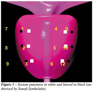
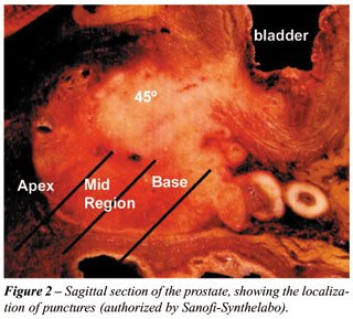
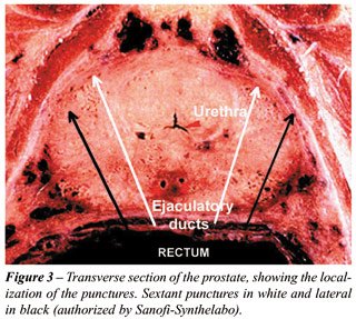
Twelve consistent prostate punctures were
performed at: a)- the parasagittal midline of the prostate (sextant punctures).
Right lobe: base, mid region, and apex (specimens 1, 2, and 3, respectively);
and Left lobe: base, mid region, and apex (specimens 4, 5, and 6 respectively);
b)- equidistant between the margin and the parasagittal midline: (lateral
punctures). Right lobe: base, mid region, and apex (specimens 7, 8, and
9, respectively); and Left lobe: base, mid region, and apex, (specimens
10, 11, and 12, respectively) (Figures-1, 2, and 3).
An additional sample was taken when there
was an ultrasonographic suspicion of neoplasm in an area, and this was
included in the systematic sampling of the region. Nevertheless, for glands
of greater volume with a hypertrophic transition zone constricting the
peripheral zone, it was determined that the surgeon should increment the
needle’s angulation intending to removing a sample of the constricted
peripheral zone and its anterior horn. Each specimen was fixed in a strip
of filter paper, and its extremity was delimited with green Indian ink.
Subsequently, it was separately packed in labeled vials containing 10%
formalin solution, representing 12 samples. All cases were analyzed by
the same pathologist, according to Prophet et al., and Bostwick &
Dundore (4,5) criteria.
For statistic analysis the parametric Student’s
“t” test of and the non-parametric “Comparisons between
2 Proportions” and “c-square” for independent samples.
RESULTS
Mean
age of selected patients in this study was 57.7 ± 9.8 years, ranging
from 41 to 80 years. The average of total PSA was 6.5 ± 1.7 ng/mL,
ranging from 2.7 to 10.0 ng/mL. Three patients presented PSA total measurement
= 4 ng/mL, of which 2 presented total PSA of 4 ng/mL and only 1 patient
with 42 years presented total PSA of 2.7 ng/mL. Prostatic mean volume
by transrectal ultrasound was 34.7 ± 8.3 cm³, the smallest
gland presenting 17.0 cm³ and the biggest 49.0 cm³.
Amidst 54 patients selected and submitted
to systematic prostate biopsy, 22 (40.7%) cases were positive for cancer,
and the mean Gleason score was 5.5, ranging from 4 to 8. Among the remainder
32 patients, 20 (62.5%) presented benign prostatic hyperplasia (BPH) associated
with chronic prostatitis, 7 (21.9%) presented only BPH, 3 (9.4%) presented
BPH, chronic prostatitis, and prostatic intra-epithelial neoplasia, 1
patient (3.1%) presented BPH associated with acute and chronic prostatitis,
and another (3.1%) presented BPH associated with prostatic intra-epithelial
neoplasia.
Among 22 patients with PCa diagnosis, 9
(40.9%) cases had cancer detected by sextant punctures and lateral punctures
as well. Individually, lateral punctures detected PCa in 11 cases (50.0%),
while sextant punctures detected only 2 cases (9.1%) of malignant tumors.
Therefore, sextant punctures were positive for PCa in 11 (50.0%) cases,
and lateral punctures in 20 (90.9%) cases (p = 0.008), primarily lateral
basal (72.7%) (p = 0.007) (Table-1).
In the analysis of the biopsy technique
performed compared to the total of patients studied, we have noticed that
sextant punctures diagnosed PCa in 20.4% of the total of patients, whilst
sextant + lateral punctures were diagnostic in 40.7% (p = 0.0367).
When the diagnostic percentage was analyzed
according to the biopsy technique used, within the group of patients with
established prostate cancer (n = 22), sextant technique would fail to
diagnose 50.0% of the patients with prostate cancer from the selected
group (p < 0.0001), while sextant + lateral peripheral punctures diagnosed
100.0% of the cases.
DISCUSSION
Patients
presenting a digital rectal exam not suggesting cancer, and persist with
biochemical suspicion of malignancy due to high PSA measures, frequently
require a second prostate biopsy.
Many of these patients, after confirming
PCa with a second biopsy, are submitted to radical prostatectomy, and
in the histological study of the surgical specimen frequently a neoplasm
of significant volume is detected, with no reasonable explanation for
the fact that the first biopsy could not detect the tumor. We observed
that there is not a consensus in literature regarding the optimal technique
for prostate biopsy. The percentage of detected tumors and false negative
results are inconsistent owing to lack of stratification. The same technique
approach was employed in all the cases without differentiating non-palpable
minimum disease from bulky tumors. Patients with PSA slightly elevated
were compared to patients with very high PSA, and generally a biopsy strategy
considering the volume of the prostatic gland was not observed. Therefore,
cases with tumors of difficult detection by biopsy were compared to cases
with tumors easily detected by only one puncture, in this way it is justified
the false negative findings described in literature, ranging from 1% to
35% (6,7).
With the idea of establishing the efficacy
in detection of PCa of the 12-punctures biopsies compared to sextant biopsies,
a patient sample with similar characteristics and with low risk for PCa
was selected. These patients presented PSA dosage £ 10 ng/mL, non-palpable
tumors and glands < 50 cm³ (8). Excluding cases with large prostates
(³ 50 cm³) had the objective of evaluating the glands in which
volume the PCa occurs with greater frequency (9), as well as using only
one standard biopsy technique approach in order not to have much variation
in prostates’ volume. This sample of patients presenting similar
features was considered ideal to compare between 12- and 6-punctures biopsies.
All conventional technical resources were used to diagnose PCa, yet punctures
in the prostate midline were avoided as in this region only 2 a 4.1% of
the tumors (7,10) are observed, and these punctures account for higher
morbidity of the procedure.
Among 240 systematically biopsied patients,
54 cases (with previously mentioned features) constituted an homogeneous
sample of patients with clinic suspicion of small volume prostate tumors,
which are difficult to detect by biopsy. Of 54 selected patients, 22 (40.9%)
presented PCa. Among the remainder 32 patients, 21 (65.6%) presented acute
and or chronic inflammatory conditions associated to BPH, 7 (21.9%) only
BPH, and 4 (12.5%) high-grade intraepithelial neoplasia.
The detected tumors index in the present
study was high, similar to the one found by Eskew et al. (7) in the so-called
5 regions biopsies, for patients with high PSA and/or palpable tumor.
This must be highlighted, given that this population is clinically considered
as presenting low risk for PCa. The diagnostic of high grade PIN was detected
in 12.5% of the patients, which is an index similar to the one reported
by Levine et al. (11). Nonetheless, Martins et al. detected high grade
PIN in 7.3% of the Brazilian population above 40 years with PSA > 4
ng/mL and digital rectal exam suspicious for PCa (12).
From a total of 22 patients with prostatic
cancer diagnosed by 12-punctures biopsy, 50% were detected by isolated
lateral punctures— these tumors would not be diagnosed by the sextant
biopsy, (p=0.008) — and 9,1% of the tumors were detected only by
the sextant punctures. The remaining tumors (40.9%) were detected by both
sextant and lateral punctures. The sextant puncture, according to literature,
fails to diagnose up to 35% of the PCa in patients with high PSA and/or
suspicious digital rectal exam without other stratifications (6,7,13).
Nevertheless Beurton et al. (14) in a protocol with one sample from patients
with similar features (stratified digital rectal exam and PSA data), reported
that sextant biopsy failed to diagnose PCa in 47% of the cases.
Various publications confirmed the existence
of false negative results from sextant biopsy; yet, the false negative
index ranged according to the employed methodology (6,13). Ballantine
Carter (10) underlined that sextant technique was idealized in a time
where diagnostic suspicion was entirely based in digital rectal exam and
sonographic findings; that in PSA era malignant lesions usually are detected
when small and still not palpable, and in this phase are found more laterally,
and may not be detected by sextant biopsy. This was actually corroborated
by Boboruglu et al. (13) report that documented the existence of PCa in
30% of the patients presenting previous negative sextant prostate biopsy
and persisted with clinical suspicion of cancer.
On the other hand, Naughton et al. (6) evaluated
244 patients with PSA between 2.5 and 20 ng/mL and/or rectal exam suspicious
for prostate cancer, detecting cancer in 26% and 27%, respectively, by
6- and 12-punctures biopsy. This was the only published study in which
the greater number of punctures failed to yield a significant increase
in the detection index of PCa. However, this protocol did not stratify
the patients for digital exam of the prostate, comparing non-palpable
tumors with extensive tumors.
The issue of the possibility of more than
6-punctures biopsies identify an excessive number of non-significant tumors
(< 0.5 cc and Gleason score £ 4) was elucidated by many authors.
Among them, Digiuseppe et al. (15) that found the presence of non-significant
disease in merely 2.8% of 3.038 radical prostatectomies studied, in patients
without previous hormonal treatment or prostate resection, showing a low
incidence of PCa without clinical meaning.
In the present study, the anatomic region
where the greater contribution for the cancer diagnostic was obtained
was the lateral regions in prostate base in 72.7% of the cases (punctures
#7 and 10), while the places where the presence of PCa was less frequently
detected were the sextant regions, also in the base of the gland (punctures
#1 and 4), in 9.1% of the cases. For none of the patients isolated basal
sextant punctures detected neoplasia, so this punctures may be excluded
from the protocol without harm. This data is in accord to the papers published
by Presti Jr. (16,17).
CONCLUSION
In the selected group of patients (PSA £ 10 ng/mL, digital examination of the prostate not suggesting malignancy, and glands < 50 cm³), the incidence of prostate cancer was high. The 12-punctures biopsies were more efficient in detecting neoplasia compared to sextant biopsies. However, it is suggested as better strategy for prostate biopsy, for patients with this features; 3 lateral punctures (basal, mid and apex), added with 2 punctures in the parasagittal midline (mid and apex), bilaterally.
REFERENCES
- Hodge KK, Mcneal JE, Terris MK, Stamey TA: Radon systematic versus directed ultrasound guided transrectal core biopsies of the prostate. J Urol. 1989;142:71-5.
- Stamey TA: Making the most out of six systematic sextant biopsies. Urology. 1995; 45: 2-12.
- Presti Jr JD, Chang JJ, Bhargava V, Shinohara K: The Optimal systematic prostate scheme should include 8 than 6 biopsies: Results of a prospective clinical trial. J Urol. 2000; 163: 163-7.
- Prophet EB, Arrington JB, Sobin LH: Laboratory methods in histotecnology. Armed Forces Institute of Pathology. American Registry of Pathology. Washington, DC. USA, 1994.
- Bostwick DG, Dundore PA: Biopsy pathology of the prostate. London UK, 1997.
- Naughtton CK, Miller DC, Mager DE, Ornstein DK, Catalona WJ: A prospective randomized trial comparing 6 versus 12 prostate biopsy cores: Impact on cancer detection. J Urol. 2000; 164: 388-92.
- Eskew LA, Bare RL, Mccullough DL: Systematic 5 region biopsy is superior to sextant method for diagnosing carcinoma of the prostate. J Urol. 1997; 157: 199-203.
- Catalona WJ, Richie JP, Ahmann FR, Hudson MA, Scardino PT, Flanigan RC: Comparison of digital rectal examination and serum prostate specific antigen in the early detection of prostate cancer: Results of a multicenter clinical trial of 6.630 men. J Urol. 1994; 151: 1283-90.
- Uzzo RG, Wei JT, Waldbaum RS, Perlmutter AP, Byrne JC, Vaughan Jr. ED: The influence of prostate size on cancer detection. Urology. 1995; 46: 831-6.
- Ballentine Carter H: Is sextant prostate biopsy the standard of the care in PSA era. AUA News. 1999; 4: 1-3.
- Levine MA, Ittman M, Melamed J, Lepor H: Two consecutive sets of transrectal ultrasound guided sextant biopsies of the prostate for the detection of prostate cancer. J Urol. 1998; 159: 471-6.
- Martins ACP, Reis RB, Suaid HJ, Maciel LMZ, Cologna AJ, Falconi RAR: Screening for carcinoma of the prostate in volunteers. Braz J Urol. 2000; 26: 516-22.
- Borboroglu PG, Comer SW, Riffenburgh RH, Amling CL: Extensive repeat transrectal ultrasound guided prostate biopsy in patients with previous benign sextant biopsies. J Urol. 2000; 163: 158-62.
- Beurton D, Barthelemy Y, Fontaine E, Chartier E, Lamotte F, Franc B: 12 systematic proatate biopsies are superior to sextant biopsies for diagnosing carcinoma: A propective randomized study. Brit J Urol. 1997; 80 (Suppl 2): 239.
- Digiuseppe JA, Sauvageot J, Epsein JI: Increasing incidence of minimal residual cancer in radical prostatectomy specimens. Am J Surg Pathol. 1997; 21: 174-8.
- Presti Jr JD: How to biopsy the prostate. AUA News. 1999; 4: 27-30.
- Presti Jr JD: Systematic biopsies of the prostate: 6 just aren’t enough. Cont Urol. 1999 ; 11: 11-7.
______________________
Received:
March, 26 2003
Accepted after revision: December, 18 2003
_______________________
Correspondence address:
Dr. Luiz Edison Slongo
Rua Portugal, 329
Curitiba, PR, 85510-280, Brazil
Fax: + 55 41 3241-1329
E-mail: slongo.uro@mps.com.br