PAINLESS
INTER EPIDIDYMAL TESTICULAR TORSION OF THE SPERMATIC CORD
(
Download pdf )
SALOMON V. ROMANO, HAIME S. HERNAN, NORBERTO FREDOTOVICH
Section of Urology, Hospital Durand, Buenos Aires, Argentina
ABSTRACT
Inter epididymal testicular torsion of the spermatic cord is extremely rare and usually diagnosed at surgery. We present an unusual case of spermatic cord torsion in a 14-year-old male patient. It is important to highlight that the torsion occurred only on the distal half of the epididymis leaving the head untwisted and edematous. In addition, the fact that this condition was painless made this case extremely rare and motivated our presentation.
Key
words: testis; epididymis; torsion
Int Braz J Urol. 2007; 33: 77-9
INTRODUCTION
Testicular
torsion is considered a surgical emergency. The testis present irreversible
damage if the torsion is not resolved within the first 6 hours. Torsion
usually occurs in young pre-puberty males, between 12 and 18 years old,
(1) even though it can be seen in other ages. The prevalence is estimated
to be 1 in 4000 patients under 25 years old.
The inter epididymal torsion of the spermatic
cord is one of the most infrequent situations. In our patient it occurred
due to the abnormal insertion between the epididymis and the testicle.
This kind of torsion is clinically undistinguishable from the typical
spermatic cord torsion and the diagnosis can only be made during surgical
exploration.
It is well known that spermatic cord torsion
is associated with intense pain. In this particular case, it was painless.
CASE REPORT
A
fourteen-year-old male patient was first seen at the urology section due
to a painless growth of his left scrotum, beginning one month before his
visit. No history of trauma or masturbation habit was refereed, but a
similar episode took place 2 years before, with complete remission after
medical treatment.
Previous evaluation in another center with
ultrasonography (US) and tumor markers assumed its etiology as inflammatory
and as a consequence treated the condition with ice, antibiotics and oral
analgesics.
At physical examination, an increased volume
and high consistence on left testicle was found, but the patient said
the size was half of what it was initially, without spontaneous or induced
pain.
The US showed a homogeneous round left testicle,
without tumor, with a para-testicular solid 16 x 21 mm hypoechoic mass
with small hydrocele (Figure-1).
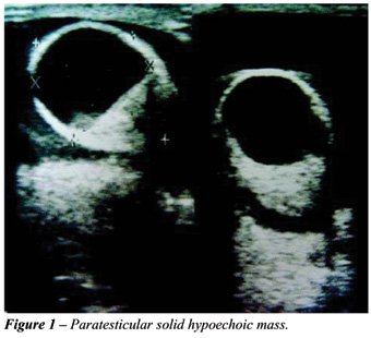
Blood flow was normal according to a color
Doppler ultrasonography. Contralateral (right) testis and epididymis were
normal.
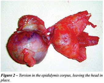
The left testicle was explored using the
inguinal approach, showing a partial intravaginal 360-degrees inter epididymal
testicular torsion of spermatic cord. The torsion site was in the epididymal
corpus, leaving the head in place (Figure-2), corresponding to the hypoechoic
mass described previously. The necrotic aspect of the testicle motivated
the orchiectomy. In the pathological study, a hemorrhagic infarction was
confirmed.
The right testicle was surgically explored
using the scrotal approach. We found a low insertion of the epididymis
in relation to the testicle, and orchiopexy was performed.
COMMENTS
Two
types of spermatic cord torsion are described in literature. In the extravaginal
torsion, the testicle and the vaginal sac turn over the spermatic cord
at the external inguinal ring, due to a lack of adherence of the tunica
vaginalis to the scrotal wall. This type of rotation can only be seen
in fetus and neonates (2). In the intravaginal torsion, there is a previous
anatomic defect. The high and narrow insertion of the tunica vaginalis
in the testicle allows it to remain free in the vaginal sac as a “bell
clapper”. This kind of defect would be bilateral and would justify
preventive orchiopexy in the contralateral testis (3). Another type of
intravaginal torsion is between the testis and the epididymis. This rare
presentation is reported only in disjunction between testis and epididymis
(4). In this type of anatomic defect, isolated epididymis torsion has
been reported. Also, torsion of the testicular and epididymal appendages
can occur. These structures can turn over their own axis and produce pain
and local inflammation, mimicking the clinical presentation of spermatic
cord torsion (3).
In the present case, the unusual presentation
with hardness and painless testicle in a young male patient, together
with unspecific complementary studies, lead us to think it was a testicular
cancer and therefore, an inguinal approach was performed.
CONFLICT OF INTEREST
None declared.
REFERENCES
- Scorer CG, Farrinfton GH: Congenital Deformities of the Testis and Epididymis. London, Butterworth and Co. 1971.
- Friedman RM, Flashner SC, Akwari OE, King LR: An experimental model of neonatal testicular torsion: evidence against an exclusively extravaginal etiology. J Urol. 1993; 150: 246-8.
- Heinen F: Escroto Agudo. Arch Argent Pediatr. 2001; 99: 554-61.
- Ravichandran S, Blades RA, Watson ME: Torsion of the epididymis: a rare cause of acute scrotum. Int J Urol. 2003; 10: 556-7.
____________________
Accepted
after revision:
July 10, 2006
_______________________
Correspondence address:
Dr. Salomon Victor Romano
25 de mayo 846
Vicente Lopez, BA, 1638, Argentina
Fax: + 54 11 4791-6735
E-mail: sromano1@arnet.com.ar
EDITORIAL COMMENT
The
case-report herein describes an anecdotal situation of painless testicular
torsion of the spermatic cord in an unusual location. The reader should
bear in mind the rarity of this clinical event. On the other hand, the
subject raised allows the editor to draw some reflections about this still
controversial issue of acute scrotum. The main differential diagnosis
of the acute scrotum includes testicular torsion and inflammatory conditions.
Color Doppler ultrasound is the current imaging modality of choice for
the radiological evaluation of acute scrotum, replacing other methods
such as nuclear scintigraphy, Doppler flowmetry and gray scale ultrasound.
Unfortunately, we cannot always rely on the exam. Bentley et al. discussed
variations in degrees of bell clapper deformity and its influence in attachments
of tunica vaginalis representing possibility of testicular blood flow
despite spermatic cord torsion (1). In their series, 4 of 14 cases had
testicular torsion confirmed intraoperatively despite a normal color Doppler
ultrasound. One should also remember that ultrasound is an operator dependent
test and a false-negative report may end catastrophically.
A
more appealing and rational algorithm for the management of acute scrotum
is also discussed in the paper of Bentley et al (1). In case of obvious
suspicion of testicular cord torsion, surgical operation is mandatory.
When there is a low index of suspicion, one should perform color Doppler
ultrasound and the diagnosis of inflammation is acceptable only in case
of increased flow, while patients with “normal flow” should
be also operated. We agree with others that 6 hours is the desirable time
form the beginning of the onset of pain to have the case resolved.
REFERENCE
1. Bentley DF, Ricchiuti DJ, Nasrallah PF, McMahon DR: Spermatic cord torsion with preserved testis perfusion: initial anatomical observations. J Urol. 2004; 172: 2373-6.
Dr. Antonio
Macedo Jr.
Division of Urology
Federal University of São Paulo, UNIFESP
São Paulo, SP, Brazil
E-mail: amcdjr@uol.com.br