IMPACT
OF OBESITY ON URETEROSCOPIC LASER LITHOTRIPSY OF URINARY TRACT CALCULI
(
Download pdf )
RICARDO NATALIN, KEITH XAVIER, ZEPHANIAH OKEKE, MANTU GUPTA
Department of Urology, Columbia University, College of Physicians and Surgeons, New York, NY, USA
ABSTRACT
Purpose:
The treatment of urinary tract stones in obese patients may differ from
the treatment of non-obese patients and their success rate varies. Our
objective was to compare ureteroscopic treatment outcomes of ureteral
and renal stones, stratified for stone size and location, between overweight,
obese and non-obese patients.
Materials and Methods: Charts were reviewed
for 500 consecutive patients presenting at our institution for renal and
ureteral stones. A total of 107 patients underwent flexible or semi-rigid
ureteroscopy with Ho:YAG laser lithotripsy and met criteria for review
and analysis.
Results: Overall, initial stone-free rates
were 91%, 97%, and 94% in normal, overweight and obese individuals respectively.
When compared to non-obese patients, there were no significant differences
(p value = 0.26; 0.50). For renal and proximal ureteral stones, the stone-free
rate in overweight and obese individuals was 94% in both groups; and a
stone-free rate of 100% was found for distal stones, also in both groups.
Conclusions: Ureteroscopic treatment of
stones in obese and overweight patients is an acceptable treatment modality,
with success rates similar to non-obese patients.
Key
words: ureter; calculi; ureteroscopy; Ho-YAG Laser; obesity
Int Braz J Urol. 2009; 35: 36-42
INTRODUCTION
Obesity
has become a major health problem in the United States and the world and
represents a chronic disease mediated by genetics, environment, metabolism,
psychosocial causes, cultural, and physiologic variables (1). The prevalence
of obesity in the United States has increased by approximately 30% from
1980 to 1994 (2). The most common method of defining obesity is the Body
Mass Index (BMI). BMI measures the height to weight ratio by taking weight
in kilograms and dividing it by height in squared meters (kg/m2).
According to the World Health Organization guidelines, a BMI of 18.5 to
25 kg/m2 is considered normal, overweight is a BMI of 25 to
29.9, obese is a BMI ≥ 30, and morbidly obese is a BMI ≥ 40
(3).
Various lithogenic risk factors are known
to be associated with obesity and increase the chance of stone formation
in these patients as hyperinsulinemia, increased BMI, hyperoxaluria, high
sodium intake, low urinary volume and hypercalciuria. Duffey et al. found
that 98% of obese patients had at least one lithogenic risk factor in
a 24-hour urine sample and 80% of them had 3 or more factors (4).
Extracorporeal shock wave lithotripsy (ESWL)
has emerged as the primary treatment of choice for renal calculi less
than 1.5-2 cm (5). ESWL has been recommended as first-line treatment of
ureteral calculi less than 1 cm, resulting in up to a 92.6% stone free
rate for proximal stones and 97.5% for mid and distal ones (6). However,
obese patients are fraught with difficulties in treating calculi by ESWL
and may not have these same high success rates as in non-obese patients.
Delakas et al. reported an increased chance of ESWL failure in obese patients
of 1.9 fold when BMI was > 30, and Muñoz et al. found a 72%
stone free rate after ESWL for these patients (7,8). In these obese patients,
a frequent factor limiting the success of ESWL is positioning the patient
so the stone can be located at the focal point of the lithotripter. Most
lithotripters have a maximum skin to stone distance of 12-14 cm for their
focal point, which can restrict the depth in which stone fragmentation
can be accomplished (9). For this reason, ESWL for obese patients may
be a sub-optimal treatment.
PCNL as a potential treatment for renal
calculi in obese patients can also be difficult. This is due to an increased
distance that needs to be traversed in order to obtain the correct access
into a calyx, making percutaneous access more difficult. Also, even if
access is obtained, normal size instruments may not be able to be used
and longer instruments including nephroscope and access sheath may be
required in an obese patient. Another potential problem during PCNL in
an obese patient is the increased anesthetic complication risk that can
ensue from the patient being in the prone position for a long period of
time.
For these previously mentioned reasons,
rigid and flexible ureteroscopy is most likely the treatment of choice
for urinary calculi in obese patients. The development of small caliber
ureteroscopes and advances in intracorporeal lithotripsy, such as ultrasound,
electrohydraulic waves, laser, and most recently the holmium: yttrium-aluminum-garnet
(Ho:YAG) laser, have permitted more successful and safer endoscopic manipulation
of ureteral calculi (10). In order to ascertain whether ureteroscopy is
more effective in obese patients, we compared outcomes data, stratified
for stone size and location, in overweight, obese, morbidly obese and
normal weight patients as defined by BMI.
MATERIALS AND METHODS
Charts
were reviewed for 500 consecutive patients treated for renal and/or ureteral
calculi at our institution over a five-year period. Inclusion criteria
for the study included all patients with radio-opaque calculi who were
treated ureteroscopically, in combination with Ho:YAG laser lithotripsy,
as primary therapy. Indications for treatment were calculi that did not
pass spontaneously or required earlier intervention because of recurrent
colic or obstruction of the upper urinary tract. Patients who had contraindications
such as pregnancy, urinary tract infection, coagulation disorders, or
previous ureteral reimplantation were excluded from the study. After a
thorough review, 107 patients met the criteria for this review.
Ureteroscopy was performed in combination
with Ho:YAG laser lithotripsy by the same surgeon (M.G.) using a small
caliber (6F) semi-rigid or flexible ureteroscope.
Distal stones were treated via a Wolf semi-rigid
ureteroscope with a 6F self-dilating tip and for proximal ureteral stones
we used the flexible ureteroscope Storz Flex-X or ACMI DUR-8 or DUR-8
Elite, depending on availability. No dilation of the ureteral orifice
was necessary because of self-dilating tip ureteroscope (for distal stones).
For proximal stones, ureteral access sheath was placed underneath the
stone (Cook Flexor, 35 cm), with size varying from 9 to 11F when using
Storz Flex-X and from 12 to 14F when using ACMI ureteroscope.
Our standard technique for ureteroscopic
treatment of ureteral calculi includes cystoscopy with retrograde pyelogram,
placement of a 0.038-inch floppy-tipped guide wire past the stone (glidewire
when necessary) to maintain access, placement of a safety wire for flexible
ureteroscopy, and ureteroscopy with Ho:YAG laser lithotripsy. Continuous
irrigation and/or intermittent manual pumping of irrigant to obtain a
clear ureteroscopic view were used where appropriate. For ureteroscopic
laser lithotripsy, a Ho:YAG laser (Trimedyne, Inc., Irvine, CA) was employed.
The Ho:YAG laser operates at a wavelength of 2100-nm and the laser frequency
was usually set between 5-10 Hz and a power of 5-10 W. Higher settings
were used to treat harder calculi. The vast majority of the patients were
treated with a 200 uH quartz fiber. Basket retrieval of stone fragments
was employed when necessary. Patients received general anesthesia at the
beginning of the procedure.
A preoperative x-ray of the kidneys, ureters,
and bladder were done in all patients, and excretory urogram (IVP), non-contrast
helical computer tomography, or sonogram were done when indicated to document
the size and location of the stone. Patients were postoperatively imaged
with radiographs, non-contrast helical computer tomography, and/or IVP
until they were stone-free or received additional treatment (0 to 3 months).
A patient was considered stone free when post operative imaging revealed
fragments of 2 mm or less. Characteristics of patient age, sex, stone
size and location, operative time, and treatment outcome were recorded
and tabulated. Average patient age and mean stone size were similar for
all groups (Table-1).
Treatment outcomes were defined as radiographic
evidence of fragmentation or complete disappearance of the stone. Retreatment
and additional procedures were also registered. All procedures were performed
on an outpatient basis.
For each of the treatment groups, 95% confidence
intervals were calculated for the overall treatment success rates. Statistical
comparison of two independent percentages was done by means of the Fisher’s
exact test (2-sided, p = 0.05). If the resulting p value was < 0.05,
the difference in the sample percentages was considered statistically
significant.
RESULTS
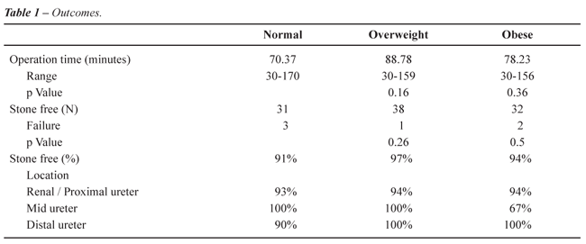
The
average patient age and mean stone size were similar for all groups (Table-2).
Mean operating time was 70.37 minutes for normal weight individuals, 88.78
minutes for overweight persons, 78.23 minutes for obese patients. These
differences were not statistically significant between groups (Table-1).

Indications for the procedure were due to
persistent pain despite analgesic medication in 51 patients, obstruction
with ultrasound revealing hydronephrosis in 24 and persistent pain associated
with evidence of obstruction in 32.
The initial stone-free rate for ureteral
calculi 1 cm or greater following treatment with ureteroscopy with Ho:YAG
laser lithotripsy was 93%. For ureteral calculi less than 1 cm, the initial
stone-free rate was 100%.
Stratified for location (Table-1), the initial
stone free rates for renal/ proximal ureteral stones ranged from 93% to
100% for all weight categories. The small numbers of patients (7 patients
in total, 1 failure) with mid-ureteral stones had stone free rates that
varied from 67% to 100%. For distal ureteral stones, the initial success
rates ranged from 90% to 100%. Neither stone size nor location appeared
to influence the efficacy of ureteroscopic treatment, since no significant
difference was observed in the stone free rates between patients with
ureteral calculi 1 cm or greater and those with calculi less than 1 cm.
Failures were due to proximal migration
of stone with inability to retrieve all fragments from lower pole in 3
patients and to residual fragments left in the ureter that failed to spontaneously
pass to the bladder in other 3 patients. No intraoperative or postoperative
complications occurred in any of the groups.
COMMENTS
When
ESWL cannot be used or is not an appropriate treatment option in the obese
patient, the next option is often ureteroscopy or percutaneous nephrolithotomy
(PCNL). El-Assmy et al. showed that PCNL in obese patients was not only
safe but that obese patients did not experience any difference in success,
operative time, or morbidity (11). Even though good results can be obtained
PCNL in the obese patient it still presents many challenges to the urologist.
The substantial amount of subcutaneous fat and increased mobility of the
kidney secondary to excess fat in the retroperitoneum make instrument
access more difficult. Also, sometimes standard PCNL equipment is not
long enough and extra-long equipment (nephroscope, etc.) has to be used
in the obese patient, making the procedure technically more difficult.
Ureteroscopic laser lithotripsy and stone extraction has been shown to
be an effective method for treating urolithiasis in morbidly obese patients
who were too large for ESWL (12). Compared to in situ ESWL, ureteroscopic
lithotripsy appears to be more effective in the treatment of proximal
ureteral calculi 1 cm or greater.
Recent technological advances, especially
in the field of optics, have allowed endoscopes to become smaller, more
flexible, and easier to introduce. Prior to the development of small caliber
ureteroscopes, the stone-free rates achieved with ureteroscopy for distal
ureteral calculi using large diameter rigid ureteroscopes (more than 10
F), ultrasonic lithotriptors, or electro hydraulic lithotriptors with
probes larger than 3 F, was greater than 90% (12,13). However, for mid-ureteral
calculi, it was in the range of 60%, and for proximal calculi, close to
50% (13). More recent contemporary series, using small diameter rigid
and flexible endoscopes as well as laser lithotriptors, have reported
success rates of greater than 90% for proximal ureteral calculi (13).
In our experience, the initial overall stone-free rate after ureteroscopic
laser lithotripsy of proximal ureteral calculi was 97%, with a stone-free
rate of 93% for calculi 1 cm or greater, which is consistent with the
success rates of other reported series.
The introduction of the Ho:YAG laser has
improved ureteroscopy stone-free rates while decreasing the risk of complications,
and thus has been employed for lithotripsy by many groups with encouraging
results. The Ho:YAG laser can fragment all types of calculi, including
hard calcium oxalate monohydrate and cystine stones, by delivering energy
through small-diameter quartz fibers that can be used through the working
channels of the smallest available ureteroscopes. It fragments stones
with an ablative effect, removing portions of the stone as dust-like particles
during the fragmentation procedure. This process allows for the treatment
of large calculi within the upper urinary tract without the burdensome
process of fragment removal. The safety and efficacy of the Ho:YAG laser
as an endoscopic lithotripter has been confirmed in other studies (14,15).
Our results show that it is possible to
achieve stone-free status even in obese patients when treating them with
ureteroscopy. Our results in fact showed higher stone-free rates in patients
with a BMI of greater than 25, although the rates are virtually the same.
One limitation of the study is the small number of patients in the morbidly
obese group. Based on these results, ureteroscopy with laser lithotripsy
should be given serious consideration in any obese patient with a stone
smaller than 2 cm. With the continued improvement in technology and scopes,
the potentially more difficult access to the ureter of obese patients,
due to body habitus reasons, can be easier overcome and stone-free rates
can approach or be equivalent to that of non-obese patients.
Long-term complication rates of ureteroscopy
range from 0.5 to 10% for larger caliber instruments (16). Complications
are rare with small caliber instruments. Our low overall complication
rate was consistent with those reported by other series. The majority
of cases may be treated without ureteral dilation and have a lower likelihood
of ureteral trauma. Thus, routine ureteral stenting following ureterscopy
and intracorporeal lithotripsy may not be necessary, thereby decreasing
morbidity (17,18).
CONCLUSIONS
Our study demonstrates that ureteroscopy is an acceptable treatment modality for all ureteral calculi and may be preferable to ESWL for obese patients. By using small caliber ureteroscopes and Ho:YAG laser lithotripsy, the target stone could be treated safely and effectively in our patients. In overweight and obese patients, results are comparable to non-obese patients. These results presented are independent of stone size and location.
CONFLICT OF INTEREST
None declared.
REFERENCES
- Pasulka PS, Bistrian BR, Benotti PN, Blackburn GL: The risks of surgery in obese patients. Ann Intern Med. 1986; 104: 540-6.
- Willett WC, Dietz WH, Colditz GA: Guidelines for healthy weight. N Engl J Med. 1999; 341: 427-34.
- Stevens J, Cai J, Pamuk ER, Williamson DF, Thun MJ, Wood JL: The effect of age on the association between body-mass index and mortality. N Engl J Med. 1998; 338: 1-7.
- Duffey BG, Pedro RN, Kriedberg C, Weiland D, Melquist J, Ikramuddin S, et al.: Lithogenic risk factors in the morbidly obese population. J Urol. 2008; 179: 1401-6.
- Jamshaid A, Ather MH, Hussain G, Khawaja KB: Single center, single operator comparative study of the effectiveness of electrohydraulic and electromagnetic lithotripters in the management of 10- to 20-mm single upper urinary tract calculi. Urology. 2008; 72: 991-5.
- Murota-Kawano A, Ohya K, Sekine H: Outpatient basis extracorporeal shock wave lithotripsy for ureter stones: efficacy of the third generation lithotripter as the first line treatment. Int J Urol. 2008; 15: 210-5.
- Delakas D, Karyotis I, Daskalopoulos G, Lianos E, Mavromanolakis E: Independent predictors of failure of shockwave lithotripsy for ureteral stones employing a second-generation lithotripter. J Endourol. 2003; 17: 201-5.
- Muñoz RD, Tirolien PP, Belhamou S, Desta M, Grimberg R, Dulys P, et al.: Treatment of reno-ureteral lithiasis with ESWL in obese patients. Apropos of 150 patients. Arch Esp Urol. 2003; 56: 933-8.
- Calvert RC, Burgess NA: Urolithiasis and obesity: metabolic and technical considerations. Curr Opin Urol. 2005; 15: 113-7.
- Preminger GM, Tiselius HG, Assimos DG, Alken P, Buck AC, Gallucci M, et al.: 2007 Guideline for the management of ureteral calculi. Eur Urol. 2007; 52: 1610-31.
- El-Assmy AM, Shokeir AA, El-Nahas AR, Shoma AM, Eraky I, El-Kenawy MR, et al.: Outcome of percutaneous nephrolithotomy: effect of body mass index. Eur Urol. 2007; 52: 199-204.
- Nguyen TA, Belis JA: Endoscopic management of urolithiasis in the morbidly obese patient. J Endourol. 1998; 12: 33-5.
- Anderson KR, Keetch DW, Albala DM, Chandhoke PS, McClennan BL, Clayman RV: Optimal therapy for the distal ureteral stone: extracorporeal shock wave lithotripsy versus ureteroscopy. J Urol. 1994; 152: 62-5.
- Jiang H, Wu Z, Ding Q: Ureteroscopy and holmium: YAG laser lithotripsy as emergency treatment for acute renal failure caused by impacted ureteral calculi. Urology. 2008; 72: 504-7.
- Farkas A, Péteri L, Lorincz L, Salah MA, Flaskó T, Varga A, et al.: Holmium:YAG laser treatment of ureteral calculi: A 5-year experience. Lasers Med Sci. 2006; 21: 170-4.
- Elashry OM, Elgamasy AK, Sabaa MA, Abo-Elenien M, Omar MA, Eltatawy HH, et al.: Ureteroscopic management of lower ureteric calculi: a 15-year single-centre experience. BJU Int. 2008; 102: 1010-7.
- Ibrahim HM, Al-Kandari AM, Shaaban HS, Elshebini YH, Shokeir AA: Role of ureteral stenting after uncomplicated ureteroscopy for distal ureteral stones: a randomized, controlled trial. J Urol. 2008; 180: 961-5.
- Cheung MC, Lee F, Yip SK, Tam PC: Outpatient holmium laser lithotripsy using semirigid ureteroscope. Is the treatment outcome affected by stone load? Eur Urol. 2001; 39: 702-8.
____________________
Accepted after revision:
November 10, 2008
_______________________
Correspondence address:
Dr. Mantu Gupta
Dept of Urology, Columbia University
Irving Pavilion, 11th Floor
161 Ft. Washington Avenue
New York, NY, 10032, USA
Fax: + 1 212 342-6870
E-mail: guptama@pol.net
EDITORIAL COMMENT
Obesity
has become a major health problem in the world. Various lithogenic risk
factors are associated with obesity, increasing the chance of stone formation
in these patients.
The
surgical treatment of kidney and ureteral stones in morbidly obese patients
remains difficult because shockwave lithotripsy may be a sub-optimal treatment
due to weight limitations and percutaneous nephrolithotomy is associated
with difficult access, anesthetic complications and a high rate of transfusion
(1).
Dash
et al. showed in a matched comparison (obese x normal) that ureteroscopic
(URS) treatment of renal calculi when matched for location and size is
as successful as and no more morbid in morbidly obese than in normal weight
patients. URS treatment of renal calculi is a safe and effective first-line
treatment for renal calculi in morbidly obese patients (2).
The
authors study demonstrates that ureteroscopy is an acceptable treatment
modality for all ureteral calculi and may be preferable to ESWL for obese
patients.
The
development of small caliber ureteroscopes and advances in intracorporeal
lithotripsy have allowed for more successful and safer endoscopic manipulation
of renal/ureteral calculi in overweight, obese, and morbidly obese patients.
REFERENCES
- Andreoni C, Afane J, Olweny E, Clayman RV: Flexible ureteroscopic lithotripsy: first-line therapy for proximal ureteral and renal calculi in the morbidly obese and superobese patient. J Endourol. 2001; 15: 493-8.
- Dash A, Schuster TG, Hollenbeck BK, Faerber GJ, Wolf JS Jr: Ureteroscopic treatment of renal calculi in morbidly obese patients: a stone-matched comparison. Urology. 2002; 60: 393-7; discussion 397.
Dr. Mauricio Rubinstein
Federal Univ. of State of Rio de Janeiro
UNIRIO
Rio de Janeiro, RJ, Brazil
E-mail: mrubins@attglobal.net
EDITORIAL COMMENT
The
authors present their experience with ureteroscopic laser lithotripsy
in obese and morbidly obese patients. The conclusion is that obesity is
not a hindrance and results are similar with those obtained in non-obese
patients.
Some
articles have been published on the outcome of percutaneous nephrolithotomy
in this group of patients and showed that results are comparable to those
obtained in non-obese (1-3). This is the first article addressing specifically
ureteroscopy in obese and results are encouraging. Since the results of
extracorporeal shock wave lithotripsy in these patients are not as good
as in non-obese, ureteroscopy could be considered the first line approach
even in proximal ureteral stones. As obesity represents a worldwide public
health problem an owing to its relationship with urolithasis, articles
comparing the various forms of treating stones in obese are welcome.
REFERENCES
- Bagrodia A, Gupta A, Ramon SD, Bensalah K, Pearle MS, Lotan Y: Impact of body mass index on cost and clinical outcome after percutaneous nephrostolithotomy. Urology. 2008: 29 (In press).
- El-Assmy AM, Shokeir AA, El-Nahas AR, Shoma AM, Eraky I, El-Kenawy MR, et al.: Outcome of percutaneous nephrolithotomy: effect of body mass index. Eur Urol. 2007; 52: 199-204.
- Nguyen TA, Belis JA: Endoscopic management of urolithiasis in the morbidly obese patient. J Endourol. 1998; 12: 33-5.
Dr.
Eduardo Mazzucchi
Division of Urology
University of Sao Paulo, USP
Sao Paulo, SP, Brazil
E-mail: mazuchi@terra.com.br