DIFFUSION
WEIGHTED IMAGING IN THE DETECTION OF UPPER URINARY TRACT UROTHELIAL TUMORS
(
Download pdf )
doi: 10.1590/S1677-55382010000100004
SHUJI NISHIZAWA, SHUN IMAI, TOSHIKAZU OKANEYA, TSUYOSHI NAKAYAMA, TAKAYUKI KAMIGAITO, TOMONORI MINAGAWA
Departments of Urology (SN, TO, TN, TK, TM) and Radiology (SI), Nagano Municipal Hospital, Nagano, Japan
ABSTRACT
Purpose:
Diffusion-weighted (DW) magnetic resonance imaging (MRI) provides information
about the biophysical properties of tissues such as cell organization
and density. DW imaging (DWI) is becoming important in the assessment
of malignant tumors. The purpose of our study was to evaluate the capability
and reliability of DWI in the evaluation of upper urinary tract urothelial
tumors.
Materials and Methods: DWI was performed in seventeen patients with upper
urinary tract urothelial tumor, previously diagnosed by either CT or retrograde
pyelography. An histological evaluation was performed after surgical resection.
Each MRI was carried out using a 1.5T superconductive magnet MRI system.
DWI images were obtained with b value of 1000 s/mm2 under normal breathing.
The apparent diffusion coefficient (ADC) values were measured.
Results: In nine patients with renal pelvis tumors and seven patients
with ureteral tumors, the lesions were shown as high-signal intensity
in the corresponding region on DWI. In one patient with carcinoma in situ
(CIS) of the ureter, the lesion was not depicted with DWI. The mean ADC
value of the tumor was 1.125 ± 0.217 x 10⁻³ mm2/s and was significantly
lower than those of the renal parenchyma (1.984 ± 0.238 x 10⁻³
mm2/s, p < 0.01) and the urine (2.941 ± 0.315 x 10⁻³ mm2/s,
p < 0.01).
Conclusions: In our study, the renal pelvic and ureteral tumors except
CIS were shown clearly with DWI. Although further studies are required,
DWI may take the place of invasive retrograde urography for detecting
tumors of the upper urinary tract.
Key
words: magnetic resonance imaging; transitional cell; neoplasm;
renal pelvis; ureter
Int Braz J Urol. 2010; 36: 18-28
INTRODUCTION
Five percent
of urothelial tumors occur from the ureter and renal pelvis or calyces,
accounting for approximately 10% of upper urinary tract neoplasms (1).
Upper urinary tract urothelial cancer is one of the most difficult lesions
to be shown by imaging studies. Moreover, it is difficult to depict ureteral
or renal pelvic small tumors. Conventionally, invasive radiography, such
as retrograde pyelo-uretrography using cystoscopy, has been the imaging
modality in detecting urothelial tumors.
Diffusion-weighted (DW) magnetic resonance imaging (MRI) is a technique
used to show water molecular diffusion in vivo. It provides information
about the biophysical properties of tissues such as cell organization
and density, microstructure, and microcirculation (2). DW imaging (DWI)
has been used in the field of neuroradiology. Recently, DWI has become
increasingly important in the assessment of malignant tumors. Several
authors have reported the usefulness of DWI in the detection of the abdominal
and pelvic malignant lesion such as prostate cancer and colon cancer (3,4).
The purpose of our study was to evaluate the efficacy and reliability
of DWI in the assessment of upper urinary tract urothelial tumors.
MATERIALS AND METHODS
Patient Population
This
was a retrospective study performed at Nagano Municipal hospital. Between
June 2003 and March 2007, seventeen patients with upper urinary tract
urothelial tumor underwent MRI examination including DWI. All patients
had upper tract urothelial tumor previously diagnosed either by computed
tomography or by retrograde pyelography. Our Institutional Ethics Committee
reviewed and approved the study protocol. Written informed consent was
obtained from all patients.
The histological study was performed after
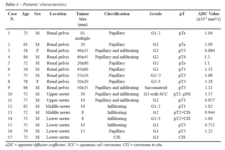
surgical resection. The patients’ characteristics are listed in
Table-1.
Just before the examination, intramuscular
or intravenous injection of 20 mg of butyl scopolamine bromide was administered
to all patients.
Imaging Protocol
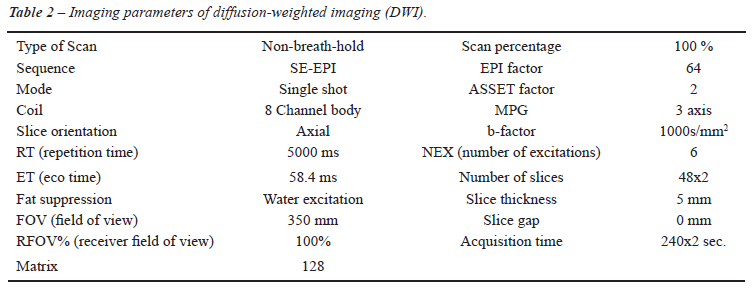
Each MRI was performed using a 1.5T superconductive magnet MRI system (Signa, Twin Speed Excite version 12.0, GE Medical Systems, Milwaukee, WI.) with maximum gradient amplitude of 40 mT/m and a maximum slew rate of 150 mT/m/second, with an 8-channel-body array coil. DW images were obtained in the axial plane under normal breathing in addition to conventional T1/T2 weighted MR images without contrast-enhanced imaging. We obtained multiple axial thin slices DWI and reconstructed 3D images and maximum intensity projection (MIP) images. The imaging parameters used for DWI are listed in Table-2.
Typically, presence of the tumor was defined
when high signal intensity appeared on DWI. Radiological diagnosis was
performed by the same radiologist (S.I.).
The apparent diffusion coefficient (ADC)
values of the tumor, the renal parenchyma and the urine in the bladder
were calculated in a circular region of interest for quantitative analysis
(Figure-1). Statistical analysis was performed by an un-paired t-test.
Results are reported as mean ± standard deviation. A p-value of
less than 0.05 was considered statistically significant.

RESULTS
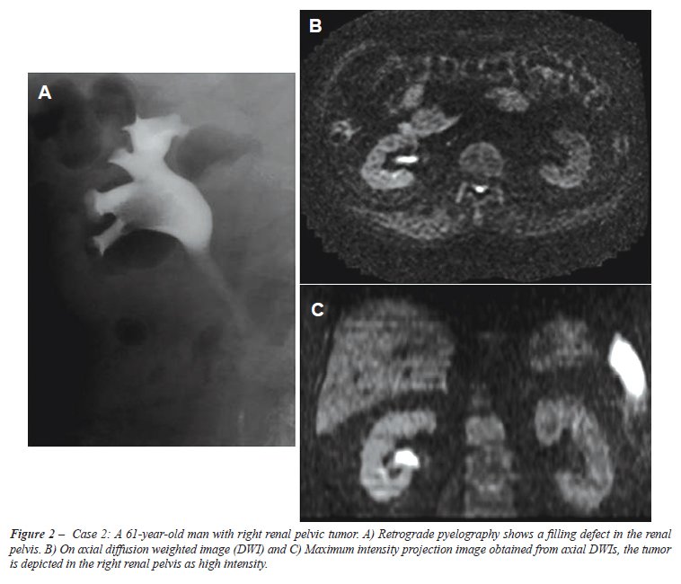
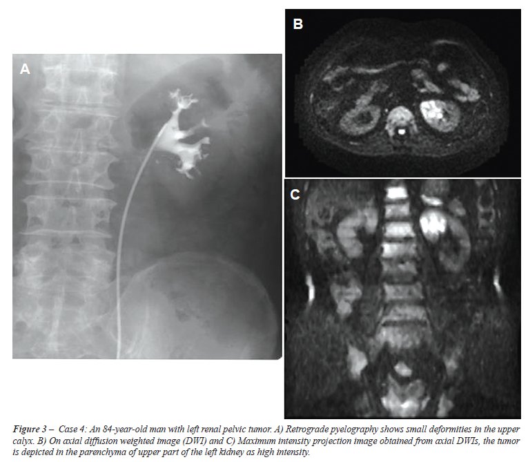
In nine patients with renal pelvic tumors, all lesions were shown as high-signal intensity in the renal pelvis or renal parenchyma on DWI (Figures-2 and 3), whereas conventional T1- and T2-weighted MRI was able to depict the lesion clearly in eight patients. In seven patients with a ureteral tumor, all tumors were depicted in the corresponding region (Figures-1 and 4). The smallest depicted tumor was approximately 8 mm in diameter. However, conventional MRI was able to depict ureteral tumor in five patients. In a patient (case 17) with carcinoma in situ (CIS) in the lower ureter and in two patients (cases 13 and 14) with associated CIS, DWI and conventional MRI failed to show the corresponding lesions. The sensitivity and positive predictive value (PPV) of DWI for detecting the tumor were 94.1% (16 of 17) and 100%, respectively. The sensitivity and PPV of conventional MRI were 76.5% (13 of 17) and 100%, respectively.
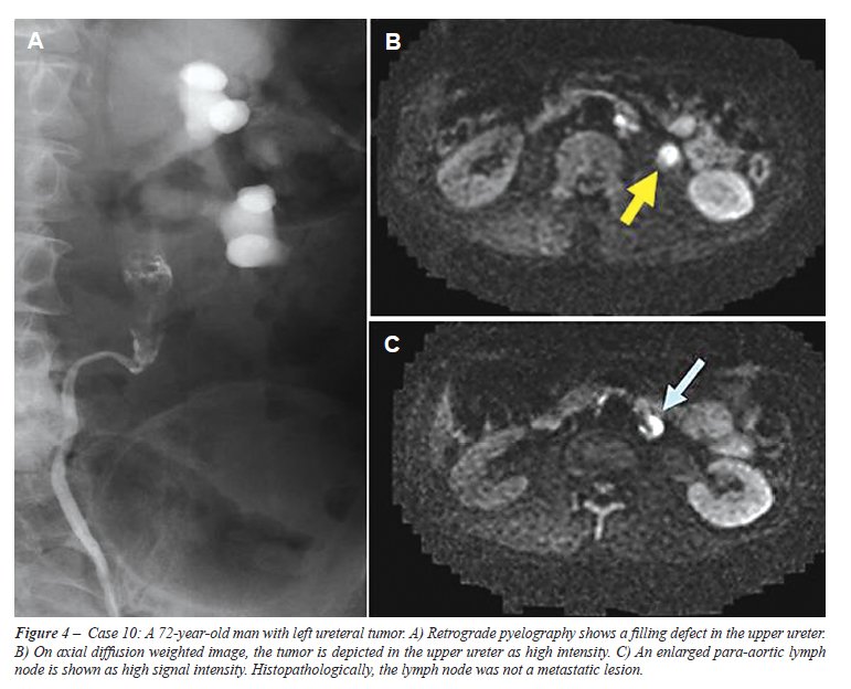
DWI showed a hyper-intense signal in several normal structures such as spleen, lymph node, spinal cord and mucus in the small intestine. Swollen lymph nodes in a patient (Figure-4, case 10) were shown as high signal intensity by DWI; however, the lymph nodes did not contain malignant cells on histopathological examination.
Histopathologically all tumors were diagnosed
as a urothelial carcinoma in the surgical specimens. The cytological tests
were negative in five patients with low-grade tumors.
The mean ADC value of the tumor was 1.125
± 0.217 x 10⁻³ mm2/s, while the values of the renal parenchyma
and the urine in the bladder were 1.984 ± 0.238 x 10⁻³ mm2/s and
2.941 ± 0.315 x10⁻³ mm2/s, respectively. The mean ADC value of
the urothelial tumor was significantly lower than those of the renal parenchyma
and the urine (p < 0.01 and p < 0.01). Regarding the tumor classification,
the mean ADC value of infiltrating tumor was significantly lower than
that of papillary tumor (0.929 ± 0.122 x 10⁻³ mm2/s and 1.245 ±
0.215 x 10⁻³ mm2/s, p < 0.05).
COMMENTS
MRI has
been infrequently used in the primary assessment of upper tract urothelial
cancer, and the MRI characteristics of this tumor have not been well described.
MRI imaging is independent of excretory function and shows multiplaner
imaging, which permits direct image acquisition in the plane of tumor
spread (5). Diffusion weighted imaging is an MRI technique and is the
only imaging method that can evaluate the diffusion process in vivo. Diffusion
is thermally induced motion of water molecules in biological tissues,
which is called Brownian motion. The speed of diffusion of water molecules
is different in the extracellular and intracellular component of the tissues.
In the intracellular component, the diffusion is relatively slow because
of the presence of cellular membranes (2). A malignant tumor often has
a larger cell diameter and denser cellularity than normal tissue and the
cell density may be indicative of tumor aggressiveness. Restriction of
water diffusion is found to be a common feature of tumors (6). Apparent
diffusion coefficient (ADC) values are quantitative expressions of diffusion
characteristics of tissues, and ADC values are related to the proportion
of extracellular and intracellular components. Since a malignant tumor
often has a larger cell diameter and cellularly denser than normal tissue
the ADC values of tumors may decrease (7). Therefore, DWI shows the tumor
as high signal intensity as well. Takahara et al. showed the potential
capability of DWI as a screening tool for malignancy-like positron emission
tomography. High-b-value DW-MRI images could be directly used for tumor
detection because of the different cellular structures of healthy and
neoplastic tissues. They reported a new DWI technique under normal breathing,
which allows acquisition of more slices with multiple signal averaging,
a higher signal-to-noise ratio, and high-quality MRI images (3). We used
multi-excitation for data acquisition under normal breathing as well.
Additional advantages of this technique are that it is completely non-invasive,
and does not require exposure to ionizing radiation. Furthermore, adding
this DWI to a routine MRI protocol requires only few minutes and does
not cause patient discomfort. DWI does not require the administration
of intravenous contrast material, which may cause allergic reaction or
renal toxicity.
DWI is available for several malignancies including abdominal and pelvic
lesions such as liver, colon, uterus and kidney and prostate cancer (8-12).
Recently Yoshida et al. reported the application of DWI for a series of
renal pelvic neoplasms (13), and Takeuchi et al. demonstrated the feasibility
of this method for the detection of ureteral tumors (14), using DWI with
diffusion gradient b-value of 800 s/mm2. In these two reports, they demonstrated
significantly lower ADC values of the tumors than of the surrounding tissues.
Several authors have recently reported the feasibility of using DWI for
the detection of a urinary bladder cancer (15-17).
In our study, the renal pelvic and ureteral tumors except CIS were clearly
shown with DWI regardless of the tumor grade. The mean ADC value of the
urothelial tumor was significantly lower than those of the renal tissue
and the urine. DWI may detect the tumor of upper urinary tract more distinctly
because urothelial tumors were surrounded by a fluid collection or urine.
We were able to demonstrate that the mean ADC value of the infiltrating
tumor was significantly lower than that of papillary tumor. This may depend
on the difference of cellular density between the tumor types. Although
Takeuchi et al. recently reported that the mean ADC value of G3 bladder
cancer was significantly lower than that of G1 and G2 tumors recently
(17), we were not able to demonstrate the difference in ADC value between
tumor grades. This may due to our small study population.
In our study, the smallest depicted tumor was approximately 8 mm in diameter.
Yoshida et al. reported they were able to obtain high signal intensity
of small renal pelvic tumors (5 mm and 7 mm in diameter) on DWI, despite
unclear conventional morphological MRI (14). DWI may provide the information
about the characteristics of a small mass even if the mass is not clearly
depicted by conventional MRI techniques. The CIS lesion in case 17 could
not be depicted either in DWI or in conventional MRI. This was also not
possible in the associated CIS lesions in the other patients. DWI may
not be able to delineate the area of CIS at present because the lesion
does not form a mass.
It might possibly be difficult to differentiate between malignant and
benign tumors such as ureteral polyp by using conventional imaging studies
and cytologic examination. Fujii et al. reported that the ADC values of
uterine endometrial benign lesion including polyp and leiomyoma were significantly
higher than that of the malignant lesion. They concluded that ADC measurement
could provide useful information in differentiating malignant from benign
uterine endometrial cavity lesions (18). Although we have no experience
with the cases of benign urinary tract tumors such as polyp or endometriosis,
DWI and ADC value may nevertheless provide the information about the property
of the mass (17,18).
An enlarged lymph node may be a false positive structure in the diagnosis
of malignant tumor with DWI. Ichikawa et al. reported that most metastatic
lymph nodes were detected because of their high signal intensity, in some
patients healthy lymph nodes also showed similarly high signal intensities
(4).
The standard work-up for the patient with hematuria consists of urinalysis
and cytologic analysis, cystoscopy and excretory urography (5). Additional
imaging is often required. The diagnosis of urothelial cancers is usually
made based on the cytological analysis of urine specimens, which are collected
on cystoscopy or retrograde pylography. These techniques are invasive
and technically demanding. The preoperative cytologic studies were negative
in five patients with low-grade tumors in our series. False negative cytologic
results may occur in cases of low-grade lesions or in which the ureter
is obstructed. MRI allows multiplanar images and MR urography can permit
localization of ureteric obstruction. As previously mentioned, several
reports have demonstrated the feasibility of DWI for the detecting upper
and lower urinary tract tumor (13-17). Although further studies are needed
to prove the value of DWI for detecting upper urinary tract tumor and
for differentiating malignant form benign urothelial tumor, MRI adding
DWI may become the first choice for imaging studies, and DWI may replace
invasive retrograde urography and imaging studies using intravenous injection
of contrast medium.
This is the third reported study on the application of DWI for the detection
of the upper urinary tract urothelial cancer. However, this study has
several limitations. It is a retrospective study, assessing only 17 patients,
and only one radiologist evaluated the images. Further studies with larger
clinical settings are necessary.
CONCLUSIONS
In our study, the renal pelvic and ureteral cancers except CIS were shown clearly with DWI regardless of the tumor grade. We demonstrated that an infiltrating tumor had lower ADC values than that of papillary tumor. Although further studies with larger clinical settings are required, DWI may replace invasive retrograde urography or conventional imaging using intravenous injection of contrast medium in detecting tumors of the upper urinary tract.
CONFLICT OF INTEREST
None declared.
REFERENCES
- Hall MC, Womack S, Sagalowsky AI, Carmody T, Erickstad MD, Roehrborn CG: Prognostic factors, recurrence, and survival in transitional cell carcinoma of the upper urinary tract: a 30-year experience in 252 patients. Urology. 1998; 52: 594-601.
- Squillaci E, Manenti G, Cova M, Di Roma M, Miano R, Palmieri G, et al.: Correlation of diffusion-weighted MR imaging with cellularity of renal tumours. Anticancer Res. 2004; 24: 4175-9.
- Takahara T, Imai Y, Yamashita T, Yasuda S, Nasu S, Van Cauteren M: Diffusion weighted whole body imaging with background body signal suppression (DWIBS): technical improvement using free breathing, STIR and high resolution 3D display. Radiat Med. 2004; 22: 275-82.
- Ichikawa T, Erturk SM, Motosugi U, Sou H, Iino H, Araki T, et al.: High-B-value diffusion-weighted MRI in colorectal cancer. AJR Am J Roentgenol. 2006; 187: 181-4.
- Browne RF, Meehan CP, Colville J, Power R, Torreggiani WC: Transitional cell carcinoma of the upper urinary tract: spectrum of imaging findings. Radiographics. 2005; 25: 1609-27.
- Sugahara T, Korogi Y, Kochi M, Ikushima I, Shigematu Y, Hirai T, et al.: Usefulness of diffusion-weighted MRI with echo-planar technique in the evaluation of cellularity in gliomas. J Magn Reson Imaging. 1999; 9: 53-60.
- Lyng H, Haraldseth O, Rofstad EK: Measurement of cell density and necrotic fraction in human melanoma xenografts by diffusion weighted magnetic resonance imaging. Magn Reson Med. 2000; 43: 828-36.
- Namimoto T, Yamashita Y, Sumi S, Tang Y, Takahashi M: Focal liver masses: characterization with diffusion-weighted echo-planar MR imaging. Radiology. 1997; 204: 739-44.
- Nasu K, Kuroki Y, Kuroki S, Murakami K, Nawano S, Moriyama N: Diffusion-weighted single shot echo planar imaging of colorectal cancer using a sensitivity-encoding technique. Jpn J Clin Oncol. 2004; 34: 620-6.
- Shimofusa R, Fujimoto H, Akamata H, Motoori K, Yamamoto S, Ueda T, et al.: Diffusion-weighted imaging of prostate cancer. J Comput Assist Tomogr. 2005; 29: 149-53.
- Charles-Edwards EM, deSouza NM: Diffusion-weighted magnetic resonance imaging and its application to cancer. Cancer Imaging. 2006; 6: 135-43.
- Squillaci E, Manenti G, Di Stefano F, Miano R, Strigari L, Simonetti G: Diffusion-weighted MR imaging in the evaluation of renal tumours. J Exp Clin Cancer Res. 2004; 23: 39-45.
- Yoshida S, Masuda H, Ishii C, Saito K, Kawakami S, Kihara K: Initial experience of functional imaging of upper urinary tract neoplasm by diffusion-weighted magnetic resonance imaging. Int J Urol. 2008; 15: 140-3.
- Takeuchi M, Matsuzaki K, Kubo H, Nishitani H: Diffusion-weighted magnetic resonance imaging of urinary epithelial cancer with upper urinary tract obstruction: preliminary results. Acta Radiol. 2008; 49: 1195-9.
- Matsuki M, Inada Y, Tatsugami F, Tanikake M, Narabayashi I, Katsuoka Y: Diffusion-weighted MR imaging for urinary bladder carcinoma: initial results. Eur Radiol. 2007; 17: 201-4.
- El-Assmy A, Abou-El-Ghar ME, Refaie HF, El-Diasty T: Diffusion-weighted MR imaging in diagnosis of superficial and invasive urinary bladder carcinoma: a preliminary prospective study. ScientificWorldJournal. 2008; 8: 364-70.
- Takeuchi M, Sasaki S, Ito M, Okada S, Takahashi S, Kawai T, et al.: Urinary bladder cancer: diffusion-weighted MR imaging--accuracy for diagnosing T stage and estimating histologic grade. Radiology. 2009; 251: 112-21.
- Fujii S, Matsusue E, Kigawa J, Sato S, Kanasaki Y, Nakanishi J, et al.: Diagnostic accuracy of the apparent diffusion coefficient in differentiating benign from malignant uterine endometrial cavity lesions: initial results. Eur Radiol. 2008; 18: 384-9.
____________________
Accepted after revision:
August 11, 2009
_______________________
Correspondence address:
Dr. Shuji Nishizawa
Nagano Municipal Hospital
1333-1 Tomitake, Nagano, 381-8551, Japan
Fax: + 81 26 295-1145
E-mail: sxnishizawa@hospital.nagano.nagano.jp
EDITORIAL COMMENT
Urothelial
carcinoma of the upper urinary tract accounts for 5% of all urothelial
carcinomas and 10% of all renal tumors. Patients with a history of urothelial
carcinoma are at high risk of developing synchronous or metachronous urothelial
carcinoma. This known multifocality requires thorough examination of the
entire urinary tract in high-risk patients (1). The diagnosis is difficult
by conventional morphological imaging, since urothelial carcinomas may
be overlooked with suboptimal examinations and even though the examination
is sufficient, small lesions may not be detected due to volume averaging
(2).
Excretory urography or computed tomography (CT) was used to evaluate high-risk
patients (3). Multiphasic CT urography offers superior detection of calculi,
urothelial tumor, and parenchymal tumor over excretory urography and allows
accurate staging of detected lesions at the same examination. However,
when the patient has a contraindication to iodinated contrast material,
retrograde pyelography or MRI is often used to image the upper urinary
tract (4). MR imaging, including MR angiography and MR urography, offers
comparable evaluation in patients who cannot tolerate iodinated contrast
material and in whom multiplanar, vascular, and collecting system imaging
is required (3,4).
Recently a new MRI technique, called diffusion-weighted MR imaging (DW-MRI)
has been applied in various abdominal diseases, especially in detecting
tumors without the need for contrast administration (5). Different from
conventional anatomical MR imaging DW-MRI provide functional information
(5). By studying molecular diffusion, the ultrastructural characteristics
of tissue can be studied in vivo through sampling water molecules and
by exploiting the natural sensitivity of MRI to the motion (2).
The research of Nishizawa et al. has shown the superiority of DW-MRI in
detecting urothelial tumors with a very high accuracy, demonstrated with
excellent images. Also in a study by Yoshida et al. two cases of highly
suspected upper urinary tract neoplasm had been detected clearly on DW-MR
images, despite unclear conventional morphological MR images (2).
Malignant masses are easily discernible against suppressed background
signal with visual assessment of DW-MR images (PET like images). This
method may be obtained after a routine abdominal MR imaging protocol approximately
in 3 to 5 minutes without an additional cost. The additional benefit of
DW-MRI is the ability to determine quantitative indices, which may be
important in the assessment of tumor cellularity, and disease response
to treatment methods and follow-up (2,5).
Further investigations will probably increase the use of functional imaging
methods and especially DW-MRI in genitourinary diseases.
REFERENCES
- Zhang J, Lefkowitz RA, Bach A: Imaging of kidney cancer. Radiol Clin North Am. 2007; 45: 119-47.
- Yoshida S, Masuda H, Ishii C, Saito K, Kawakami S, Kihara K: Initial experience of functional imaging of upper urinary tract neoplasm by diffusion-weighted magnetic resonance imaging. Int J Urol. 2008; 15: 140-3.
- Takahashi N, Kawashima A, Glockner JF, Hartman RP, Leibovich BC, Brau AC, ET AL.: Small (<2-cm) upper-tract urothelial carcinoma: evaluation with gadolinium-enhanced three-dimensional spoiled gradient-recalled echo MR urography. Radiology. 2008; 247: 451-7.
- Browne RF, Meehan CP, Colville J, Power R, Torreggiani WC: Transitional cell carcinoma of the upper urinary tract: spectrum of imaging findings. Radiographics. 2005; 25: 1609-27.
- Manenti G, Di Roma M, Mancino S, Bartolucci DA, Palmieri G, Mastrangeli R, et al.: Malignant renal neoplasms: correlation between ADC values and cellularity in diffusion weighted magnetic resonance imaging at 3 T. Radiol Med. 2008; 113: 199-213.
Dr.
Ozgur Kilickesmez
Department of Radiology
Yeditepe University Hospital
Istanbul, Turkey
E-mail:okilickesmez@yahoo.com
EDITORIAL COMMENT
A combination
of morphological imaging modalities using iodinated radiocontrast media,
such as excretory urography and computed tomography with urinary cytologic
examination, has been used for diagnosing upper urinary tract cancer (UUTC).
However, these media may cause renal insufficiency. Contrast-induced nephropathy
due to iodinated radiocontrast media is currently the third most common
cause of acute renal failure in hospitalized patients (1). Diffusion-weighted
magnetic resonance imaging (DWI) is a functional imaging technique with
no contrast agent and is applicable to patients with allergies against
contrast agents or existing renal insufficiency. Furthermore, the addition
of DWI to a routine MRI examination can be readily adopted for most current
clinical MRI scanners with only a few minutes and no additional equipment.
DWI has been reported to be a useful technique to detect UUTC in a noninvasive
manner because of clear contrast between high signal intensity of UUTC
and well-restrained signal intensity of the surrounding tissue (2). This
study has confirmed the above points. The location (pelvis or ureter)
and the size of the UUTC seem to have little impact on diagnostic potentiality
because of good contrast between the tumor and the surrounding tissues,
even if the tumor burden was small (8 mm in this paper, 5 mm in our experience).
The impact in the degree of diffusion within the tumor on the estimation
of grade or depth has recently been shown on DWI in some malignancies,
including bladder cancer (3). In this study, ADC values of the infiltrating
tumor were significantly lower than that of papillary tumor. However,
overlap among ADC values between them seems to exist. Also, overlap among
ADC values for tumor, renal parenchyma and collecting system exists. Further
investigations should be performed to clarify the clinical importance
of ADC values in evaluating tumor aggressiveness.
The current study also showed the limitation of DWI technique in assessing
the UUTC. Depicting carcinoma in situ (CIS) lesions is also challenging
in DWI, as well as in other conventional imaging modalities. The DWI contrast
reflects molecular diffusion, not the presence of existing cancer cells.
Therefore, it remains challenging to distinguish a malignant disease from
nonmalignant lesions, such as benign neoplasms, hematomas, abscesses and
inflammatory condition.
We have to keep in mind that we could not gain anatomical information
from DWI. Addition of DWI to anatomical imaging increases the accuracy
of MRI to UUTC.
REFERENCES
- Solomon R: Contrast-medium-induced acute renal failure. Kidney Int. 1998; 53: 230-42. Erratum in: Kidney Int 1998; 53: 818. published erratum of serious dosage error appears in Kidney Int 1998; 53: 1109.
- Yoshida S, Masuda H, Ishii C, Saito K, Kawakami S, Kihara K: Initial experience of functional imaging of upper urinary tract neoplasm by diffusion-weighted magnetic resonance imaging. Int J Urol. 2008; 15: 140-3.
- Abou-El-Ghar ME, El-Assmy A, Refaie HF, El-Diasty T: Bladder cancer: diagnosis with diffusion-weighted MR imaging in patients with gross hematuria. Radiology. 2009; 251: 415-21.
Dr.
Hitoshi Masuda
Department of Urology
Tokyo Medical and
Dental University Graduate School
Tokyo, Japan
E-mail: hi-masu.uro@tmd.ac.jp