LAPAROSCOPIC
URETERAL REIMPLANT FOR URETERAL STRICTURE
(
Download pdf )
doi: 10.1590/S1677-55382010000010006
RODRIGO S. Q. SOARES, RUBENS A. DE ABREU JR, JOSE E. F. TAVORA
Department of Urology, Hospital dos Servidores do Estado de Minas Gerais, IPSEMG, Belo Horizonte, Minas Gerais, Brazil
ABSTRACT
Purpose:
Evaluate the initial experience of laparoscopic ureteral reimplant for
ureteral stenosis.
Materials and Methods: From January 2004
to June 2008, 10 patients underwent 11 laparoscopic reconstruction surgeries
for ureteral stenosis. Seven cases of stenosis of the distal ureter, two
at the level of iliac vessels, a case of bilateral distal stenosis and
one in the medium third. Eight ureteroneocystotomies were performed by
extravesical technique with anti-reflux mechanism, two cases of vesical
reimplant with Boari technique and one case using the psoas hitch technique.
Results: The average surgical time was 166
minutes (115-245 min), mean blood loss was 162 mL (100-210 mL) and the
average hospital stay was 2.9 days (2-4 days). There were two complications:
a lesion of the sigmoid colon identified peroperatively and treated with
laparoscopic sutures with good evolution, and a case of ureteral stone
obstruction at the 30th day postoperative, treated by laser ureterolitotripsy.
All patients had resolution of the stenosis at an average follow-up period
of 18 months (3-54 months).
Conclusion: Laparoscopic surgery represents
a feasible, safe and low morbidity technique for ureteral reimplant in
ureteral stenosis.
Key
words: ureter; stricture; reconstruction; laparoscopy
Int Braz J Urol. 2010; 36: 38-43
INTRODUCTION
The main causes for ureteral stricture are surgical traumas, impacted
ureteral stones, extrinsic compression, tumor and congenital or idiopathic
disorders. Ureteral stenoses are the most frequent complications observed
in pelvic surgery. Currently, endourological, gynecological and laparoscopic
procedures are also reasons for referral for a large number of cases (1).
Treatments focus on the anatomic aspects
of stenosis, such as length of the lesion, complexity of obstruction and
vascularization of the ureter. Partial and segmental stenoses can be treated
by endoscopic procedures such as dilation or internal ureterotomy with
placement of double J catheter with good follow-up results. Reconstruction
technique procedures are needed for total complex stenosis.
In the last decades, open surgeries have
been performed for these types of pathologies. With the advancement of
technology, the laparoscopic ureter-vesical reimplant was introduced in
1994 by Reddy and Evans to correct vesicoureteral reflux (2). In the literature,
major series have been published with similar results (3,4).
We report our experience with laparoscopic
ureteral reimplant in ureteral stenoses of different etiologies.
MATERIALS AND METHODS
Ten
patients (8 females and 2 males) underwent 11 laparoscopic ureteral reimplants
due to ureteral stenosis, at our hospital, from January 2004 to June 2008.
Four patients had stenosis after open surgery
and 4 had ureteral stenosis resulting from ureteral stone endoscopic procedure
complications. The remaining two patients had an idiopathic congenital
bilateral ureteral stenosis and an extrinsic ureteral compression by the
ovarian vein (ovarian vein syndrome). In one patient after abdominal hysterectomy,
the ureteral stricture extended to the mid ureter, caused by ischemic
and inflammatory reaction. In all patients, an abdominal CT scan confirmed
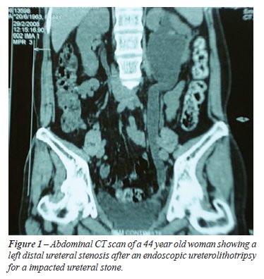
the localization and the length of the ureteral stricture (Figure-1).
Endoscopic treatment was carried out in
all cases except in one patient with idiopathic bilateral ureteral stenosis
and another with ureteral compression by the ovarian vein.
Two of these procedures were interrupted
due to complete stricture lesion post hysterectomy. In four cases, the
dilation with a balloon catheter was chosen, as well as the placement
of a double J stent for six weeks. In two patients with stenosis post
ureteral calculi, a laser ureterotomy was performed and a double-J catheter
was left indwelling for 6 weeks.
Table-1 shows the characteristics of these
cases.

Technique
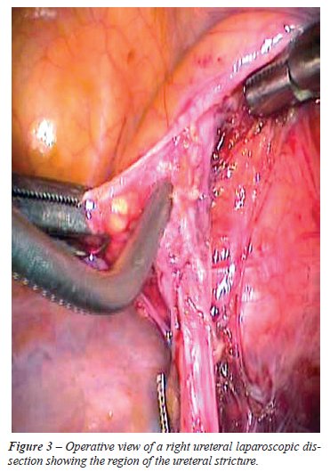
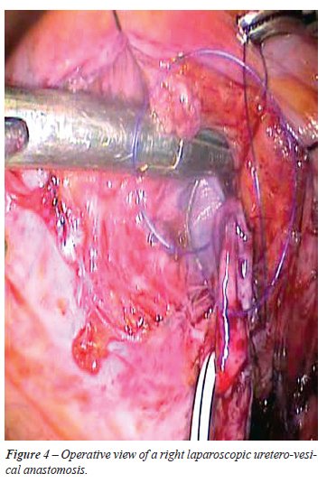
All patients underwent transperitoneal video laparoscopic surgery. The patient is placed in a flat dorsal Trendelenburg position and the surgery is performed using the four pelvic trocar technique (Figure-2). The surgery is carried out by opening the Toldt fascia, followed by the identification and dissection of the ureter in the area close to the stenosis (Figure-3).

The ureter is transected near the area of
the stenosis and spatulated. The vesical dome is fixed to the wall with
a stitch for a better exposition. The detrusor muscle is opened lengthwise
for approximately 3 cm to expose the vesical mucosa. The vesical mucosa
is opened and the posterior ureterovesical anastomosis is performed with
separated vicryl 4.0 sutures (Figure-4).
A double J catheter is placed through one
of the trocars. The anastomosis is completed and the detrusor muscle is
closed by a continuous suture for anti-reflux tunnel.
In cases of tension due to the high ureteral
stenosis, the ureteroneocystostomy with a psoas hitch muscle or Boari
Flap technique is carried out. In the middle of this opening, a stitch
with vicryl 4.0 is tightened, pulling the bladder to facilitate the anastomosis
to the edge of the ureteral stump. Anastomosis is completed with simple
stitches and the bladder is sewn lengthwise. The fixation of the vesical
part in the greater psoas muscle is also performed with vicryl 3-0 sutures.
As soon as the detrusor closing is completed, the bladder is filled with
200 mL of physiologic serum to evaluate overflowing. The cavity is drained
with either a Penrose or a tubular suction drain (Figure-2).
RESULTS
The average patient age was 44.7 years. The average surgical time was 166 min. (115-245 min.), the average amount of bleeding was 162 mL (100-210 mL) and the mean hospital stay was 2.9 days (2 - 4 days). In one of the patients, with stenosis after ureterovesical reimplant by ureteral reflux, there was a sigmoid colon lesion during dissection of the ureter and it was sutured laparoscopically, with good results. In another patient with reimplant due to a secondary stenosis, after ileocolectomy, there was a migration of a kidney stone to the ureter on the 30th day post surgery, and a transureteroscopic laser ureterolitotripsy was carried out, with good evolution (Table-2).

On average, the Penrose/tubular drain was
removed on the second day post surgery. The double-J catheter was removed
4 weeks post surgery.
All patients were followed-up using ultrasonography and cystourethrography
3 months after the surgery, with a mean follow-up period of 18 months
(3 - 54 months), and finally, all of them proved to be asymptomatic and
without evidence of obstruction or reflux.
COMMENTS
With
the improvement of the minimal invasive treatment in urological and gynecological
disorders, like laparoscopic pelvic surgery or endoscopic ureteral procedures,
a large number of complications have been reported in the learning curve
of these procedure such as ureteral damage (5).
Ureteral stenosis has also been described
as a consequence of several etiologies. Malignancy, radiotherapy, ischemia,
retroperitoneal fibrosis, endometriosis, infection (tuberculosis), congenital
and idiopathic disorders are seldom attributed in the large series.
Diagnosis is rarely confirmed by using imaging procedures. When planning
surgery, an excretory urography, CT scan, retrograde pyelography or magnetic
resonance imaging can be performed in order to determine all the characteristics
of the lesion. It is advisable to carry out an ureteroscopy with cytology
and biopsy in cases of gross hematuria and suspected lesion to avoid malignancy.
The recommended approach for each ureteral
lesion has to be determined following its diagnosis and localization.
The endoscopic treatment by dilation or by ureterotomy represents a good
alternative for segmental or partial stenosis with good results. However,
reconstruction surgeries represent the main choice for complex situations
or for failure in more conservative treatment.
Traditionally, ureteral lesion reconstruction
is performed by open surgery. The first case of laparoscopic ureteral
management of ureteral injury was first described in a woman who underwent
pelvic endometriosis treatment by Gomel and James, in 1991 (6). The first
laparoscopic ureterovesical reimplant was performed in 1994, by Reddy
and Evans to correct a vesicoureteral reflux (2).
Laparoscopy offers advantages of a minimum
invasive procedure and a wide access to the entire urinary system. Currently,
it represents an alternative in ureteral reconstruction surgery.
The ideal time to perform this reconstruction
remains controversial. Some authors recommend a minimum time of 6 weeks
after the injury prior to carring out a new surgical operation in cases
of lesions caused by surgical trauma, in order to allow maximum resolution
of the inflammatory process. In one of our cases, characterized by ureteral
lesions after vaginal hysterectomy, the laparoscopic reimplant was performed
15 days after hysterectomy without any technical difficulties and with
good results. In our experience, in cases of ureteral lesions in vaginal
and endoscopic surgeries, the laparoscopic access represents a good option
that can be performed immediately.
The most common surgical choice for treatment
of distal ureteral stenoses is ureteral reimplant (ureteroneocystostomy).
It can be performed by extra or intra-vesical technique using Politano-Leadbetter,
Lich-Gregoir, the Boari technique (Boari’s flap) or psoas-hitch
technique in cases of major stenoses. In the literature, the performance
of reimplant with the Boari or psoas-hitch technique is described with
favorable results and low occurrence of reflux (7-9). In these cases,
the laparoscopic access offers advantages such as mobilization of the
bladder, ureter and kidney, making the anastomosis easier and without
tension and/or adequate size of the vesical flap. We did not experience
any difficulty when performing this procedure in 3 of our patients and
none of them presented vesicoureteral reflux post-surgery.
Data show similar results between an open
and laparoscopic ureteroneocystostomy in cases of ureteral stenoses with
low morbidity for the last laparoscopic procedure (10,11). Recently, several
reported studies on robotic ureteroneocystostomy have been published showing
successful results similar to those obtained with the laparoscopic technique
(12,13). Ureteroneocystostomy has also been described using transumbilical
endoscopic single port technique (NOTES) (14).
In the present study, an endoscopic procedure was carried out before the
decision to apply the laparoscopic technique for all patients. Although
the endoscopic treatment represents an attractive alternative, we believe
that for the cases of complete ureteral stenosis or late diagnosis, the
ureteral reimplant represents a definitive treatment. However, an attempt
to perform endoscopic dilation or ureterotomy should be considered with
caution for ureteral stenosis. A laparoscopic procedure is feasible, practical
and cost effective for trained laparoscopic urologists.
CONCLUSION
Ureteral lesion is a common affection that has been increasing due to pelvic endourologic, laparoscopic and open procedures. Results show that the laparoscopic ureteral reimplant is an effective alternative with similar results compared to open technique, with minimum morbidity. Laparoscopic ureteral reimplant can be an excellent choice in treatments of distal ureteral stenosis.
CONFLICT OF INTEREST
None declared.
REFERENCES
- Parpala-Spårman T, Paananen I, Santala M, Ohtonen P, Hellström P: Increasing numbers of ureteric injuries after the introduction of laparoscopic surgery. Scand J Urol Nephrol. 2008; 42: 422-7.
- Reddy PK, Evans RM: Laparoscopic ureteroneocystostomy. J Urol. 1994; 152: 2057-9.
- Ehrlich RM, Gershman A, Fuchs G: Laparoscopic vesicoureteroplasty in children: initial case reports. Urology. 1994; 43: 255-61.
- Yohannes P, Gershbaum D, Rotariu PE, Smith AD, Lee BR: Management of ureteral stricture disease during laparoscopic ureteroneocystostomy. J Endourol. 2001; 15: 839-43.
- Ostrzenski A, Radolinski B, Ostrzenska KM: A review of laparoscopic ureteral injury in pelvic surgery. Obstet Gynecol Surv. 2003; 58: 794-9.
- Gomel V, James C: Intraoperative management of ureteral injury during operative laparoscopy. Fertil Steril. 1991; 55: 416-9.
- Castillo OA, Litvak JP, Kerkebe M, Olivares R, Urena RD: Early experience with the laparoscopic boari flap at a single institution. J Urol. 2005; 173: 862-5.
- Modi P, Gupta R, Rizvi SJ: Laparoscopic ureteroneocystostomy and psoas hitch for post-hysterectomy ureterovaginal fistula. J Urol. 2008; 180: 615-7.
- Sievert DK, Nagele U, Stenzl A: Laparoscopic ureteroneocystostomy and psoas hitch for post-hysterectomy ureterovaginal fistula. Int Braz J Urol 2008; 34: 530-2.
- Rassweiler JJ, Gözen AS, Erdogru T, Sugiono M, Teber D: Ureteral reimplantation for management of ureteral strictures: a retrospective comparison of laparoscopic and open techniques. Eur Urol. 2007; 51: 512-22; discussion 522-3.
- Simmons MN, Gill IS, Fergany AF, Kaouk JH, Desai MM: Laparoscopic ureteral reconstruction for benign stricture disease. Urology. 2007; 69: 280-4.
- Yohannes P, Chiou RK, Pelinkovic D: Rapid communication: pure robot-assisted laparoscopic ureteral reimplantation for ureteral stricture disease: case report. J Endourol. 2003; 17: 891-3.
- Casale P, Patel RP, Kolon TF: Nerve sparing robotic extravesical ureteral reimplantation. J Urol. 2008; 179: 1987-9; discussion 1990.
- Desai MM, Stein R, Rao P, Canes D, Aron M, Rao PP, et al.: Embryonic natural orifice transumbilical endoscopic surgery (E-NOTES) for advanced reconstruction: initial experience. Urology. 2009; 73: 182-7.
____________________
Accepted after revision:
August 25, 2009
_______________________
Correspondence address:
Dr. Rodrigo S. Quintela Soares
Rua Ceará, 450, CTC São Lucas
Belo Horizonte, MG, 30150-310, Brazil
E-mail: quintelarod@yahoo.com
EDITORIAL COMMENT
Lower
ureter is involved not only in primary diseases of ureter and bladder
but secondarily, in diseases of colon and genital organs of the female.
It is prudent to establish the pathology prior to consider for the operative
approach. In this series, one patient had involvement of the ureter due
to Crohn’s disease and laparoscopic ureteral reimplantation was
performed successfully. Inflammatory conditions often require disease
control prior to subjecting patient for such surgery.
Dissection of the diseased lower segment
of ureter is often difficult and vascularity could be precarious. In such
circumstances, no attempt should be made to dissect deep down into the
pelvis. Ureter should be divided just above the lesion and decision of
ureteral reimplantation with or without additional procedure like psoas
hitch or Boari bladder flap reconstruction could be planned so that tension
free anastomosis is achieved. Regular use of psoas hitch provides good
intramural length of ureter into bladder giving anti-reflux mechanism.
Dr. Pranjal
Modi
Institute of Kidney Diseases & Research Center
Civil Hospital Campus
Ahmedabad, Gujarat, India.
E-mail: dr_pranjal@yahoo.com