MANAGEMENT OF URETHRAL STRICTURES WITH ACUCISE CATHETER
RICARDO J. DUARTE, ANUAR I. MITRE, AMILCAR M. GIRON, SAMI ARAP
Division of Urology, School of Medicine, University of São Paulo, USP, São Paulo, SP, Brazil
ABSTRACT
Purpose:
To evaluate the use of a cutting balloon catheter (Acucise catheter) for
the management of urethral strictures.
Material and Methods: Twenty male patients
with urethral stricture were treated; of these, 13 had undergone previous
treatment unsuccessfully. The patients presented with a weak urinary stream,
voiding symptoms, maximum urinary flow lower than 15 ml/s, and the retrograde
and urinary urethrocystography indicated a urethral stricture less than
20 mm in length. Location of the stenosis and consequent positioning of
the balloon were assessed through urethroscopy and fluoroscopy. The metallic
guide wire was placed at the 12 o’clock position and an electrocautery
incision made. Clinical criteria, results of urinary flowmetry and the
urethrocystography – prior to treatment and after six months were
classified as: improved, unchanged and worse.
Results: Eighty-five percent of the patients
reported clinical improvement following the internal urethrotomy with
the Acucise catheter. According to the evaluation by urinary flowmetry,
in six patients (30%) results were considered good, in 4 (20%) the outcome
was fair and in 10 (50%), poor. However, 71.4% of the 7 patients that
had not undergo previous treatment evidenced good and fair outcomes. In
75% percent of the patients there was a radiological improvement and no
cases of worsening of conditions were found.
Conclusions: The use of the Acucise catheter
proved to be simple and safe, and it may be considered favorably as a
new therapeutic option.
Key words:
urethra; urethral stricture; urethrotomy; Acucise catheter
Braz J Urol, 27: 358-366, 2001
INTRODCTION
Despite
the advances, urethral stenosis still represents one of the most common
and challenging medical problems (1). Reports of treatment for urethral
strictures can be found in Hindu texts dating back to 6 centuries before
Christ (2). However, less invasive management methods, with lower rates
of recurrence continue to be investigated.
Urethral strictures are basically treated
by various techniques including urethral dilations (3-5), cold knife internal
urethrotomies (6,7), laser internal urethrotomy (8-10); self-expandable
prosthesis (11,12) urethroplasties with a primary termino-terminal anastomosis
(13,14) or substitution urethral reconstruction using skin flaps or grafts
in one or two-stage repairs (15-19).
Urethral dilation is the oldest method used
for the treatment of urethral stricture. However, several authors and
patients may prefer to treat urethral stenosis with periodic dilations
performed in the hospital, in the office or at home as self-catheterization.
The drawback of this approach is possible lesions to the epithelium with
increased fibrosis. Urethral balloon dilations have been indicated as
advantageous because they promote a uniform dilation and cause little
local trauma (5,20).
Cold knife urethrotomy has been widely employed
(6). However, stenosis recurrence rates have been high with this method
(21), up to 82% of cases (22). Stricture recurrence rates following internal
urethrotomy are equivalent to those seen with urethral dilations (23,24).
Based on satisfactory clinical results obtained
with the use of the Acucise cutting balloon catheter (Applied Medical
Technologies, Laguna Hills, CA) for the treatment of pyeloureteral junction
and ureteral stenosis the authors realized the use of the Acucise catheter
for management of urethral stenosis disease.
The method not described before has the
advantage of combining the principles of balloon dilation with an incision
using the Acucise catheter. The urethral incision is uniform, limited
to the diameter of the balloon and to the length of the metallic wire
of the catheter.
MATERIAL AND METHODS
Between
December 1997 and October 1998, 20 male patients with partial stenosis
of urethra no longer than 20 mm and with maximum urine flow under 15 ml/s
were submitted to internal urethrotomy with an Acucise catheter. Patient
age ranged from 15 to 83 years, mean 59.5 years. Seventeen patients (85%)
were Caucasian (white) and 3 were LatiNegro (15%). The most frequent complaints
were a weak urinary stream (90%) and voiding symptoms (85%). Time of disease
from onset of symptoms to surgery ranged from 6 to 144 months, mean 37
months.
Only 7 (35%) of the 20 patients studied
had not been submitted to any previous urethral treatment. In the remaining
13 patients, 5 (25%) had undergone cold knife internal urethrotomy and
9 (45%) had been submitted unsuccessfully to various methods of treatment
for stenosis of urethra. Regarding location, data evidenced: there were
14 (70%) cases of strictures of the bulbar urethra, 5 patients (25%) had
a penile urethra stricture and there was one case of stricture of the
membranous urethra (5%) (Table-1). As for extent, findings indicated:
up to 5 mm, 4 patients (20%); between 6 and 10 mm, 13 patients (65%) and
from 11 to 20 mm, 3 patients (15%) ( Table-2). The most commonly detected
etiology was iatrogenic: there were 13 (65%); 2 cases of traumatic stenosis
(10%); in 4 patients (20%) it was not possible to determine the etiology,
and in 1 patient (4%) stenosis occurred following neourethroplasty (Table-3).
Previous treatment for urethral stenosis in these patients included: a
single cold knife internal urethrotomy in 5 patients (25%); internal urethrotomy
followed by periodic dilations in other 5 patients (25%); 1 patient had
been managed with dilations only; 2 patients had been submitted to a termino-terminal
urethroplasty followed by periodic dilations. Seven patients had not been
submitted to any treatment prior to the procedure using the Acucise catheter
(35%) (Table-4).


Eighteen patients were given spinal anesthesia
and in 2 patients sedation was used. All patients were given 1 g of intravenous
cephalotine at the beginning of the procedure. Surgery was performed with
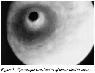
the patient in the lithotomy position and it started with a retrograde
urethrography using a fluoroscope with a “C-arm” at a 60o angle
relatively to the patient. A urethroscopy (Figure -1) was carried out
next for identification of the stenosis location (21F cystoscope). A 0.028F
guide wire was passed to the stricture and taken along the urethra as
far as the bladder. An Acucise catheter was passed over the guide wire
to the stenosis site. The metallic wire of the Acucise catheter was directed
toward the most anterior part of the urethra, at the 12 o’clock position
(Figure-2). At this time, the balloon (Figure-3) was inflated using 2.2
ml of contrast material; under fluoroscopy it was possible to observe
the constriction ring between the extremities of the balloon. The electrocautery
was then used after being regulated at 75 watts for 5 seconds. During
the incision fluoroscopy allowed the surgeon to follow the disappearance
of the constriction ring caused by the urethral stenosis. The balloon
was maintained inflated for 10 minutes for hemostatic purposes (Figure-4).
After that, the balloon was deflated, the catheter removed and the cystoscope
introduced into the bladder (Figure-5). An 18F urethral Foley catheter
was then inserted and left in place for a period of 10 days postoperatively.
Average time of procedure was 45 minutes. The patients were discharged
from the hospital on the same day of the procedure. During the monthly
follow-up data were collected from patient information as to their ability
to urinate and from the urinary flowmetry and, after a six-month period,
from the retrograde and urinary urethrocystography.


Success
of internal urethrotomy using an Acucise catheter for treatment of urethral
stenosis was assessed according to the following criteria: patient information
(improved, unchanged and worse) and urinary flowmetry (maximum urine flow:
good > 15 ml/s; fair: 10 to 15 ml/s and poor: < 10 ml/s). Retrograde
and urinary urethrocystographies carried out prior to the treatment and
six months after the procedure were compared and classified as: improved
(enlarged caliber of the stenosis and reduction of the dilation proximally),
unchanged and worse.


The variables were analyzed statistically
(Friedman and Mann-Whitney non-parametric test), and the rate considered
significant was 0.05 (p).
RESULTS
Concerning
pre and postoperative signs and symptoms, results of the internal urethrotomy
with Acucise evidenced improvement in 17 cases (85%), 2 cases remained
unchanged (10%) and in one condition grew worse (5%).
Considering the maximum urine flow, and
comparing pre and postoperative results one month after the internal urethrotomy
using the Acucise catheter, data revealed an average increase from 7.5
ml/s to 13.4 ml/s. During the six-month follow-up period the increase
in the maximum urine flow remained statistically stable. The maximum urine
flow was considered good (Max Q > 15 ml/s) in 6 patients (30%) and
fair (Max Q 10 to 15 ml/s) in 4 patients (20%). In 10 patients (50%) results
were poor (Max Q < 10 ml/s). However, in the patients that had not
been submitted to any previous treatment (7 patients) success rates were
good (57.1%) and fair (14.3%) in 71.4% of cases. Comparing outcomes in
patients without previous treatment with the stricture recurrence rates
a statistical tendency was observed to a less favorable evolution in the
operated cases (p = 0.0842). Results were also compared regarding extent
of the urethral stenosis. The 4 patients with a stricture up to 5 mm were
compared with the other 16 cases and no statistical difference could be
found in these two groups. Likewise, the results in patients with iatrogenic
urethral stenosis were compared with the non-iatrogenic cases and no statistical
difference was evidenced between the 2 groups. With regard to location,
in this study the stricture occurred most frequently in the area of the
bulbar urethra: 14 cases; as to other locations, penile or membranous,
no significant statistical difference was found.
Results of the internal urethrotomy with
Acucise were evaluated by a retrograde and urinary urethrocystography
performed 6 months following the procedure. The radiographic study revealed
improvement in 16 cases (75%); 4 cases remained unchanged (25%).
A noted postoperative complication was fever
in 3 patients after catheter removal; these patients were successfully
treated on an outpatient basis with norfloxacin. Outcome in these cases
was poor. Bleeding, edema or urinary incontinence was not observed.
The
development of more effective and lasting techniques for the treatment
of urethral strictures by means of minimally invasive procedures continues
to represent an important area of research.
Despite being widely employed, cold knife
internal urethrotomy as a treatment modality for urethral stenosis is
related to high rates of stricture recurrence (25) to the point that some
authors consider urethral dilation equivalent in efficiency to this procedure,
but with lower costs (26). In this sense, in order to improve results
of urethral dilations several authors have recommended the use of dilating
balloons; the feasibility of this method has been demonstrated, and it
is associated with high success rates (5,20).
The use of dilating balloons attached to
a cutting wire, or the Acucise catheter, for the treatment of urethral
stenosis was not previously described.
During the preliminary stage of the present
study the authors discussed potential risk of lesion to the sphincter
using the Acucise urethrotomy catheter in the area of the bulbomembranous
urethra. However, this complication has been considered a remote possibility
due to the diameter, limited to 24F, of the balloon when inflated. In
fact, in this study no patients developed urinary incontinence. As in
observations made by Giannakopoulos et al. (1997) (27) no complications
were observed with the Acucise catheter relative to the use of electric
current. The incision via metallic wire is linear, uniform and limited
to the 3 cm of the balloon length; tissue lesions beyond these limits
or in depth injuries are therefore unlikely. No occurrences of extravasation
of the irrigation fluid or bacteriemia were observed in the patients treated.
Fernandes et al. (1993) (28) considers that the use of balloons for the
treatment of urethral stenosis has the advantage of promoting a lower
absorption of fluids.
As for results, based on information given
by the patients, the authors could observe higher success rates (85%)
than the good and fair rates indicated by the urinary flowmetry (50%).
The urine flow measures used in the investigation of the low urinary tract
can reveal variations relative to urinary volume, sex, age and position
taken by the patient.
The six-month follow-up was considered too
short; however, most stricture recurrences take place within this period
(22,25,29,30). A study based on the pre and postoperative retrograde and
urinary urethrocystographies indicated results considered better in 15
cases (75%).
When results from the clinical evaluation
and flowmetry and urethrocystography were compared it was observed that
there was a greater correlation between the clinical and the radiographic
evaluation (88.2%) than between the clinical evaluation and the urinary
flowmetry (58.8%). There was also a small correlation between the urethrocystography
and the urinary flowmetry (66.6%).
In the present study only 7 patients (35%)
had not been submitted to any previous treatment for urethral stenosis;
in 5 of them good and fair results were achieved (71.4%). On the other
hand, in 13 patients with recurrent stenosis the evaluation by urinary
flowmetry revealed a failure rate of 65%. The medical literature reports
that patients with recurrent stenosis are also considered of worse prognosis
for endourologic treatment (21).
The cost of the Acucise catheter must be
taken into account. Each catheter was used at least 5 times and resterilized
with glutaraldeide, decreasing its cost. Furthermore, reduction in costs
as a whole is achieved with the overall smaller time of the procedure
and the lower rates of complications, and probable recurrence rates of
stenosis. As this is an initial study, the Acucise catheters were reused
after being sterilized. In the future, with the advances in technology,
less expensive adequate catheters can be developed. In this case, the
catheters will be used only once.
In fact, this is a first study using balloon
and a cutting wire with the advantage that this technique is very easy
to perform, safe because is a linear cut limited to 24F without irrigation,
and less traumatic to the urethra. The cost may be reduced with new developments
of the appropriate catheter.
Management of urethral stenosis by internal urethrotomy using the Acucise catheter proved to be a simple and safe procedure, and can be considered a new minimally invasive therapeutic option. The risks of complications are few and no bleeding neither incontinence was observed. This can be a new and beneficial therapeutic alternative. Further studies are necessary with longer follow-up and comparing it with other outpatient procedures.
REFERENCES
- Holm-Nielsen A, Shultz A, Moller-Pederson V: Direct vision internal urethrotomy: a critical review of 365 operations. Br J Urol, 56: 308-312, 1984.
- Attwater HL: The history of urethral stricture. Br J Urol, 15: 39-51, 1943.
- Devereux PC, Burfiel GD: Prolonged follow-up of urethral stricture treated by intermittent dilation. Br J Urol, 42: 321-329, 1970.
- Newman LH, Stone NN, Chircus JH, Kramer HC: Recurrent urethral stricture disease management by clean intermittent self-catheterization. J Urol, 144: 1142-1143, 1990.
- Levine LA, Engebrecht BP: Adjuvant home urethral balloon dilatation for recalcitrant urethral stricture. J Urol, 158: 818-821, 1997.
- Sachse H: Zur behandlung der harnrohrenstriktur: Die transuretrale schlizung unter sicht mit scharfem schmitt. Fortchr Med, 92: 12, 1974.
- Sacknoff EF, Kerr WS Jr: Direct vision cold knife urethrotomy. J Urol, 123: 492-496, 1980.
- Bulow H, Bulow U, Frohmuller HGW: Transurethral laser urethrotomy in man: preliminary report. J Urol, 121: 286-287, 1979.
- Smith JA Jr, Dixon JA: Neodymium:YAG laser for treatment of benign urethral strictures. J Urol, 131: 1080-1081, 1984.
- Becker HC, Miller J, Noske HD, Klask JP, Weidner W: Transurethral laser urethrotomy with argin laser: experience with 900 urethrotomies in 450 patients from 1978 to 1993. Urol Int, 55: 150-153, 1993.
- Milroy EJ, Chapple CR, Cooper JE, Eldin A, Wallsten H, Seddon AM, Rowles PM: A new treatment for urethral strictures. Lancet, 1 (8600): 1424-1427, 1988.
- Yachia D, Beyar M: Temporary implanted urethral coil stent for the treatment of recurrent urethral strictures: a preliminary report. J Urol, 146: 1001-1004, 1991.
- Arap S, Lucon AM, Mitre AI, Wroclawski ER, Glina S, Shan CJ, de Góes GM: Correção do estreitamento completo de uretra bulbar e membranosa através de uretroplastia termino-terminal. Rev Hosp Clin Fac Med São Paulo, 41: 31-35, 1986.
- Jordan GH: Anterior urethral reconstruction: concepts and concerns. Contemp Urol, 10: 80-96, 1998.
- Orandi A: One-stage urethroplasty: 4 years follow-up. J Urol, 107: 977-980, 1972.
- Quartey JKM: One-stage penile/preputial cutaneous island flap urethroplasty for urethral stricture: a preliminary report. J Urol, 129: 284-287, 1983.
- Mitre AI, Arap S, Lucon AM: Preputial island flap in extensive urethral stricture repair. World J Urol, 10: 94-99, 1992.
- Mundy AR: Early experience with use of bucal mucosa for substitution urethroplasty. Br J Urol, 77: 2A, Supplement 1, (Abstract), 1996.
- Barbagli G, Selli C, Tosco A: Reoperative surgery for recurrent strictures of the penile and bulbous urethra. J Urol, 156: 76-77, 1996.
- Fishman IJ: Experience with a hydraulic balloon urethral dilator for office and self dilation. J Urol, 147: 287A, 1992.
- Jordan GH, Schlossberg SM, Devine CJ: Surgery of the Penis and Urethra. In: Walsh PC, Retik AB, Vaughan ED Jr (eds.). Campbell’s Urology. 7th ed. Philadelphia, Saunders, vol.3, pp 3316-3394, 1998.
- Pansadoro V, Emiliozzi P: Internal urethrotomy in the management of anterior urethral strictures: long-term follow-up. J Urol, 156: 78-79, 1996.
- Stormont TJ, Suman VJ, Oesterling JE: Newly diagnosed bulbar urethral strictures: etiology and outcome of various treatments. J Urol, 150: 1725-1728, 1993.
- Ziprin P, Wheeler J, Davies G, Stepherson TP: The long-term follow-up of urethroplasty for non-traumatic urethral strictures. Br J Urol, 77: 2A, Supplement 1, (Abstract), 1996.
- Gibod L, Le Portz B: Endoscopic urethrotomy: does it live up to its promises? J Urol, 127: 433-435, 1982.
- Webster GD: Endoscopy and dilation of urethral defects and strictures (Editorial). J Urol, 157: 102-103, 1997.
- Giannakopoulos X, Grammeniatis E, Gartzios A, Tsoumanis P, Kammenos A: Sachse urethrotomy versus endoscopic urethrotomy plus transurethral resection of the fibrous callus (Guillemin’s technique) in the treatment of urethral stricture. Urology, 49: 243-247, 1997.
- Fernandez AF, Esteban AR, Banuelos MJR, Gil FJ, Franco MR, Ardanza A, Otero MG: Dilatación hidráulica de estenosis de uretra bajo control ecográfico. Un nuevo enfoque. Arch Esp Urol, 46: 40-42, 1993.
- Walter PC, Parson CL, Schmidt JD: Direct vision internal urethrotomy in the management of urethral stricture. J Urol, 123: 497-499, 1980.
- Seenkamp JW, Heyns CF, Kock MLS: Internal urethrotomy versus dilation as treatment for male urethral strictures: a prospective, randomized comparison. J Urol, 157: 98-101, 1997.
_______________________
Received: January 31, 2001
Accepted after revision: July 19, 2001
________________________
Correspondence address:
Dr. Ricardo Jordão Duarte
Caixa Postal 11273-9
São Paulo, SP, 05422-970, Brazil
Fax: + + (55) (11) 3064-7013
E-mail: ricjordao@uol.com.br
EDITORIAL COMMENT - I
This
article represents an original approach to urethral stenosis. However,
there are some controversial aspects like the fluoroscopic control of
the Acucise position and its relation to the urethral sphincter (membranous
and bulbar urethra).
Cold knife urethrotomy has a high recurrence
rate in cases of intense scar tissue down the spongy tissue. Also the
urethral balloon dilation is not able to solve this problem. Therefore,
how the combination of urethral dilation and electrocautery will work
across the periurethral scar tissue is indeed not clear.
Another intriguing point is the reuse of
the Acucise for so many times without any technical problem with the device.
The last but not the least is the high price
of the Acucise catheter, even with repeated sterilization and reuse of
the device.
Dr. Nelson
Rodrigues Netto, Jr.
Professor and Chairman of Urology
University of Campinas, Unicamp
Campinas, São Paulo, Brazil
EDITORIAL COMMENT - II
In
this study, the authors describe their technique of Acucise incision of
benign urethral strictures. Their results were fair overall, but do provide
an interesting use of the Acucise device.
Following Acucise incision, 85% of the patients
reported clinical improvement in their voiding parameters. By uroflometry,
50% of patients had either a good or fair improvement. Moreover, 75% of
the patients demonstrated radiological improvement following Acucise incision.
There were no significant complications in the patients treated with the
Acucise device. The authors conclude that the Acucise catheter can provide
a simple and safe method of treating benign urethral strictures.
The authors comment briefly on the cost
of the Acucise catheter. While the authors have decreased the cost of
the device by repeat sterilization and reuse of the device, this technique
would not be allowed in many operating rooms across the world. In fact,
the Acucise device is quite expensive costing greater than $1500 US and
therefore if only single use were allowed, the procedure would be cost
prohibitive.
My overall concern of this particular study
is the expense and potential problems related to reuse of a clearly disposable
device. It is one thing to reuse balloons or catheters, which do not rely
on electrical current for their proper performance. However, reuse of
the Acucise with repeat sterilization may indeed cause problems with the
electrical current and the cutting capabilities of the device.
Dr. Glenn
M. Preminger
Professor of Urological Surgery
Duke University Medical Center
Durham, North Caroline, USA
EDITORIAL COMMENT - III
This
study is the first report of a series of urethral strictures treated with
a cutting balloon catheter (Acucise catheter). Of the 20 patients treated,
of whom 13 had undergone prior treatment for urethral stricture, 85% reported
clinical improvement but by objective urinary flowmetry the results were
considered good in only 30%. The authors’ conclusion was favorable
towards the technique.
Although in some settings one-time use medical
devices are resterilized and reused, this is generally limited to devices
with simple contours and without complex interfaces. Wires, catheters,
and dilators can likely be resterilized safely, but to apply resterilization
to a cutting balloon catheter, with its complex shape and internal surfaces,
might expose the surgeon and patient to significant risk of failure of
the sterilization or the equipment. This practice should not be recommended
without further testing. In addition, the final contentions that the cutting
balloon catheter provides shorter procedure time, lower complication rate,
and lower rate of recurrence are not at all supported by data in the manuscript.
An additional disadvantage of the technique described is the need for
fluoroscopy, which adds considerably to the instrumentation burden of
the urethrotomy. In summary, the authors have not provided data that are
in any way suggestive that internal urethrotomy with a cutting balloon
catheter would be superior or even equivalent to other standard techniques.
Dr. J.
Stuart Wolf, Jr.
Professor of Urological Surgery
University of Michigan
Ann Arbor, Michigan, USA