PEYRONIES DISEASE
TULIO M. GRAZIOTTIN, JULIO RESPLANDE, SHAHRAM S. GHOLAMI, TOM F. LUE
Department of Urology, University of California School of Medicine, San Francisco, California, USA
ABSTRACT
Peyronie’s disease can be considered an exaggeration of the wound repair process and is linked to penile trauma. Although a better understanding of the pathophysiology of the disease was achieved recently, the best alternative to treat the patients remains a dilemma. Pain, shortening, plaque and erectile dysfunction are clinical characteristics of this disease, and solution to patients complains should steer the therapy. Non-surgical treatment is offered to patients with pain, plaque or deformity of less than one year. Surgical treatment should be delayed until the process become stabilized. Surgery is indicated in patients with stable and severe deformity of more than one year of duration and for patients who have penile shortening, narrowing or indentation, or a combination of the above that preclude normal sexual intercourse. If the patient is also impotent and fails to respond to non-surgical treatment, we recommend a penile prosthesis insertion.
Key words:
penis; penile induration; Peyronie’s disease; impotence
Braz J Urol, 27: 326-340, 2001
INCIDENCE AND BACKGROUND
Peyronie’s
disease (indurato penis plastica) is characterized by the formation of
fibrous plaques within the tunica albuginea. In 1561, Fallopius first
reported the disease, which later bore the name of the surgeon to King
Louis XV of France, Francois Gigot De la Peyronie, who popularized the
disease entity in 1743. Peyronie’s disease is estimated to affect
0.4 to 3.5% of adult male patients worldwide (1-4). Autopsy studies have
suggested a much higher incidence of subclinical plaques or fibrotic lesions
noted in the penis. These plaques impede expansion of the tunica during
erection resulting in penile bending. In some extreme cases, these plaques
may induce a collar-like or an hourglass-like appearance in the erect
penis with dense calcified areas.
Peyronie’s disease has been reported
to occur in association with Dupuytren’s contractures, plantar fascial
contractures, tympanosclerosis as well as trauma, urethral instrumentation,
diabetes, gout, Pagets’ disease, and the use of beta- blockers (5).
This condition can occur in a familial pattern (6). There is a 10 to 40%
chance that the descendent of a patient with Dupuytren’s contracture
will develop that problem, and a 15% chance that a man so afflicted will
develop Peyronie’s disease.
Contemporary thinking suggests Peyronie’s
disease represents a localized aberration of the wound healing process.
Pathologically, Peyronie’s plaques begin with fibrin deposition and
end up like scars. Clinical data, anatomical pathology, and bioengineering
analysis all implicate trauma as an initiation factor in Peyronie’s
disease (7,8). Fibrin deposition is recognized as one of the initial consequences
of microvascular injury, and it may be the precursor to Peyronie’s
plaque formation (9).
The search for a genetic link for Peyronie’s
disease has yet to identify a genetically predisposed population. However,
there are reports associating this condition and Paget’s disease
of the bone, Dupuytren’s contracture, and specific HLA subtypes (6,10-12).
Studies of Peyronie’s patients have implicated an autoimmune component
(13,14). It is likely that a certain proportion of men in this age group
respond to mechanical tunical stress and microvascular trauma with an
aberrant or hyperactive wound healing response (7,8,15,16). Thus, there
may be a subpopulation whose genetic background is such that response
to wound healing predisposes development of Peyronie’s plaques.
PATHOLOGY AND BASIC SCIENCE
Pathologically,
Peyronie’s disease is associated with perivascular round cell infiltration,
which can be found within the tunica albuginea (17). Fibrin deposition,
presumably from microvascular injury, has also been found in relation
to Peyronie’s plaques, but not in normal or scarred tunica from individuals
without Peyronie’s disease (9). Plaques consist of dense collagenous
connective tissue with reduced and fragmented elastic fibers. In about
one third of patients, radiologically or sonographically demonstrable
dystrophic calcifications are present (18). The scar tissue of Peyronie’s
disease contains excessive amounts of type III collagen, which renders
it particularly responsive to the wound contraction process (19).
One of the most likely causes of Peyronie’s
disease may be repeated tunical mechanical stress and microvascular trauma.
Excessive bending during erection or blunt trauma to the erect penis may
result in bleeding into the subtunical spaces or tunical delamination
at the point where the septum integrates into the inner circular layer
of the tunica albuginea (7,8,15). Such microvascular trauma may come from
sexual intercourse; either with the woman on top or an accident during
penetration where the man misses the vagina and injures the penis. Microvascular
trauma or subtunical bleeding can result in fluid and fibrinogen in the
subtunical layers. The resulting fibrin deposits may be key in the initiation
of a wound healing response, which encompasses pain, hematoma, and subsequent
inflammatory response with recruitment of macrophages and neutrophils
(9,16,20). These cells, in response to clot formation, release a variety
of cytokines, autocoids and vasoactive factors, which may precipitate
a fibrotic reaction. The unique anatomy of the tunica albuginea with its
multiple sublayers of dense fibrous tissue and hypovascularity may «trap»
the inflammatory reaction. This may prolong the process to months or years
and therefore foster the formation of Peyronie’s plaque.
There has yet to be a detailed examination
of the cell types involved in the pathogenesis of Peyronie’s disease.
Transforming growth factor-b1 has a pleotropic effect on fibroblast function
by increasing transcription and synthesis of collagen, proteoglycans and
fibronectin while also increasing synthesis of tissue inhibitors of collagenase,
which prevents connective tissue breakdown. A role for TGF-b1 has been
proposed in the pathogenesis of Peyronie’s disease (21-23). Finally,
in the later stages of healing, the connective tissue is remodeled by
specific collagenases and proteases. In Peyronie’s disease, defects
in overproduction of collagen and other tissue remodeling mechanisms may
result in an inability to resolve the injury and in plaque formation.
NATURAL HISTORY
AND
PRESENTATION
In
most cases, onset is associated with an active phase, consisting of painful
erections, a palpable plaque and bending of the penis. Up to a third of
patients with Peyronie’s disease present with painless curvature.
Whether the onset of deformity associated with the active phase is gradual
or sudden, pain usually resolves and the pathologic process itself seems
to stabilize after 12 to 18 months. A relatively quiescent secondary phase
follows, which is characterized clinically by painless stable deformity,
and pathologically by mature scar. Earlier report characterized Peyronie’s
disease as a process of gradual spontaneous resolution (24). More recent
data do not support the above conclusion (25). Painful erection almost
always resolves with time; penile deformity usually does not.
The reported incidence of erectile dysfunction
in Peyronie’s disease is variable. Bystrom & Rubio reported that
52% of 106 patients had coital difficulties and 17% had poor penile rigidity
distal to the plaque (26). However, only 8% of patients described coital
difficulties at the initial presentation suggesting that this was probably
a late feature of the disease. Stecker & Devine found abnormal nocturnal
penile tumescence in 29% of patients with Peyronie’s disease with
suspected organic impotence although in only 5% of patients could the
Peyronie’s disease plaque be the sole cause of the dysfunction (27).
Other series have reported an incidence of erectile dysfunction of 19%
(28). Amin et al. discovered that out of 208 patients investigated routinely
by color Doppler ultrasound for erectile dysfunction, 20% had undiagnosed
Peyronie’s disease (29).
It is clear, therefore, that erectile dysfunction
in Peyronie’s disease is common and is usually due to 1 or more of
4 factors; psychological or performance anxiety, severe penile deformity,
a flail penis, or impaired penile vascular function (30). The deformity
of the penis may so severe that penetration is difficult, painful or impossible.
This is more likely to occur if the deformity is in a ventral or lateral
direction, where deviation from the normal angle of vaginal entry is maximal.
There is a small group of patients with extensive Peyronie’s disease
who have circumferential plaques and a degree of cavernous fibrosis causing
a flail penis. Tumescence is absent from this segment and if extensive
it may result in a hinge effect and an unstable penis. Erectile dysfunction
may be due to concomitant vascular disease that occurs in 30% of patients
with Peyronie’s disease (31) or to veno-occlusive dysfunction (32,33).
Most studies have used both color Doppler ultrasound and cavernosometry
to investigate the impaired erection in Peyronie’s disease. Lopez
& Jarow showed that out of 76 patients, 36% had arterial disease and
59% had veno-occlusive dysfunction (34). Others have also suggested there
is a mixture of arteriogenic and venogenic factors (33,35). It is thought
that the venous leakage may occur through the emissary veins that pass
near the Peyronie’s plaque into the dorsal vein of the penis. The
reduced compliance of the tunica albuginea of the plaque prevents the
normal compression of these veins during erection and therefore does not
compress the venous channels.
Peyronie’s disease typically presents
with one of the following 4 complaints: painful erection, penile deformity
or shortening during erection, presence of a plaque or induration on the
shaft of the penis, or erectile dysfunction. Almost all patients have
either a well-defined plaque or an area of induration that is palpable
on physical examination which 38-62% of the patients are unaware of (26,28,36,37).
The plaque is usually located on the dorsal surface of the penis with
a corresponding dorsal penile deformity. Lateral and ventral sited plaques
are not as common but result in more coital difficulty, as there is a
greater deviation from the natural coital angle. Penile pain may be present
with erection or during sexual intercourse. The pain is not severe in
nature but may interfere with sexual function. Spontaneous improvement
in pain usually occurs as the inflammation settles.
DIAGNOSIS
Peyronie’s
disease is usually apparent by patient history and physical examination.
The medical history should include time and mode of onset (sudden or gradual),
course of disease (stable or progressive), history of penile surgery,
urethral instrumentation or trauma, medication or drug abuse and family
history of Peyronie’s disease or Dupuytren’s contracture. Risk
factors for erectile dysfunction should also be obtained.
A detailed psychosexual history should include
penile rigidity during erection, shortening, induration, hourglass constriction,
or pain with or without erection. Other important information should also
be determined such as ability to have intercourse, adequacy of erection
(rigidity and duration), frequency of intercourse, libido, and psychological
impact. A photograph of patient’s erect penis to identify the extent,
direction, and character of erectile distortion is helpful.
Examination of the penis is facilitated
by its gentle stretching. This will help identify the size, location and
consistence of plaques which may be helpful in determine the stage of
the disease and monitoring its progression. The patient should also be
examined for the presence of Dupuytren’s or plantar fascial contractures.
Further diagnostic studies should include photography or drawing of the
erect penis after intracavernous injection or vacuum erection device.
The stretch length of the penis should also be documented.
Many patients have mild symptoms and reassurance,
particularly that the palpable lump is not cancer, is all that is necessary.
The majority of patients with Peyronie’s disease may be managed without
vascular investigation. Penile curvature, especially in young patients,
may cause severe psychological distress, which may need to be corrected.
Patients usually give an accurate description of their deformity to within
10-20º (38,39). However, when planning a surgical correction of the
deformity, documentation of the deformity during erection either by intracavernosal
injection of a vasoactive agent or a vacuum device is very helpful.
When the site and size of the Peyronie’s
plaque needs to be assessed, ultrasound usually will suffice and is particularly
helpful in monitoring the progress of medical treatment (2). Patients
who also complain of impaired erections, further evaluation is essential.
Color duplex sonography performed before and after intracavernous injection
of a vasoactive agent allows for assessment of the structure of the corpus
cavernosum, tunica albuginea and the cavernous arterial and venous function
(40). Color duplex ultrasound is also excellent in detecting collateral
arterial connections between dorsal, cavernosal and spongiosal arteries.
Dynamic infusion cavernosometry can be used as an adjunct to duplex ultrasound
to confirm the diagnosis of veno-occlusive dysfunction (41). Finally,
in rare cases MRI can be used for detailed evaluation of penile anatomy
prior to surgical intervention.
During the evaluation of patients with Peyronie’s
disease, other causes of bending and induration of the penis must also
be considered. These differential diagnoses include: congenital curvature
of the penis, chordee with or without hypospadias, penile dorsal vein
thrombosis, cavernosal fibrosis secondary to local trauma, leukemic infiltration
of the corpora cavernosa, ventral curvature secondary to urethral instrumentation,
benign or malignant primary or secondary tumors, late syphilitic lesion,
and penile infiltration with lymphogranuloma venereum.
NON-SURGICAL TREATMENT
The
initial approach to treatment should be conservative. Many patients with
a minor curvature and normal erectile function can be given reassurance
with no invasive diagnostic tests or treatments. Medical management is
indicated for patients with a greater degree of curvature or symptoms.
Non-surgical treatments can be divided into systemic, local, or intra-lesional
therapies.
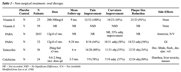
Vitamin E is a commonly used oral therapy
for Peyronie’s disease. In 1948, Scott & Scardino (42) reported
a beneficial effect on treatment of 23 patients with vitamin E, a tocopherol
with antioxidant properties. The proposed dose was 200 to 300 mg per day.
In 1990, Gelbard et al. compared the effects of the vitamin E treatment
and natural progression of the Peyronie’s disease (25). They noted
no significant differences between the 2 groups with respect to pain,
bend, ability for intercourse, and over-all perception of disease progression.
At this time however, vitamin E continues to be the primary mode of treatment
due to its mild side effects and low cost, despite the lack of controlled
study showing its benefits.
Potassium aminobenzoate (Potaba) has been
studied for the oral treatment of Peyronie’s disease (43,44). Its
mechanism of action, though not well understood, is decreasing fibrogenesis
by decreasing serotonin through increased tissue utilization of oxygen
and increased activity of monoamine oxidase. The most extensive review
of this therapy of 2653 patients has found it to be successful in 57%
of patients (45). The dosage is 20 grams a day for 3 months. Due to Potaba’s
modest results in literature, relatively high cost, and high side effect,
enthusiasm for the use of this medication is cautiously guarded.
Several small, uncontrolled, short-term
reports suggest mild to moderate benefit using oral Tamoxifen (46). It
has been suggested that Tamoxifen facilitates the release of transforming
growth factor-beta (TGF-b) from fibroblasts (47). TGF-b has been shown
to play a central role in regulating immune response, inflammation and
tissue repair by deactivating macrophages and T lymphocytes. Tamoxifen
results in a reduced inflammatory response and, therefore, diminished
angiogenesis and fibrogenesis (48). Tamoxifen dosage is 20 mg twice a
day with minimal reported side effects of gastrointestinal distress and
alopecia.
Colchicine therapy is the most recently
reported oral therapy for Peyronie’s disease. Akkus et al., in an
uncontrolled study, showed a decrease in plaques size and an improvement
in penile curvature in approximately 50% of the 24 patients they treated
(49). The main side effect of colchicine is gastrointestinal upset with
diarrhea reported in 33% of subjects. Recommended dosing is 0.6 to 1.2
mg daily during the first week of treatment followed by an increase to
1.8 to 2.4 mg for three months. Colchicine is an anti-microtubular agent,
which inhibits the proliferation of inflammatory cells and fibroblasts.
It can also increase collagenase activity and reduce collagen synthesis
(50-52). We currently use colchicine as our first line agent in the treatment
of acutely acquired Peyronie’s disease.
Based upon anti-inflammatory properties,
as well as decreased collagen synthesis by unclear mechanisms, steroids
have been used as an intralesional therapy for Peyronie’s disease.
Several short-term non-controlled subjective studies have reported good
responses using various steroids (37,53-55). Nevertheless, we do not recommend
intralesional steroids in the treatment of Peyronie’s disease. This
type of therapy has many local side effects including local tissue atrophy
and skin thinning while offering only an inconsistent improvement in well-established
curvature. Steroid injections ultimately make surgery more complex due
to the difficulty in subsequent separation of tissue planes.
In a randomized placebo controlled study,
purified intralesional clostridial collagenase was shown to have some
benefit over placebo for mild to moderate degrees of Peyronie’s disease.
In more severe curvature however, the response to treatment was not statistically
significant (56). Its mechanism of action is via altering collagen content
of penile plaque. This drug is currently being evaluated for approval
for use in the United States of America.
Orgotein, an anti-inflammatory metalloprotein
with pronounced superoxide dismutase activity, has been used by several
groups in Europe (57-60). Reports of subjective benefits are as high as
80-90%. It is not available in USA and has been taken off the market in
several European countries due to its significant toxicity.
The potential use of interferons as an intralesional
therapy for Peyronie’s disease has been demonstrated (61). In fibroblasts
derived from Peyronie’s plaques, the addition of interferons decreased
the rate of proliferation in a dose-dependent fashion, decreased the production
of extracellular collagen, and increased the production of collagenase.
Several clinical trials of intralesional interferon for Peyronie’s
disease have been published (62-64). Objective improvement in deformity
was considered small with mean improvement of 20 degrees. Patients with
small plaques (< 4 cm) were more likely to have a better response.
All patients experience brief influenza-like side effects.
The calcium channel blocker verapamil was
first reported as an intralesional therapy by Levine et al. and Rehman
et al. (65-67). Penile shaft narrowing, decreased in 100% of patients,
but curvature improved in only 42%. Fifty-eight percent reported that
their sexual performance had improved. Overall, 83% noted that the disease
had arrested or improved with no recurrence of symptoms or deformity within
the eight months follow-up period. Verapamil and other calcium channel
blockers affect cytokine expression associated with early phases of wound
healing and inflammation and increase the proteolytic activity of collagenase
(68). Matrix remodeling is enhanced by human fibroblasts in burn scars
and vascular smooth muscle cells in vitro (49,69). Goals of this treatment
are to stabilize disease process and reactivate a more normal remodeling
process, yielding gradual improvement in deformity. Multiple doses, 10
mg injected every 2-4 weeks for 12 weeks, are given over time. The main
side effect is ecchymosis. This is currently the most frequently used
intralesional therapy for Peyronie’s disease.
Iontophoresis was examined as a means of
enhancing topical delivery of verapamil (10 mg) and dexamethasone (4 mg)
with a local electric field in 15 patients with Peyronie’s disease.
At 5 months follow-up, penile pain resolved in 66%, curvature improved
in 53% and plaque size reduced or softened in 40% of cases (70). Local
penile lithotripsy has also been proposed as topical therapy for Peyronie’s
disease with limited numbers of patients reporting subjective results
(71). The rationale for this approach is not known. Topical verapamil
cream has also been advocated for Peyronie’s disease. However, we
cannot comment on its use because of the lack of analyzable data.
Overall, therapeutic advances in Peyronie’s
disease have not resulted in a reliable cure. This may be due to an incomplete
understanding of the basic pathophysiology of this disease and the lack
of an animal model for study. Recent advances in the understanding of
disorders of wound healing have allowed forward strides in the understanding
of this disease and offer new therapies, such as the injection of calcium
antagonists and interferon. Recent reports of the involvement of TGF-b
in human Peyronie’s disease and the induction of Peyronie’s
like condition by injecting TGF-b into the rat’s tunica albuginea
may help provide a new strategy in combating the disease.
SURGICAL TREATMENT
The
indications for surgical correction include: severe curvature, narrowing,
or indentation of more than one year’s duration, sexual difficulty
or partner discomfort because of deformity, or severe penile shortening.
Surgical correction of penile curvature is reserved for those who fail
conservative measures. There is a considerable variation in the deformity
that makes penetration difficult. In young men particularly with congenital
deformities the bend causes more psychological distress than physical
disability and it may be necessary to correct curvature as little as 20-30
degrees. In contrast, an older man with a stable relationship and partner
is better able to cope with a more severe degree of deformity. Of note,
a ventral deformity causes more difficulty in vaginal penetration than
a dorsal or lateral one.
Prior to surgery, a detailed evaluation
of penile vascular and erectile function is highly recommended. Reconstructive
surgery is not recommended in the acute phase of the disease. In the past,
many penile implants have been performed in patients with normal penile
rigidity to treat severe curvature. In the current era, penile implants
should be reserved for Peyronie’s patients who have severe erectile
dysfunction that does not respond to non-surgical erectile dysfunction
therapy. The surgical treatments for penile curvature are classified into
3 different categories: tunical shortening procedures, tunical lengthening
procedures, and prosthetic procedures.
Shortening procedures are reconstructive
techniques performed on the convex surface of the penis at the site opposite
to the penile plaque. These procedures are the easiest to perform and
require the least expertise. Patient selection is extremely important.
Shortening procedures are most appropriate for patients with useful erections,
adequate penile length, and without hourglass deformity. Reed Nesbit first
described the correction of congenital erectile deformities by shortening
the opposite side of the penis by the excision of an ellipse of tunica
albuginea (72). The Nesbit technique was re-introduced for Peyronie’s
disease in 1979 (73). In a review of 359 men operated upon between 1977
and 1992, 295 (82%) had good results and were able to have intercourse
after correction with this technique (74). A literature review has confirmed
these favorable results (30). The overall results of the Nesbit procedure
have improved in the operations performed after 1985. This is thought
to be due to better patient selection (74). With increasing time after
the operation, there is a decrease in satisfaction with the results of
the Nesbit procedure. The most common complication of this procedure as
with all shortening procedures is loss of penile length. This complication,
however, does not preclude the great majority of men from having sexual
intercourse. Others complications reported include erectile dysfunction,
penile hematoma, penile narrowing or indentation, urethral injury, herniation,
suture granuloma, numbness and phimosis (74).
A modification of the Nesbit procedure was
described by Lemberger et al. (10) and further refined by Yachia (75).
The approach is similar to the Nesbit procedure but instead of removing
an ellipse of tunica, a long longitudinal or multiple smaller longitudinal
incisions are made in the corpora cavernosa. These incisions are then
closed horizontally in a Heineke-Mikulicz fashion in order to correct
the angle of penile curvature. Many authors claim a high percentage of
good results with this technique reporting satisfaction rates between
79-95% (10,75-81). The complications of this procedure are similar to
those reported for the Nesbit technique.
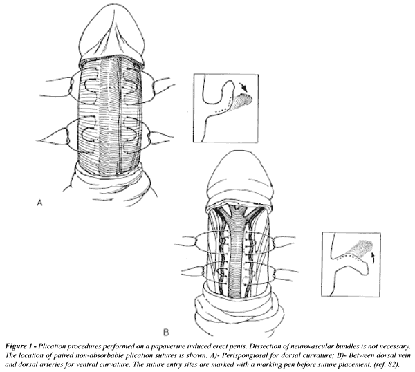
Wedge resection or incision of the tunica
requires extensive dissection of the neurovascular bundle or the corpus
spongiosum. A simplified approach for correcting penile curvature is to
perform plication on an erect penis produced by intracavernous injection
of papaverine or alprostadil (82). Plication is a simple, outpatient procedure,
which takes approximately 30 minutes under local anesthesia. Tissue incisions
or removal or dissection of the neurovascular bundle or urethra is not
necessary (Figure-1). Some authors described high recurrence rates and
poor results with prolonged follow-up. The literature reports significant
variation in results ranging from 38-100% satisfactory results (83-87).
Gholami et al. recently reported 98% satisfaction rate with over 90 patients
followed for one to five years (88). Complications of plication surgery
include loss of penile length, phimosis, penile narrowing, erectile dysfunction,
suture granuloma and palpable suture lumps on penis (85,86,89).
Tunical lengthening procedures use reconstructive
techniques to correct penile curvature while restoring the length of the
curved, shortened penis. This surgery is performed on the concave, diseased
side of the penis and requires plaque excision or incision with grafting.
Lengthening of the tunica with graft placement is indicated in patients
with severe curvature resulting in a shortened and deformed penis with
penile narrowing or hourglass deformities. Lengthening can also be done
on patients that present with recurrent curvature after other surgical
procedures. These procedures are the most difficult to perform and require
the greatest expertise. Few investigators have experience with more than
50 patients. The replacement of diseased tunica albuginea in Peyronie’s
disease was largely unsuccessful until the use of dermal graft first described
by Bystrom & Rubio (26) in Scandinavia and by Devine & Horton
(90) in the United States. Subsequently, many autologous tissues (temporalis
fascia, dura mater, tunica vaginalis and dorsal or saphenous vein), cadaveric
tissue (dermis, fascia or pericardium) and synthetic materials (Dacron
and Gortex) have been used with different results. Excision of the plaque
has been the standard approach. However, it is now known that the pathologic
process of Peyronie’s disease extends far beyond the plaque and removing
a large area of tunica albuginea may impair erectile function (90-92).
A review of the literature shows that there is great variability in the
outcome of plaque excision with the most common problem being erectile
dysfunction (26,32,92). Due to the high incidence of erectile dysfunction,
contracture of the graft, late recurrence and poor long terms results,
the excision of the plaque and grafting is less performed today (93).
In 1991, Gelbard & Hayden (94) proposed
plaque incision and grafting rather than excision, decreasing some of
the complications of excising the tunica albuginea. The less the tunica
and its underlying erectile tissue are altered, the better the postoperative
erectile rigidity. No perfect graft material has been described for replacing
the diseased tunica albuginea. Lue et al. report on their experience with
the use of saphenous vein graft in 112 patients with Peyronie’s disease
(Figure-2). Successful straightening was accomplished in 95% of the patients
with 13% of potent men complaining of decrease in erectile function (95,96).

Patients with Peyronie’s disease and
impotence, which does not respond to medical management, are usually treated
with penile prostheses with or without excision or incision of the plaque.
Literature review (91,93,97,98) shows excellent results provided men have
realistic expectations. Although there may be some intrusion of the plaque
on the corporal bodies, this usually does not cause any difficulty in
the implantation. Historically, a penile prosthesis would be performed
on any patient who had a penile deformity but without erectile dysfunction.
With advances in the medical treatment of erectile dysfunction, prosthesis
surgery is now reserved as the final treatment option or in those with
severe erectile dysfunction. In most patients with mild to moderate curvature,
insertion of a penile prosthesis tends to straighten the penis and no
additional procedures are necessary. However, in cases of severe deformity,
incision and grafting the diseased tunica albuginea with synthetic material
grafts or cadaveric tissue may be necessary during prosthesis placement.
The use of operative molding of the penis over the prosthesis is helpful
in order to give good correction of the deformity (99).
CONCLUSION
Peyronie’s
disease remains one of the most perplexing diseases in urology. With continued
basic research in wound healing and scarring, our understanding and management
of this frustrating disease will improve. Initial treatment of Peyronie’s
disease should be conservative with expectant therapy and medical management.
Once the penile curvature and plaque have stabilized, patients with severe
deformity may be offered surgery depending on their symptoms and complaints.
Patient selection is key to proper treatment. Less experienced surgeons
should limit themselves to medical management or simple surgical management
of the disease including plication or Nesbit procedures. Tunical lengthening
procedures or complicated penile prosthesis should be reserved for surgeons
with familiarity and expertise in this type of reconstruction. Education
of the pathogenesis and natural history of the disease will allow the
patient and his partner to make an informed decision in regards to his
treatment options and expected outcomes.
_____________________________________
Drs. T.M. Graziottin and Respland are fellows
sponsored by CAPES, Brazil

REFERENCES
- Carson C, Jordan G, Gelbard M: Peyronie’s disease: new concepts in etiology, diagnosis and treatment. Contemp Urol, 11: 44, 1999.
- Lindsay MB, Schain DM, Grambsch P, Benson RC, Beard CM, Kurland LT: The incidence of Peyronie’s disease in Rochester, Minnesota, 1950 through 1984. J Urol, 146: 1007-1009, 1991.
- Schwarzer U, Klotz T, Braun M, Wassmer G, Englemann U: Prevalence of Peyronie’s disease: results of an 8,000 men survey. J Urol, 163: 167, 2000.
- Smith BH: Subclinical Peyronie’s disease. Am J Clin Pathol, 52: 385-390, 1969.
- Carrieri MP, Serraino D, Palmiotto F, Nucci G, Sasso F: A case-control study on risk factors for Peyronie’s disease. J Clin Epidemiol, 51: 511-515, 1998.
- Nyberg LM Jr., Bias WB, Hochberg MC, Walsh PC: Identification of an inherited form of Peyronie’s disease with autosomal dominant inheritance and association with Dupuytren’s contracture and histocompatibility B7 cross-reacting antigens. J Urol, 128: 48-51, 1982.
- Devine CJ Jr., Somers KD, Jordan SG, Schlossberg SM: Proposal: trauma as the cause of the Peyronie’s lesion. J Urol, 157: 285-290, 1997.
- Jarow JP, Lowe FC: Penile trauma: an etiologic factor in Peyronie’s disease and erectile dysfunction. J Urol, 158: 1388-1390, 1997.
- Somers KD, Dawson DM: Fibrin deposition in Peyronie’s disease plaque. J Urol, 157: 311-315, 1997.
- Lemberger RJ, Bishop MC, Bates CP: Nesbit’s operation for Peyronie’s disease. Br J Urol, 56: 721-723, 1984.
- Lyles KW, Gold DT, Newton RA, Parekh S, Shipp KM, Pieper CF, Krishan R, Carson CC: Peyronie’s disease is associated with Paget’s disease of bone. J Bone Miner Res, 12: 929-934, 1997.
- Ralph DJ, Schwartz G, Moore W, Pryor JP, Ebringer A, Bottazzo GF: The genetic and bacteriological aspects of Peyronie’s disease. J Urol, 157: 291-294, 1997.
- Schiavino D, Sasso F, Nucera E, Alcini E, Gulino G, Milani A, Patriarca G: Immunologic findings in Peyronie’s disease: a controlled study. Urology, 50: 764-768, 1997.
- Stewart S, Malto M, Sandberg L, Colburn KK: Increased serum levels of anti-elastin antibodies in patients with Peyronie’s disease. J Urol, 152: 105-106, 1994.
- Devine CJ Jr., Horton CE: Peyronie’s disease. Clin Plast Surg, 15: 405-409, 1988.
- Diegelmann RF: Cellular and biochemical aspects of normal and abnormal wound healing: an overview. J Urol, 157: 298-302, 1997.
- Davis CJ Jr.: The microscopic pathology of Peyronie’s disease. J Urol, 157: 282-284, 1997.
- Gelbard MK: Dystrophic penile calcification in Peyronie’s disease. J Urol, 139: 738-740, 1988.
- Ehrlich HP: Scar contracture: cellular and connective tissue aspects in Peyronie’s disease. J Urol, 157: 316-319, 1997.
- Van de Water L: Mechanisms by which fibrin and fibronectin appear in healing wounds: implications for Peyronie’s disease. J Urol, 157: 306-310, 1997.
- El-Sakka AI, Hassan MU, Nunes L, Bhatnagar RS, Yen TS, Lue TF: Histological and ultrastructural alterations in an animal model of Peyronie’s disease. Br J Urol, 81: 445-452, 1998.
- El-Sakka AI, Hassoba HM, Chui RM, Bhatnagar RS, Dahiya R, Lue TF: An animal model of Peyronie’s-like condition associated with an increase of transforming growth factor beta mRNA and protein expression. J Urol, 158: 2284-2290, 1997.
- El-Sakka AI, Hassoba HM, Pillarisetty RJ, Dahiya R, Lue TF: Peyronie’s disease is associated with an increase in transforming growth factor-beta protein expression. J Urol, 158: 1391-1394, 1997.
- Williams JL, Thomas GG: The natural history of Peyronie’s disease. J Urol, 103: 75-76, 1970.
- Gelbard MK, Dorey F, James K: The natural history of Peyronie’s disease. J Urol, 144: 1376-1379, 1990.
- Bystrom J, Rubio C: Induratio penis plastica Peyronie’s disease: clinical features and etiology. Scand J Urol Nephrol, 10: 12-20, 1976.
- Stecker JF Jr., Devine CJ Jr.: Evaluation of erectile dysfunction in patients with Peyronie’s disease. J Urol, 132: 680-681, 1984.
- Furlow WL, Swenson HE Jr., Lee RE: Peyronie’s disease: a study of its natural history and treatment with orthovoltage radiotherapy. J Urol, 114: 69-71, 1975.
- Amin Z, Patel U, Friedman EP, Vale JA, Kirby R, Lees WR: Colour Doppler and duplex ultrasound assessment of Peyronie’s disease in impotent men. Br J Radiol, 66: 398-402, 1993.
- Pryor JP: Peyronie’s disease and impotence. Acta Urol Belg, 56: 317-321, 1988.
- Chilton CP, Castle WM, Westwood CA, Pryor JP: Factors associated in the aetiology of peyronie’s disease. Br J Urol, 54: 748-750, 1982.
- Gasior B, Levine F, Howannesian A, Krane R, Goldstein I: Plaque-associated corporal veno-occlusive dysfunction in idiopathic Peyronie’s disease: a pharmacocavernosomatic and pharmacocavernosographic study. World J Urol, 8: 90-96, 1990.
- Montorsi F, Guazzoni G, Bergamaschi F, Consonni P, Rigatti P, Pizzini G, Miani A: Vascular abnormalities in Peyronie’s disease: the role of color Doppler sonography. J Urol, 151: 373-375, 1994.
- Lopez JA, Jarow JP: Penile vascular evaluation of men with Peyronie’s disease. J Urol, 149: 53-55, 1993.
- Levine LA, Coogan CL: Penile vascular assessment using color duplex sonography in men with Peyronie’s disease. J Urol, 155: 1270-1273, 1996.
- Burford CE, Glen JE, Burford EH: Fibrous cavernositis: further observation with report of 31 additional cases. J Urol, 49: 350-356, 1943.
- Williams G, Green NA: The non-surgical treatment of Peyronie’s disease. Br J Urol, 52: 392-395, 1980.
- Desai KM, Gingell JC: Out-patient assessment of penile curvature. Br J Urol, 60: 470-471, 1987.
- Kelami A: Classification of congenital and acquired penile deviation. Urol Int, 38: 229-233, 1983.
- Ralph DJ, Hughes T, Lees WR, Pryor JP: Pre-operative assessment of Peyronie’s disease using colour Doppler sonography. Br J Urol, 69: 629-632, 1992.
- Jordan GH, Angermeier KW: Preoperative evaluation of erectile function with dynamic infusion cavernosometry/cavernosography in patients undergoing surgery for Peyronie’s disease: correlation with postoperative results. J Urol, 150: 1138-1142, 1993.
- Scott W, Scardino P: A new concept in the treatment of Peyronie’s disease. South Med J, 41: 173, 1948.
- Carson CC: Potassium para-aminobenzoate for the treatment of Peyronie’s disease: is it effective? Tech Urol, 3: 135-139, 1997.
- Zarafonatis C, Horrax T: Treatment of Peyronie’s disease with POTABA. J Urol, 81: 770-772, 1953.
- Hasche-Klunder R: Treatment of peyronie’s disease with para-aminobenzoacidic potassium (POTABA). Urologe, 17: 224-227, 1978.
- Ralph DJ, Brooks MD, Bottazzo GF, Pryor JP: The treatment of Peyronie’s disease with tamoxifen. Br J Urol, 70: 648-651, 1992.
- Colleta A, Wakefield L, Howell F: Anti-oestrogens induce the secretion of active transforming growth factor beta from human fetal fibroblasts. Br J Cancer, 62: 405-409, 1990.
- Wahl S, McCartney-Francis N, Mergenhagen S: Inflammatory and immunomodulatory roles of TGF-b. Immunol Today, 10: 258-261, 1989.
- Akkus E, Carrier S, Rehman J, Breza J, Kadioglu A, Lue TF: Is colchicine effective in Peyronie’s disease? A pilot study. Urology, 44: 291-295, 1994.
- Diegelmann R, Peterkofsky B: Inhibition of collagen secretion from bone and cultured fibroblasts by microtubular disruptive drugs. Proc Natl Acad Sci USA, 69: 892-896, 1972.
- Ehrlich H, Bornstein P: Microtubules in transcellular movement of procollagen. Nature, 238: 257-260, 1972.
- Harris EJ, Krane S: Effects of colchicine on collagenase in culture of rheumatoid synovium. Arthritis Rheum, 14: 669-684, 1971.
- Bodner H, Howard AH, Kaplan JH: Peyronie’s disease: cortisone-hyaluronidase-hydrocortisone therapy. J Urol, 134: 400, 1954.
- Tearsley G: Peyronie’s diease: the new approach. J Urol, 105: 523, 1954.
- Winter CC, Khanna R: Peyronie’s disease: results with dermo-jet injection of dexamethasone. J Urol, 114: 898-900, 1975.
- Gelbard MK, James K, Riach P, Dorey F: Collagenase versus placebo in the treatment of Peyronie’s disease: a double-blind study. J Urol, 149: 56-58, 1993.
- Bartsch G, Menander-Huber KB, Huber W, Marberger H: Orgotein, a new drug for the treatment of Peyronie’s disease. Eur J Rheumatol Inflamm, 4: 250-259, 1981.
- Gustafson H, Johansson B, Edsmyr F: Peyronie’s disease: experience of local treatment with Orgotein. Eur Urol, 7: 346-348, 1981.
- Primus G: Orgotein in the treatment of plastic induration of the penis (Peyronie’s disease). Int Urol Nephrol, 25: 169-172, 1993.
- Verges J, Chateau A: New therapy for Peyronie’s disease: superoxide dismutase by ionization. Comparison with an earlier classical series. Ann Urol, 22: 143-144, 1988.
- Duncan MR, Berman B, Nseyo UO: Regulation of the proliferation and biosynthetic activities of cultured human Peyronie’s disease fibroblasts by interferons-alpha, -beta and -gamma. Scand J Urol Nephrol, 25: 89-94, 1991.
- Judge IS, Wisniewski ZS: Intralesional interferon in the treatment of Peyronie’s disease: a pilot study. Br J Urol, 79: 40-42, 1997.
- Kahari VM, Heino J, Vuorio T, Vuorio E: Interferon-alfa and interferon-gama reduce excessive collagen synthesis and procollagen m RNA levels of scleroderma fibroblasts in culture. Biochem Biophys Acta, 968: 45-50, 1988.
- Wegner HE, Andresen R, Knispel HH, Miller K: Local interferon-alpha 2b is not an effective treatment in early-stage Peyronie’s disease. Eur Urol, 32: 190-193, 1997.
- Levine LA: Treatment of Peyronie’s disease with intralesional verapamil injection. J Urol, 158: 1395-1399, 1997.
- Levine LA, Merrick PF, Lee RC: Intralesional verapamil injection for the treatment of Peyronie’s disease. J Urol, 151: 1522-1524, 1994.
- Rehman J, Benet A, Melman A: Use of intralesional verapamil to dissolve Peyronie’s disease plaque: a long-term single-blind study. Urology, 51: 620-626, 1998.
- Roth M, Eickelberg O, Kohler E, Erne P, Block LH: Ca2+ channel blockers modulate metabolism of collagens within the extracellular matrix. Proc Natl Acad Sci, 93: 5478-5482, 1996.
- Lee RC, Ping J: Calcium antagonists retard extracellular matrix production in connective tissue equivalent. J Surg Res, 49: 463, 1990.
- Treffiletti S, Annoscia S, Montefiore F, Boccafoschi C: Iontophoresis in the conservative treatment of Peyronie’s disease: preliminary experience. Arch Ital Urol Androl, 69: 323-327, 1997.
- Bellorofonte C, Ruoppolo M, Tura M, Zaatar C, Tombolini P, Menchini Fabris GF: Possibility of using the piezoelectric lithotriptor in the treatment of severe cavernous fibrosis. Arch Ital Urol Nefrol Androl, 61: 417-422, 1989.
- Nesbit RH: Congenital curvature of the phallus: report of three cases with description of the corrective operation. J Urol, 93: 230, 1965.
- Pryor JP, Fitzpatrick JM: A new approach to the correction of the penile deformity in Peyronie’s disease. J Urol, 122: 622-623, 1979.
- Ralph DJ, al-Akraa M, Pryor JP: The Nesbit operation for Peyronie’s disease: 16-year experience. J Urol, 154: 1362-1363, 1995.
- Yachia D: Modified corporoplasty for the treatment of penile curvature. J Urol, 143: 80-82, 1990.
- Ebbehoj J, Metz P: New operation for « krummerik» (penile curvature). Urology, 26: 76-78, 1985.
- Geertsen UA, Brok KE, Andersen B, Nielsen HV: Peyronie curvature treated by plication of the penile fasciae. Br J Urol, 77: 733-735, 1996.
- Kelami A: Congenital penile deviation and its treatment with the Nesbit-Kelami technique. Br J Urol, 60: 261-263, 1987.
- Licht MR, Lewis RW: Modified Nesbit procedure for the treatment of Peyronie’s disease: a comparative outcome analysis. J Urol, 158: 460-463, 1997.
- Porst H: Congenital and Acquired Penile Deviations and Penile Fractures. In: Porst H (ed.), Penile Disorders. Berlin, Springer-Verlang, pp. 37-56, 1997.
- Rehman J, Benet A, Minsky LS, Melman A: Results of surgical treatment for abnormal penile curvature: Peyronie’s disease and congenital deviation by modified Nesbit plication (tunical shaving and plication). J Urol, 157: 1288-1291, 1997.
- Donatucci CF, Lue TF: Correction of penile deformity assisted by intracavernous injection of papaverine. J Urol, 147: 1108-1110, 1992.
- Claes H, Baert L: Corporeal plication for surgical correction in Peyronie’s disease improves rigidity. Int J Impot Res, 7: 119-122, 1995.
- Essed E, Schroeder FH: New surgical treatment for Peyronie disease. Urology, 25: 582-587, 1985.
- Klevmark B, Andersen M, Schultz A, Talseth T: Congenital and acquired curvature of the penis treated surgically by plication of the tunica albuginea. Br J Urol, 74: 501-506, 1994.
- Poulsen J, Kirkeby HJ: Treatment of penile curvature: a retrospective study of 175 patients operated with plication of the tunica albuginea or with the Nesbit procedure. Br J Urol, 75: 370-374, 1995.
- Thiounn N, Missirliu A, Zerbib M, Larrouy M, Dje K, Flam T, Debre B: Corporeal plication for surgical correction of penile curvature: experience with 60 patients. Eur Urol, 33: 401-404, 1998.
- Gholami S, Goharderakhshan RZ, Lue TF: Personal Communication. May, 2000.
- Nooter RI, Bosch JL, Schroder FH: Peyronie’s disease and congenital penile curvature: long-term results of operative treatment with the plication procedure. Br J Urol, 74: 497-500, 1994.
- Devine CJ Jr., Horton CE: Surgical treatment of Peyronie’s disease with a dermal graff. J Urol, 111: 44-49, 1974.
- Carson CC: Penile prosthesis implantation in the treatment of Peyronie’s disease. Int J Impot Res, 10: 125-128, 1998.
- Dalkin BL, Carter MF: Venogenic impotence following dermal graft repair for Peyronie’s disease. J Urol, 146: 849-851, 1991.
- Pryor J: The Management of Peyronie’s Disease. In: Porst H (ed.). Penile Disorders. Berlin, Springer-Verlang, pp. 35-56, 1997.
- Gelbard MK, Hayden B: Expanding contractures of the tunica albuginea due to Peyronie’s disease with temporalis fascia free grafts. J Urol, 145: 772-776, 1991.
- El-Sakka AI, Rashwan HM, Lue TF: Venous patch graft for Peyronie’s disease. Part II: outcome analysis. J Urol, 160: 2050-2053, 1998.
- Lue TF, El-Sakka AI: Venous patch graft for Peyronie’s disease. Part I: technique. J Urol, 160: 2047-2049, 1998.
- Eigner EB, Kabalin JN, Kessler R: Penile implants in the treatment of Peyronie’s disease. J Urol, 145: 69-71, 1991.
- Montague DK, Angermeier KW, Lakin MM, Ingleright BJ: AMS 3-piece inflatable penile prosthesis implantation in men with Peyronie’s disease: comparison of CX and Ultrex cylinders. J Urol, 156: 1633-1635, 1996.
- Wilson SK, Delk JR: A new treatment for Peyronie’s disease: modeling the penis over an inflatable penile prosthesis. J Urol, 152: 1121-1123, 1994.
- Lue TF, Gelbard MK, Gueglio G, Jordan GH, Levine LA, Moreland R, Pryor J, Ralph D, Yachia D: Peyronie’s disease. In: Jardin A, Wagner G, Khoury S, Giuliano F, Padma-Nathan H, Rosen R (eds.). Erectile Dysfunction. Procedings of the first international consultation on erectile dysfunction co-sponsored by WHO and ICED. Oxford, Health Publication Ltd., pp. 437-475, 2000
______________________
Received: March 12, 2001
Accepted: April 22, 2001
_______________________
Correspondence address:
Dr. Tom F. Lue
Department of Urology
University of California, San Francisco
533 Parnassus Ave, U-575
San Francisco, CA, 94143-0738, USA
Fax: + + (1) (415) 476-8849
E-mail: tlue@urol.ucsf.edu