VOLUME-WEIGHTED
MEAN GLOMERULAR VOLUME IN SPONTANEOUSLY HYPERTENSIVE RATS TREATED WITH
DIFFERENT DOSES OF SPIRONOLACTONE
(
Download pdf )
LEILA M. M. PEREIRA, DANIELLE L. QUERIDO, ÁGATA C. M. MADEIRA, CARLOS A. MANDARIM-DE-LACERDA
Laboratory of Morphometry and Cardiovascular Morphology, Biomedical Center, State University of Rio de Janeiro (UERJ), RJ, Brazil
ABSTRACT
Objectives:
Ascertain alterations in volume-weighted glomerular volume in monotherapy
of spontaneously hypertensive rats with different doses of spironolactone.
Material and Methods: We have studied young
adult male spontaneously hypertensive rats (SHR) 20 weeks old, divided
in 4 groups of 5 animals each: Controls, spironolactone lower dose (5mg/kg/day),
spironolactone average dose (10mg/kg/day), and spironolactone higher dose
(30mg/kg/day) dissolved in drinking water (for 12 weeks). Serum albumin,
blood urea nitrogen (BUN), and creatinine, and the volume-weighted mean
glomerular volume (VWGV) were measured.
Results: Common progress of blood pressure
(BP) in young adult SHR was greatly altered by spironolactone treatment,
by either attenuation or reversion of BP increasing tendency. Biochemical
evaluation of kidney function indicated normal levels for albumin and
creatinine, and high levels for BUN in SHR in all groups. Thickened intrarenal
arteries and venous stasis were observed in all groups. Treated SHR spironolactone
average and higher doses glomeruli presented smaller and more regular
profiles; glomerular hypertrophy with glomerular tuft gross disarray in
controls and treated SHR spironolactone lower dose was observed. VWGV
was significantly greater in control and treated SHR spironolactone lower
dose than in treated SHR spironolactone average and higher doses.
Conclusions: Monotherapy with spironolactone
may affect glomerular size and shape in a dose-dependent way; spironolactone
showed a significant effect in preserving VWGV, and may be used associated
with other drugs in antihypertensive therapy to prevent secondary effects
of hypertension in the kidney.
Key words:
kidney; glomerulus; hypertension; rat; drug therapy; spironolactone; stereology
Int Braz J Urol. 2002; 28: 356-62
INTRODUCTION
Major
target organs of hypertension are blood vessels, heart, brain, and kidneys.
Secondary effects of hypertension on these organs account for morbidity
and mortality data associated with hypertension. Systemic hypertension
is often associated with a variety of renal diseases, and is a risk factor
for renal failure. It is now clear that management of systemic blood pressure
is an important means for treating progressive renal disease (1).
It
has become increasingly evident that kidneys play a central role in pathogenesis
of hypertension in a number of experimental models, including spontaneously
hypertensive rats (SHR). Kidneys play an important role in long-term regulation
of blood pressure via pressure-natriuresis relation. Pressure-natriuresis
relation is dysfunctional in SHR, requiring higher renal perfusion pressure
to discharge normal sodium and water volumes (2). Aldosterone (ALDO) plays
an essential role in regulating body sodium and potassium homeostasis
by acting on epithelial tissues such as kidney and colon (3).
In antihypertensive therapy, a revival of
the effects of using spironolactone on blood pressure is due to the protective
effect on heart, regarding left ventricular hypertrophy, and on kidneys,
resulting in proteinuria reduction. Side effects are expected to be smaller
when using individual drugs in monotherapy (spironolactone doses required
for a truly effective antifibrotic regimen are not sufficient to satisfactorily
decrease blood pressure in man in many cases, and is accompanied by the
well-known side effects of ALDO antagonists as potentially serious hyperkalemia)
(4). Organ protective effects of spironolactone may explain the prognostic
value of anti-ALDO therapy in patients with severe chronic heart failure
evaluated in Randomized Aldactone™ (spironolactone) Evaluation
Study for Congestive Heart Failure (RALES) mortality trial (5).
Glomerular morphology is altered in a variety
of diseases states (6). Estimating renal glomerular volume is a useful
technique with clinical, diagnostic, and prognostic relevance in several
conditions, including renal artery stenosis (7), and glomerulosclerosis
(8-11). The purpose of the present study is to determine alterations in
the volume-weighted glomerular volume when treating SHR in monotherapy
with different doses of spironolactone.
MATERIALS AND METHODS
Young
adult male SHR, 20 weeks old, weight 302 ± 28 g (mean ±
standard deviation [SD]), were selected in this study. They have an initial
systolic blood pressure (BP) of 151 ± 2 mmHg and were obtained
from a colony maintained in our Laboratory of Morphometry & Cardiovascular
Morphology (www2.uerj.br/~lmmc),
Rio de Janeiro. They were separated into 4 groups of 5 animals each according
to the following design: a) Control group – SHR manipulated and sacrificed
as the animals of the experimental groups, but they only received water
and food ad libitum; b) Spironolactone, lower dose group
- SHR received spironolactone, 5mg/kg/day dissolved in drinking water
(7a-(acetylthio)-17a-hydroxy-3-oxopregn-4-ene-21-carboxylic acid g-lactone)
(Sigma Chemical Co., St. Louis, Lot 77H1291); c)
Spironolactone, average dose group - SHR received spironolactone, 10mg/kg/day
dissolved in drinking water; d) Spironolactone, higher dose group - SHR
received spironolactone, 30mg/kg/day dissolved in drinking water.
The Animal Experimentation Ethics Committee
of the State University of Rio de Janeiro approved all protocols.
Animal care according to the “Guide for the Care Use of Laboratory
Animals” published by US National Institutes of Health
(NIH) (Publication No. 85-23, revised 1996). Animals were individually
lodged in the animal room, maintained at a temperature of 20-23°C,
humidity of 55-65%, and 12-h light/dark cycle (artificial lights, 7–19h).
SHR were fed a standard diet (Nuvilab™, Rio de Janeiro, Brazil) and
had free access to fresh water. After the acclimatization period, animals
were maintained alive and treated for 12 weeks. BP was weekly verified
in conscious SHR using a non-invasive method of tail-cuff plethysmography
(RTBP1007, Kent Scientific Co, Litchfield, CT, USA).
At euthanasia (morning of day 91), animals
were deeply anaesthetized, blood sample was collected directly from the
heart, and then they were sacrificed (heart injection of 3 ml KCl at 10%).
Serum albumin, BUN, and creatinine were measured (Central Laboratory
of the University Hospital).
Tissue
Processing
The left kidney was studied, divided into
2 halves and then placed 48h at room temperature in fixative (freshly
prepared 4% w/v formaldehyde in 0.1M phosphate buffer pH 7.2), embedded
in Paraplast Plus™ and sectioned using a systematic uniformly random
sampled, sections were 3-µm thick and stained with Masson trichrome
and picro-sirius red.
Stereology
and statistical analysis
Estimate of volume-weighted mean glomerular
volume (VWGV) was made through the “point-sampled intercepts method”
(12). Five microscopic fields were analyzed per section, 3 sections per
kidney, and 5 animals per group (75 fields per group). A test-system consisting
of parallel lines associated with test points was superposed on each field.
The direction of the lines on the sample was determined by lottery. For
each point inside the unbiased counting frame, which hits a glomerulus
intercept through the point, measurement of the intercept length was performed
using a 32mm long logarithmic rule composed of a series of 15 classes,
where width of any class is approximately 17% larger than that of the
preceding class (13). Each individual intercept was cubed, and the mean
of all values was multiplied by p/3 in every case to obtain VWGV (14-15).
Differences among groups were tested by analysis of variance, and multiple
comparison Newman-Keuls test (16).
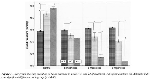
RESULTS
Results
are summarized in Table-1 and Figures-1 and 2. Figure-1 shows BP variation
in week 1, 7, and 12, for all groups. SHR had moderate hypertension at
the beginning of the study, and no significant differences among the groups
at this time. BP in control SHR increased after the first 4 weeks of experimentation,
reaching the value of 177 ± 3 mmHg after 10 weeks, and then stabilized.
However, this usual increasing of BP in young adult SHR was greatly altered
by spironolactone treatment, with either attenuation or reversion of BP
increasing tendency. SHR treated with spironolactone lower dose had a
slight BP decrease (140 ± 1 mmHg in the last 2 weeks of experimentation).
For SHR treated with spironolactone average and higher doses BP had an
accentuated decrease, which was more evident after week 7 of experimentation.
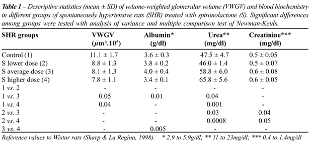
Biochemical evaluation of kidney function
indicated normal levels for albumin and creatinine, and high levels for
BUN in SHR in all groups. Serum albumin level was 11% higher in SHR treated
with spironolactone average dose than in controls; this level was 15%
lower in treated SHR higher dose than in SHR receiving spironolactone
higher dose. BUN had no difference between control and spironolactone
lower dose SHR. Creatinine and BUN were significantly more elevated in
SHR treated with spironolactone average and higher doses.
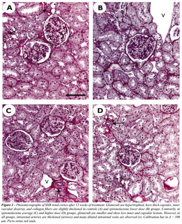
Renal cortical structure was quite similar
in control and treated SHR spironolactone lower dose, and thickness of
the interstitial collagen fibers surrounding the tubuli was slightly larger
than that observed in SHR spironolactone average and higher dose groups.
Thickened intrarenal arteries (mainly affecting the tunica media thickness)
and venous stasis were observed in all groups. Glomerular size and shape
was the major microscopic difference among the groups. In treated SHR
using spironolactone average and higher doses glomeruli had small and
more regular profiles; in control and treated SHR spironolactone lower
dose glomerular hypertrophy with glomerular tuft gross disarray was observed
(Figure-2). The VWGV was significantly higher in control and treated SHR
spironolactone lower dose than in treated SHR spironolactone average and
higher doses.
DISCUSSION
The
present report studied SHR since they were young adults treated with spironolactone
monotherapy (varied doses). The study was extended for 12 weeks and BP,
biochemical kidney functional tests, and VWGV were diversely affected
by different treatments. Briefly, the effect of the spironolactone on
the typical SHR BP increasing tendency and on VWGV was dose-dependent.
This effect was modest on the BP using a spironolactone dose of 5mg/kg/day,
but it was pronounced using a dose of 30mg/kg/day.
The rat normal range for biochemical evaluation
of kidney function is wide (17). BUN normally increases in high-protein
diet or increased protein catabolism from gastrointestinal bleeding, corticosteroids,
tissue trauma, burns, or tetracycline. It normally decreases in low-protein
diet, or decreased protein catabolism from liver disease or cachexia.
Like urea, creatinine is freely filtered at the glomerulus. Creatinine
excretion is dependent on filtration. Increased serum creatinine occurs
in increased creatinine or creatinine intake from a recent meal or the
use of creatinine supplements for bodybuilding. Decreased serum creatinine
occurs in decreased creatinine intake or generation from diminished muscle
mass associated with cachexia, aging, or a low protein intake (18). BUN
high levels found in all groups can be explained by the high-protein content
of Nuvilab™ diet. Albumin and creatinine were both normal in all
groups.
Glomeruli had considerable and general alterations
in rats submitted to a nitric oxide synthesis blockade (a model of systemic
hypertension), characterized by global or segmental glomerular sclerosis.
Renal parenchyma showed only some glomeruli presenting atrophic structure,
tubular atrophy, and extensive fibrosis, and VWGV was 100% greater in
these rats than in controls (11,15). Some previous studies considered
monotherapy with spironolactone efficient to prevent or reduce cardiac
fibrosis, even without simultaneous BP reduction.
These studies used different spironolactone
dose and via of administration, time of observations, as well as different
experimental models or human individuals to support their conclusions.
In equivalency to a 350g rat, spironolactone dose normally varied from
0.13 to 17.5mg/day (this study used 1.75 to 10.5mg/day), but Brilla (5)
and Lacolley et al. (19) used an extreme dose of 200mg/kg/day (equivalent
to 70mg/day in a 350g rat). Therefore, it yields confuse experimental
group denominations, like “low dose” or “high dose”
used in some studies.
Antihypertensive therapy usually associates
different drugs. Combination of spironolactone and ACE inhibitors must
be administered only in the absence of hyperkalemia and significant renal
dysfunction, and under careful monitoring of potassium levels and renal
function (20). There is an advantage in the combination of spironolactone
and blockade of calcium channels, because this drug can prevent organ
fibrosis by attenuating the number of fribrogenetic potential of extracellular
matrix producing myofibroblasts at sites of repair (21). This is possible
because AngII and ALDO may synergistically operate on intracellular calcium,
and an increased intracellular free calcium levels are associated with
proliferation of fibroblasts (22).
Finally, present results suggest that monotherapy
with spironolactone may affect glomerular size and shape in a dose-dependent
way; spironolactone showed a significant effect in preservation of VWGV
and can be used associated with other drugs in antihypertensive therapy
to prevent kidney secondary effects of hypertension.
________________________________
Supported by CNPq and FAPERJ grants.
Luciene O. Sampaio and Thatiany S.
Marinho provided technical assistance.
REFERENCES
- Frohlich ED, Apstein C, Armstrong ML, Cohn JN, Cutler JA, Devereux RB, et al.: Target organ consequences in hypertension: pathogenesis and prevention. Hypertension. 1991; 18: I-143-I-145.
- Kost Jr CK, Li P, Willians S, Jackson EK: Renal vascular responses to angiotensin II in conscious spontaneously hypertensive and normotensive rats. J Cardiovasc Pharmacol.1998; 31: 854-61.
- Delcayre C, Silvestre JS: Aldosterone and the heart: towards a physiological function? Cardiovasc Res. 1999; 43:7-12.
- Maish B, Brilla C, Kruse T: Directions in antihypertensive treatment – our future from the past. Eur Heart J. 1995; 16 (Suppl C): 74-83.
- Brilla CG: Aldosterone and myocardial fibrosis in heart failure. Herz. 2000; 25: 299-306.
- Cahil MM, Kett MM, McCausland JE, Alcorn D, Bertram J: Glomerular stereology: Why, what and how to measure glomerular structure. Nephology. 1996; 2: 305-13.
- Marcussen N: Atubular glomeruli and the structural basis for chronic renal failure. Lab Invest. 1992; 66: 265-84.
- Fogo A, Hawkins EP, Berry PL, Glick AD, Ichikawa I: Glomerular hypertrophy in minimal change disease predicts subsequent progression to focal glomerular sclerosis. Kidney Int. 1990; 38: 115-23.
- Irzyniec T, Mall G, Greber D, Ritz E: Beneficial effect of nifedipine and moxonidine on glomerulosclesrosis in spontaneously hypertensive rats- a micromorphometric study. Am J Hypertens. 1992; 5: 437-43.
- Perico N, Detcheva N, Khalil EL, Remuzzi G: Cyclosporine induces glomerulosclerosis:three-dimensional definition of the lesions in rat model of renal transplant. Kidney Int. 1996; 49: 1283-88.
- Pereira LMM, Mandarim-de-Lacerda CA: Glomerular profile numerical density per área and mean glomerular volume in rats submitted to nitric oxide synthese blockade. Histol Histopathol. 2001; 16: 15-20.
- Gundersen HJG, Jensen EB: Stereological estimation of the volume-weighted mean volume of arbitrary particles observed on random sections. J Microsc. 1985; 138: 127-42.
- Sørensen FB: Stereological estimation of the mean and variance of nuclear volume from vertical section. J Microsc. 1989; 162: 203-29.
- Mandarim-de-Lacerda CA: Estereologia e urologia: volume nuclear médio ponderado na classificação e prognóstico de tumores. Braz J Urol. 1999; 25: 286-290.
- Mandarim-de-Lacerda CA, Pereira LMM: Renal cortical remodelling by NOs blockers in rats is prevented by ACE inhibitor and calcium channel blocker. J Cell Mol Med. 2001; 5: 276-83.
- Zar JH: Biostatistical analysis. Upper Saddle River, Prentice-Hall,1999; p. 663.
- Sharp PE, La Regina MC: The laboratory rat. Boca Raton, CRC Press, 1998; p. 214.
- Wilcox CS: Evaluation of Kidney Function: Biochemical and Nuclear Medicine Tests. In: Tischer CC, Wilcox CS (eds.). Nephrology & Hypertension. Philadelphia, Lippincott Williams & Wilkins,1999; pp.19-22.
- Lacolley P, Safar MR, Lucet B, Ledudal K, Labat C, Benetos A: Prevention of aortic and cardiac fibrosis by spironolactone in old normotensive rats. J Am Coll Cardiol. 2001; 37: 662-7.
- Zannad F: Angiontensin-converting enzyme inhibitor and spironolactone combination therapy. New objectives in congestive heart failure treatment. Am J Cardiol. 1993; 71: 24A-39A.
- Ramires FJA, Sun Y, Weber KT: Myocardial fibrosis associated with ALDO or angiotensin II administration: attenuation by calcium channel blockade. J Mol Cell Cardiol. 1998; 30: 475-83.
- Sadoshima J, Izumo S: Molecular characterization of angiotensin II-induced hypertrophy of cardiac myocytes and hyperplasia of cardiac fibroblasts. Critical role of the AT1 receptor subtype. Cir Res. 1993, 73: 413-23.
______________________
Received: January 8, 2002
Accepted after revision: February 15, 2002
_______________________
Correspondence address:
Dra. Leila Maria Meirelles Pereira
Lab. de Morfometria & Morfologia Cardiovascular, UERJ
Av. 28 de Setembro, 87 fundos
Rio de Janeiro, RJ, 20551-030, Brazil
Fax: + 55 21 2587-6416
E-mail: lmaria@uerj.br