VARIABLES
PREDICTIVE OF VOIDING DISFUNCTION FOLLOWING APONEUROTIC sling SURGERY:
MULTIVARIATE ANALYSIS
(
Download pdf )
SÍLVIO H.M. DE ALMEIDA, ÉMERSON GREGÓRIO, SAWLA EL SAYED, FREDERICO C. FRAGA, HORÁCIO A. MOREIRA, MARCO A.F. RODRIGUES
Sector of Urology, Department of Surgery, State University of Londrina, Paraná, Brazil
ABSTRACT
Introduction:
Aponeurotic sling surgeries can evolve with obstruction or voiding dysfunction
in 5 to 20% of patients. There are few studies on factors that could possibly
predispose to voiding difficulties or urinary retention. The objective
of this work is to identify these potential clinical or urodynamic factors.
Materials and Methods: Records from 130
patients who underwent aponeurotic sling surgeries were reviewed. All
patients underwent a throughout urodynamic study during pre-operative
investigation. The variables studied were age above 65 years, previous
pelvic surgeries, concomitant surgeries, post-voiding residue higher than
100 mL, vesical obstruction (according to Blaivas-Groutz nomogram) and
urinary flow under 12 mL/s. Post-voiding residue was assessed on the seventh
post-operative day through vesical catheterization. Recovering of spontaneous
voiding after 7 post-operative days or with a residue higher than 100
mL, was regarded as voiding dysfunction. Univariate analysis was performed
with qui-square test and Fisher’s exact test, and multivariate analysis
was performed by logistic regression with a = 5%.
Results: Age in the studied group ranged
from 41 to 83 years (mean 56.7 years), with 69 (53%) patients having urethral
hypermobility and 61 (47%) having intrinsic urethral lesion. Normal voiding
occurred in 97 (75.6 %) women with 7 post-operative days. The only significant
variable in the univariate (p = 0.014) and multivariate (p = 0.017) analysis
was post-voiding residue higher than 100 mL.
Conclusion: Pre-operative presence of a
post-voiding residual urine higher than 100 mL was the only variable predictive
of voiding dysfunction.
Key
words: urinary incontinence; surgery; voiding dysfunction; urethra;
bladder
Int Braz J Urol. 2004; 30: 302-6
INTRODUCTION
Pubovaginal
sling surgery has been used for treating urinary incontinence due to sphincteric
lesion for decades and with good results (1). Some years ago, some reports
showed up demonstrating good results for all types of female stress urinary
incontinence as well (2-4). This major indication of the technique has
been accompanied by incidences of prolonged urinary retention of 5 to
20% (4-6).
The urethrovesical junction undergoes, due
to the fibrotic process, a change in position during the postoperative
period for synthetic and autologous slings (7). Such change could exert
an obstructive effect on the urethra, increasing voiding pressure and
decreasing the urinary flow, which could explain the outcome with retention.
However, videourodynamic studies have demonstrated that a band carefully
place with no tension, would not have an obstructive effect (8,9).
A number of factors are suggested as carrying
a risk for voiding dysfunction, such as advanced age, concomitant surgeries,
previous surgeries for urinary incontinence, urinary flow below 12 mL/s,
large post-voiding residual urine and voiding with low contractility or
with Valsalva’s maneuver, among others (6,10-13).
The objective of this study is to analyze
the importance of some of these clinical and urodynamic factors for predicting
the difficulty to obtain normal voiding following aponeurotic sling surgeries,
using multivariate analysis.
MATERIALS AND METHODS
One
hundred and thirty patients with urinary incontinence due to urethral
hypermobility or intrinsic sphincteric lesion, who underwent sling surgery
with aponeurosis of the rectus muscle of abdomen, between 1998 and 2003,
had their medical records reviewed. All patients underwent a throughout
urodynamic study with 2 vesical fillings previously to surgery.
The exam was performed according to the
standards of the International Continence Society, using 0.9% saline solution
at a temperature of 37° Celsius, with a 7F two-way urethral catheter
and a 10F rectal catheter (14).
The abdominal leak point pressure under
stress was measured at half cystometric capacity, in upright position,
with the presence of a 7F two-way catheter inside the urethra, considering
the lowest value for vesical pressure in the absence of detrusor contraction
(15). Patients were initially asked to perform repeated Valsalva’s
maneuvers for 3 times. In the absence of leakage with Valsalva, the patient
was asked to cough. Those patients with abdominal leak point pressure
under stress above 60 cm of H2O were regarded as having urinary incontinence
with some degree of urethral hypermobility.
The same surgeon performed all surgeries,
using segments of aponeurosis of the rectus abdominal muscle measuring
approximately 2 cm x 7 cm and with Pfanestiel-type incisions in the abdomen
and elliptical incision in the vagina. A forceps was interposed between
the urethra and the tape at the moment of tying the threads, in order
to avoid any compression over the urethra.
The clinical variables analyzed were age
above 65 years, performance of previous surgery for urinary incontinence,
performance of other pelvic procedure concomitantly to the surgery, presence
of debilitating neuropathic or chronic diseases, and the mechanism of
urinary incontinence (urethral hypermobility or sphincteric lesion).
The pre-operative urodynamic variables studied
were peak free urinary flow below 12 mL/s, residual urine on voiding cystometry
(without abdominal stress) higher than 100 mL, and the presence of obstruction
superior or equal to moderate type in the Blaivas-Groutz nomogram for
female obstruction (16).
On the first post-operative day, the bladder
was filled, the catheter was removed and voiding was observed for 6 hours.
In the absence of retention, patients were discharged from the hospital.
If clinically required, they remained at the hospital, however without
catheter.
In case of retention, patients were catheterized
again for 3 days, when the process of catheter removal was repeated. On
the seventh post-operative day, post-voiding residual urine was assessed
through vesical catheterization, in all patients. Late voiding was considered
when there was a post-voiding residual urine higher than 100 mL 7 days
after surgery. The variables were studied through univariate analysis
(qui-square and Fisher’s exact tests) and multivariate analysis
with logistic regression.
RESULTS
Age
in the group under study ranged from 41 to 83 years (mean 56.7 years),
with 69 (53%) patients having urethral hypermobility and 61 (47%) having
urethral lesion, and 97 (75.6%) presented normal voiding within 7 days
postoperatively. Three patients (2.3%) required urethrolysis due to prolonged
retention.
Table-1 shows the frequencies for each variable.
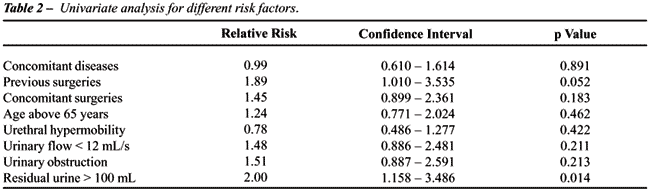
Table-2 demonstrates the results obtained in univariate analysis, with
the only significant variable being post-voiding residual urine, which
was also observed in the multivariate analysis (p = 0.017).
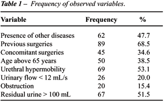
A post-voiding residual urine superior to
100 mL occurred in 67 patients, and 16 of them were obstructed, 22 had
large prolapses, 15 presented contractility deficiency, and in 14 it was
not possible to suppose any cause for the increased residual urine.
COMMENTS
Even
if videourodynamics does not demonstrate obstruction following sling surgeries,
the literature stresses that decreasing the tape tension reduces the risk
of voiding dysfunction, but reduces therapeutic efficacy as well. Flood
et al.(17) compared the presence of early voiding dysfunction in 2 groups
where the only variable was tape tension. Voiding efficiency (smallest
post-voiding residue) was significantly lowest in the group of tension-free
tapes, however the failure indexes (any leakage at 3 months after surgery)
were also significantly higher in this group (17). Petrou & Broderick
demonstrated that urethral position changes in a retropubic direction
after surgery, and that occurs progressively as the remodeling of aponeurotic
tape takes place (18). Such change would lead to the necessity of voiding
adaptation, which would be more efficient and prompter depending on each
patient’s functional characteristics.
Voiding with weak detrusor contraction or
with Valsalva’s maneuver has been associated with a higher risk
of urinary retention and even surgical failure (12,19,20). Miller et al.
(12) observed that of 21 women that voided without contraction on the
pre-operative test, 4 (23%) presented postoperative urinary retention,
versus none among other 48 women with normal contraction. Still in the
same study, no patient with contraction superior to 12 cm of H2O presented
retention.
Among the parameters tested for voiding
dysfunction, only the post-voiding residue was a significant factor. However,
the authors stress that the small sample limits the conclusions of the
study (12).
Voiding residue can be an indicator of voiding
efficiency, either achieved by Valsalva’s maneuver or by effective
detrusor contraction. Its pre-operative presence, due to loss of contractility,
obstruction, or both, can mean a demonstration of such efficiency loss,
and consequently, a risk factor for post-operative voiding dysfunction.
We could not find in literature another
work that studied, exclusively in sling surgeries, the risk factors for
voiding dysfunction using multivariate statistical analysis. Kobak et
al. (13) studied 3 groups of patients undergoing Burch surgery, anterior
colporrhaphy and vaginal wall sling with multivariate analysis, and observed
that advanced age, previous cystopexy, larger vesical volume on the first
voiding desire and high post-voiding residual urine were risk factors
for postoperative voiding dysfunction. The authors did not associate pre-operative
voiding mechanism, intensity of contraction and use of Valsalva’s
maneuver, with risk of voiding dysfunction. The closest comparison to
our group of patients would be only those 34 sling surgeries performed
in this study, even if they were made on the vaginal wall. However, the
type of surgery was not stratified by the authors (13).
Advanced age is the clinical information
most frequently related to the risk of urinary retention following aponeurotic
sling surgeries and even following “tension-free vaginal tape”
(TVT), probably due to the higher risk of dysfunctional pelvic nervous
plexuses and detrusor muscle (6,19). In this work, clinical factors were
not predictive of voiding difficulties, reinforcing the theory that pre-operative
urodynamic results are more important.
There is no universally accepted urodynamic
criterion for diagnosing vesical obstruction in women. We used the Blaivas-Groutz
nomogram, which classifies the obstruction levels in non-obstructed, slightly
obstructed, moderately and severely obstructed (6). However this nomogram
has not been shown able to predict postoperative dysfunction. In a randomized
study between Burch surgery and TVT, it was observed that the nomogram
did not show differences either between patients with objective cure of
incontinence, failure or voiding dysfunction in both groups (20).
The methodology used in trials, usually
retrospective, with limited statistical methods and samples, as well as
different definitions of urinary retention and voiding dysfunction, grouping
different types of surgery, explain the discordant results found in literature.
Though we have not studied the voiding mechanism and the presence of involuntary
contractions, the statistical analysis, the sample volume, and the selection
of patients who underwent surgeries with aponeurotic slings only, strengthen
the results of this work.
Urethral obstruction probably is not the
only causal agent, both for achieving surgical success and for postoperative
voiding dysfunction. Factors related to voiding dynamic and efficacy and
to changes in the periurethral collagen, may act as well. Our results
reinforce the notion that the pre-operative presence of significant post-voiding
residual urine is not a contra-indication for performing the aponeurotic
sling; however, it alerts the surgeon to the risk of any difficulty concerning
the adaptation to a new voiding dynamics and consequently the recovery
of normal voiding.
CONCLUSION
Voiding residual urine above 100 mL was the only variable predictive of voiding dysfunction in the postoperative period of aponeurotic sling surgery in a multivariate analysis.
REFERENCES
- McGuire EJ, Bennett CJ, Konnak JA, Sonda LP, Savastano JA: Experience with pubovaginal slings for urinary incontinence at the University of Michigan. J Urol. 1987; 138: 525-6.
- Chaikin DC, Rosenthal J, Blaivas JG: Pubovaginal sling for all types of stress urinary incontinence: long-term analysis. J Urol. 1998; 160: 1312-6.
- Silva-Filho AL, Triginelli SA, Noviello MB, Santos-Filho AS, Pires CR, Cunha-Melo JR: Pubovaginal sling in the treatment of stress urinary incontinence for hypermobility and intrinsic sphincteric deficiency. Int Braz J Urol. 2003; 29: 540-44.
- Morgan TO Jr, Westney OL, McGuire EJ: Pubovaginal sling: 4-year outcome analysis and quality of life assessment. J Urol. 2000; 163: 1845-8.
- Chan PT, Fournier C, Corcos J: Short-term complications of pubovaginal sling procedure for genuine stress incontinence in women. Urology. 2000; 55: 207-11.
- Iglesia CB, Shott S, Fenner DE, Brubaker L: Effect of preoperative voiding mechanism on success rate of autologous rectus fascia suburethral sling procedure. Obstet Gynecol. 1998; 91: 577-81.
- Mclennan MT, Clifford FM, Cannon S: The position of the urethrovesical junction after incontinence surgery: early postoperative changes. Int Urogynecol J. 2004; 15: 44-8.
- Klutke JJ, Klutke CG, Bergman J, Elia G: Urodynamics changes in voiding after anti-incontinence surgery: an insight into the mechanism of cure. Urology. 1999; 54: 1003-7.
- Kuo HC. Comparison of video urodynamic results after the pubovaginal sling procedure using rectus fascia and polypropylene mesh for stress urinary incontinence. J Urol 2001; 165: 163-8.
- Nitti VC, TU LM, Guitlin J: Diagnosing bladder outlet obstruction in women. J Urol. 1999; 161: 1535-40.
- Cormier L, Ferchaud J, Galas JM, Guillemin F, Mangin P: Diagnosis of female bladder outlet obstruction and relevance of the parameter area under the curve of detrusor pressure during voiding: preliminary results. J Urol. 2002; 167: 2083-7.
- Miller EA, Amundsen CL, Toh KL, Flynn BJ, Webster GD: Preoperative urodynamic evaluation may predict voiding dysfunction in women undergoing pubovaginal sling. J Urol. 2003; 169: 2234-7.
- Kobak WH, Walters MD, Piedmonte MR: Determinants of voiding after three types of incontinence surgery: a multivariable analysis. Obstet Gynecol. 2001; 97: 86-91.
- Schafer W, Abrams P, Liao L, Mattiasson A, Pesce F, Spangberg A, et al.: Good urodynamic practices: uroflowmetry, filling cystometry, and pressure-flow studies. Neurourol Urodyn. 2002; 21: 261-74.
- Abrams P, Cardozo L, Fall M, Griffiths D, Rosier P, Ulmsten U, et al.: The standardisation of terminology of lower urinary tract function: report from the standardisation sub-committee of the international continence society. Am J Obstet. Gynecol. 2002; 187: 116-126.
- Blaivas JG, Groutz A: Bladder outlet obstruction nomogram, for women with lower tract symptomatology. Neurourol. Urodyn. 2000; 19: 553-64.
- Flood H, Muratib S, Shah A, Etisham M, Khan MS: Early voiding dysfunction and efficacy after pubovaginal sling: the role of sling tension. Aust N Z J Surg. 1998; 69: A121.
- Petrou SP, Broderick GA. A sling is not just a backboard of urethral support. J Pelvic Surg. 2001; 7: 11-14.
- Mutone N, Brizendine E, Hale D: Factors that influence voiding function after the tension-free vaginal tape procedure for stress urinary incontinence. Am J Obstet Gynecol. 2003; 188: 1477-81.
- Wang AC, Chen MC: Comparison of tension-free vaginal taping versus modified Burch colposuspension on urethral obstruction: a randomized controlled trial. Neurourol Urodyn. 2003; 22: 185-90.
____________________
Received:
April 14, 2004
Accepted after revision: July 7, 2004
_______________________
Correspondence address:
Dr. Sílvio Henrique Maia de Almeida
Rua Francisco Marcelino da Silva 270
Londrina, PR, 86047-160, Brazil
Fax: + 55 43 3342-9148
E-mail: salmeida@sercomtel.com.br