SPONTANEOUS
NEPHROCUTANEOUS FISTULA
(
Download pdf )
ALBERTO A. ANTUNES, ADRIANO A. CALADO, EVANDRO FALCÃO
Service of Urology, Getúlio Vargas Hospital, Recife, Pernambuco, Brazil
ABSTRACT
Spontaneous
renal fistula to the skin is rare. The majority of cases develop in patients
with antecedents of previous renal surgery, renal trauma, renal tumors,
and chronic urinary tract infection with abscess formation.
We report the case of a 62-year old woman,
who complained of urine leakage through the skin in the lumbar region
for 2 years. She underwent a fistulography that revealed drainage of contrast
agent to the collecting system and images suggesting renal lithiasis on
this side. The patient underwent simple nephrectomy on this side and evolved
without intercurrences in the post-operative period.
Currently, the occurrence of spontaneous
renal and perirenal abscesses is extremely rare, except in patients with
diabetes, neoplasias and immunodepression in general.
Key
words: kidney; lithiasis; fistula; lumbar region; nephrectomy
Int Braz J Urol. 2004; 30: 316-8
INTRODUCTION
Spontaneous
renal fistula to adjacent organs is not an uncommon phenomenon, however
the spontaneous communication between kidney and skin is rare and few
cases are described in the literature (1-3). The occurrence of spontaneous
fistulas in patients without surgical history is rare (3). All cases reported
in the literature are associated with chronic urinary tract infection
and nephrolithiasis.
The authors report one more case of this
rare complication of lithiasis-induced chronic pyelonephritis.
CASE REPORT
A
62-year old woman was admitted to the urology service reporting urine
leakage from the skin in the lumbar region for 2 years. She referred local
inflammatory process with drainage of purulent secretion at the onset
of the clinical picture. There was no report of previous pyelonephritis.
The physical examination evidenced a fistulous orifice in skin on left
lumbar region (Figure-1). Urine culture was negative. The patient denied
diabetes or past history of local trauma.

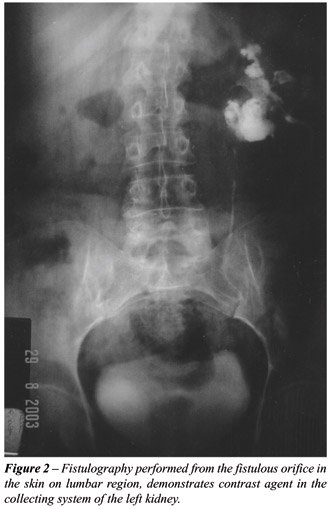
A fistulography was performed, revealing
drainage of the contrast agent to the collecting system, and images suggesting
renal lithiasis on this side (Figure-2). Renal scintigraphy with DMSA
revealed relative renal function of only 5% on the left side. The contralateral
kidney was normal.
Patient underwent left lumbotomy, where
an atrophic kidney was found, with adhesions to adjacent structures. Then
a simple left nephrectomy was performed.
The pathological examination of the surgical
specimen revealed chronic pyelonephritis associated with multiple renal
calcifications. Testing for tuberculosis in the renal tissue was negative.
Patient evolved without intercurrences and was discharged from the hospital
on the seventh postoperative day.
COMMENTS
Renal
fistulas usually are complications of surgical procedures on the kidney,
renal trauma, tumors, and chronic urinary tract infections with formation
of perirenal abscess (1). Such abscesses can derive from organs that are
adjacent to the kidney, as well as from the kidney itself, by extension
of urinary infection to the adjacent tissues, either by contiguity or
by lymphatic route. In other occasions, abscesses can originate from an
urinoma or urinary pseudocyst, that arise as result of external or surgical
trauma on the kidney, promoting loss of continuity between it and the
surrounding tissues (2).
Currently, the occurrence of renal and perirenal
abscesses is rare, except patients with diabetes, with neoplasias or immunodepression
in general. The outcome of these abscesses, when left untreated, is unforeseeable
(2).
Fistulas can develop between the kidney
and the pleural cavity, lungs and bronchia, bowel, and skin. However,
the latter are rare, and whenever they occur, they typically involve patients
with a past history of renal surgery (1).
The majority of fistulas presents spontaneous
drainage through the lumbar region following those points with lowest
resistance, such as the lumbar triangle (Petit) and the lumbar quadrilateral
(Grynfeld), establishing a fistulous pathway that communicates the perirenal
tissues and collecting system with the external environment (2). The association
with infectious renal stones is frequent and has occurred in all cases
described in the literature (1-3). The patient in this case had a staghorn
stone in the involved kidney.
Therapeutic approaches must be based on
the renal function and on the patient’s ability to tolerate the
surgical procedure, and can include total nephrectomy, partial nephrectomy
or isolated antibiotic therapy (3). In the present case, the patient evolved
without postoperative intercurrences and was free of symptoms.
REFERENCES
- Bryniak SR.: Primary spontaneous renocutaneous fistula. Urology. 1983; 21: 516-7.
- Sarmiento RC, Blasco CF, Herrera FF, Chica RA, Ostale GJ: Spontaneous nephrocutaneous fistula. Report of a case and review of the literature. Arch Esp Urol. 1990; 43: 411-3.
- Singer AJ: Spontaneous nephrocutaneous fistula. Urology. 2002; 60: 1109-10.
_____________________
Received: March 5, 2004
Accepted after revision: July 21, 2004
________________________
Correspondence address:
Dr. Alberto Azoubel Antunes
Rua 3 de maio, 17 / 31
São Paulo, SP, 04044-020, Brazil
Fone: + 55 11 55735385
E-mail: betoazoubel@yahoo.com.br