LAPAROSCOPIC
SURGERY FOR TREATMENT OF INCISIONAL LUMBAR HERNIA
(
Download pdf )
M. TOBIAS-MACHADO, FREDDY J. RINCON, MARCO T. LASMAR, JOAO P. ZAMBON, ROBERTO V. JULIANO, ERIC R. WROCLAWSKI
Section of Urology, ABC Medical School, Santo Andre, Sao Paulo, Brazil
ABSTRACT
Objective:
To present results obtained with laparoscopic correction of incisional
lumbar hernia in patients with minimum follow-up of 1 year.
Materials and Methods: We prospectively
studied 7 patients diagnosed with incisional lumbar hernia after physical
examination and computerized tomography. We used laparoscopic transperitoneal
access through 3 ports. One polypropylene mesh was introduced in the abdominal
cavity and fixed by titanium clamps to the margins of the hernia ring
following release of the peritoneum.
Results: All cases were successfully completed
with no conversion required. Mean surgical time was 120 minutes and discharge
from hospital occurred between the 1st and the 2nd postoperative days.
There were no intraoperative complications or hernia recurrence in any
case. Postoperatively, we had 2 minor complications: one case of seroma
that resolved spontaneously after 60 days and one patient presenting lumbar
pain that persisted until the 3rd postoperative month. The return to usual
activities occurred on average 3 weeks following intervention. Of the
7 patients, 6 were satisfied with the esthetical and functional effect
produced by the procedure.
Conclusions: The surgical correction of
incisional lumbar hernia by laparoscopic access is an excellent option
for a minimally invasive treatment, with adequate long-term results.
Key
words: lumbar region; hernia; surgical procedures, operative;
laparoscopy
Int Braz J Urol. 2005; 31: 309-14
INTRODUCTION
Lumbar
hernias are not common, with 2 weak sites existing in the region: the
superior (Grynfeltt-Lesshalft) and inferior (Petit) lumbar triangle. All
others are known as diffuse lumbar hernias, which are usually associated
with conventional extraperitoneal lumbar access (1).
Some surgical repair procedures have been
described; the most frequently used being either the open technique with
primary closure or the use of prosthetic material. Open surgery requires
a large incision and extensive exposure and dissection of the herniated
area. Additionally, the margins of the hernia ring are poorly defined
and often require a peritoneal opening for establishing its limits (2,3).
Despite the wide use of the laparoscopic
techniques for treating ventral abdominal hernias, a few services have
reported sporadic cases using the laparoscopic approach for correction
of lumbar defects. Preliminary results suggest that this technique shows
advantages concerning patient recovery, especially in shorter hospital
stays and prompter returns to normal activities (3-6).
This study aims to present and discuss the
long-term results of the laparoscopic repair for incisional lumbar hernias.
MATERIALS AND METHODS
Patient
Selection and Follow-up
From January 2002 to January 2004, we prospectively
studied 7 patients with incisional lumbar hernias who had undergone previous
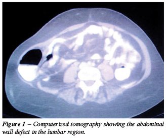
lumbotomies. Diagnosis was obtained by physical examination, including
palpation of the ring’s margins, and documented through computerized
tomography (Figure-1). Patients with any formal contraindication for laparoscopic
surgery, coagulation disorders or connective tissue disease were excluded
from the study.
Variables pertaining to patients were described
and analyzed, such as age, gender, body mass index (BMI), cause of previous
lumbar incision such as data relative to the procedure and patient’s
outcome, such as surgical time, blood loss, analgesic requirements, complications,
conversion rate, hospital stay, recovery time until returning to normal
activities, and functional and esthetic features.
All included patients were followed up by
our outpatient service 7, 30, 90, 180 days and finally 1 year following
surgery, when a patient satisfaction questionnaire was applied and a control
computerized tomography was performed to objectively document the results.
The minimal follow-up time for including the results in this study was
1 year.
Surgical
Time
Laparoscopic repair with transperitoneal
access was used in all cases. Antibiotic prophylaxis was performed with
cefalotin. Patients were placed in right or left lateral decubitus according
to the side of herniation and the table was inclined 60 degrees. The first
10-mm Hasson trocar was inserted through the umbilical incision under
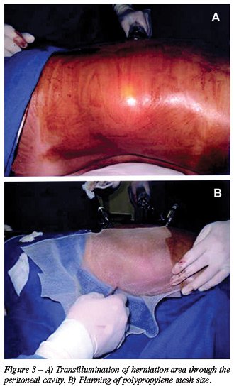
direct viewing (Figure-2). The cavity was then insufflated through the
access trocar with CO2, until a tension of 15 mm Hg was reached. Immediately
afterwards, the 0 degree optics was introduced and the cavity was inspected
to check for the presence of the hernia ring. The herniation area was
transilluminated through the peritoneal cavity in order to plan the proper
size of the polypropylene mesh (Figure-3).
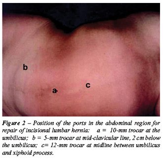
The second 5-mm port was placed under direct
viewing at the mid-clavicular line 2 cm below the umbilical scar, and
the third 12-mm port (suited for the stapler) was placed at the midline
between the navel and the xiphoid process (Figure-2).
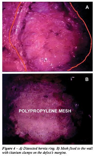
The peritoneum was released while medially
withdrawing the colon that is typically included in the defect in order
to expose the entire hernia ring (Figure-4). External palpation of the
wall can help to accurately delimitate the defect. The surgical table
must allow the patient to be arranged in many different positions for
complete dissection of the defect. Subsequently, the mesh was inserted
into the cavity through the 12-mm trocar and fixed on the wall by an articulate
hernia “stapler” using titanium clamps at the margins of the
defect (Figure-4). Fixation limits are paravertebral musculature posteriorly,
the costal arch superiorly, the iliac spine inferiorly and the abdominal
wall musculature anteriorly. During this procedure, CO2 tension was reduced
to 7-10 mm Hg in order to make the fixation of the mesh easier. Next,
the entire mesh was covered by the previously dissected peritoneum and
clamped to the wall to prevent it contacting the intestinal loops. Finally,
the cavity was reviewed, the ports were removed and the incisions were
closed. No drain was left close to the mesh.
RESULTS
Mean
age was 52 years (40 - 65), with BMI from 20-25 (5 cases) and 26-30 (2
cases).
The wall defects ranged in size from 6 x
8 cm to 10 x 15 cm (mean 8 x 12 cm).
Three patients were male and 4 were female,
with 4 cases occurring on the left side and 3 cases on the right side.
In relation to the surgery that caused the previous lumbar incision, there
were 3 cases of nephrectomy for kidney donation, 2 cases of nephrectomy
due to renal tumor, 1 case of nephrectomy due to hydronephrosis and 1
case of pyelolithotomy.
All procedures were successfully completed
by laparoscopic access. During laparoscopic inspection it was possible
to distinctively assess the size of the hernia ring and anatomical structures
involved in the hernial defect in all patients. The polypropylene mesh
was easily inserted into the cavity and fixed by titanium clamps to the
ring margins through the 12-mm port.
Surgical time ranged from 90 to 150 minutes
(mean 120). There were no intraoperative complications and mean blood
loss was 70 mL (50 - 80). Analgesia was obtained using only dipyrone on
the first postoperative day in 6 cases. Discharge from hospital occurred
on average 12 to 36 hours (mean 24) following surgery. Patients returned
to their usual activities 2 to 5 weeks after surgery (mean 3).
As far as postoperative complications were
concerned, we found 2 minor complications, specifically one case of seroma
that resolved spontaneously after 60 days and one female patient presented
lumbar pain that lasted until the 3rd postoperative month. This case,
which was interpreted as neuropathic pain, required treatment with major
analgesics, tricyclic anti-depressants and corticoids for symptom improvement.
Probably, a clamp used for fixating the mesh was applied to some nervous
bundle at the posterior abdominal wall.
We did not observe a recurrence of hernia
in any of the patients during a mean follow-up of 12 months.
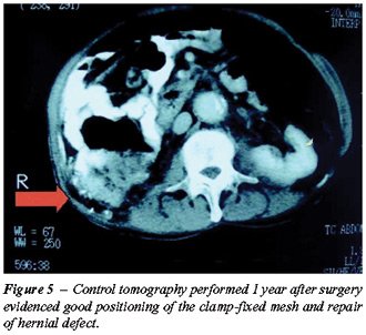
The control tomography performed 1 year
after the surgery revealed good positioning of the mesh that had been
fixed by clamps and repair of the defect in all patients (Figure-5). The
esthetic and functional aspect of the defect as reported by the patient
was very adequate in 6 out of 7 cases when compared with the preoperative
aspect. One patient who presented muscular atonia at the incision’s
anterior portion before surgery was partly satisfied with the esthetic
result.
COMMENTS
Lumbar
hernias are relatively rare, with a little more than 300 cases found in
the literature (1,2,7). They can be classified into congenital (10 - 20%)
or acquired (80 - 90%) hernias. Acquired hernias are divided into 2 types
– spontaneous and traumatic (incisional) (1,2,4).
The present study included patients with
acquired traumatic lumbar hernias secondary to lumbar incision for conventional
renal surgery. Though the classical lumbotomy is still largely used in
our country, the increasing use of laparoscopic and percutaneous surgery
for treating surgical conditions of the kidneys and adrenal glands will
certainly reduce the occurrence of such complications.
In general, lumbar hernias are diagnosed
using clinical criteria (6). The main complaint of patient is the perception
of a reducible tumor with solid consistency in the incision area, which
can be accompanied by lumbar discomfort. Recent publications describe
the importance of computerized tomography to identify the hernia, demonstrating
in detail the anatomy and differential diagnosis with other pathologies
(1,6,8). The computerized tomography was an important diagnostic method
for identifying, confirming and objectively documenting the hernia in
this study.
When untreated, lumbar hernia can reach
gigantic proportions, thus increasing the risk of incarceration (25%)
and strangulation (8%) (6). The hernial content can include the epiploon,
small or large bowel, spleen and the kidney itself (1).
If permitted by the patient’s general
condition, the lumbar hernia always has surgical indication with several
techniques being described in the literature. Due to its rarity, there
is no standardized technique. The difficulty in delimiting the margins
of the fascial defect, the weakness of the involved structures, the participation
of a bone element, and the surgeon’s expertise are all elements
taken into account during surgical planning (1,6).
The open technique for reconstruction of
lumbar hernias requires a large incision, which is often associated with
more severe pain, a longer convalescence period and increased morbidity
(1,4). For the conventional repair of such hernias, natural structures
from the region itself or synthetic materials (made of polypropylene or
polytetrafluoroethylene) can be used. Results described for surgery without
mesh have been poor, probably due to the low tensile quality of the local
tissues, which is why the repair with synthetic material has been preferred
(1,4,5).
With the intention of reducing the morbidity
observed with the conventional technique while maintaining the results
from open surgery with mesh, the laparoscopic access has been recently
described.
Using the expertise in repair of ventral
hernias that has been accumulated in many centers, the same principles
could be applied to lumbar hernias as well. Initial experiences have shown
significant advantages of the laparoscopic approach over conventional
surgery. The majority of studies describing this technique has reported
low morbidity, less significant pain and earlier returns to normal activities
(2,4,6). Other studies have confirmed that this access promotes optimal
visualization of the ring’s limits, is safe and simple, and is considered
a minimally invasive procedure (2,3,9).
The repair of lumbar hernia by laparoscopic
approach was first published in 1997 by Heniford et al. (7). The following
year, Arca et al. (4) published the first results from experience with
7 patients with lumbar hernias treated by the laparoscopic approach. The
authors concluded that there was improved visualization of the anatomical
defects, reduced hospital stay, and no recurrence in this sample during
a 15-month period.
In the present study, we observed an excellent
exposure of structures and achieved perfect anatomical visualization of
the hernia ring. There was little postoperative pain, reduced mean hospital
stay, and the return to usual activities occurred promptly. During the
12-month follow-up period, no recurrence of herniation was evidenced.
In one case, the posterior hernial defect was repaired, but patient satisfaction
was not completely achieved due to atonia of the abdominal wall secondary
to a nervous lesion occurring after the lumbotomy.
Among the small number of published studies
on laparoscopic repair of lumbar hernias, none of them has described significant
complications (2-7). Comparative studies between the open and laparoscopic
approach reported in the literature refer only to the surgical treatment
of ventral incisional hernias. There are no such studies involving lumbar
herniation, which, in a certain way, does not allow us to definitely conclude
which access is best (10,11). Our impression, however, is that the laparoscopic
repair seems to have advantages concerning the visualization of the hernial
defect and the postoperative recovery.
CONCLUSIONS
The laparoscopic repair of incisional lumbar hernia is a minimally invasive procedure with moderate complexity, which promotes adequate functional and esthetic results. It provides excellent exposure and definition of the wall defect limits, mild postoperative pain, short hospital stay and early return to normal activities. If comparative studies confirm the superiority of the laparoscopic approach in relation to the open technique, the laparoscopic procedure could become the method of choice for repair of lumbar hernias.
REFERENCES
- Moreno-Egea A, Torralba-Martinez JA, Morales G, Fernandez T, Girela E, Aguayo-Albasini JL: Open vs laparoscopic repair of secondary lumbar hernias: a prospective nonrandomized study. Surg Endosc. 2005; 19: 184-7.
- Sakarya A, Aydede H, Erhan MY, Kara E, Ilkgul O, Yavuz C: Laparoscopic repair of acquired lumbar hernia. Surg Endosc. 2003; 17: 1494.
- Maeda K, Kanehira E, Shino H, Yamamura K: Laparoscopic tension-free hernioplasty for lumbar hernia. Surg Endosc. 2003; 17: 1497.
- Arca MJ, Heniford BT, Pokorny R, Wilson MA, Mayes J, Gagner M: Laparoscopic repair of lumbar hernias. J Am Coll Surg. 1998; 187: 147-52.
- Bickel A, Haj M, Eitan A: Laparoscopic management of lumbar hernia. Surg Endosc. 1997; 11: 1129-30.
- Shekarriz B, Graziottin TM, Gholami S, Lu HF, Yamada H, Duh QY, et al.: Transperitoneal preperitoneal laparoscopic lumbar incisional hernorrhaphy. J Urol. 2001; 166: 1267-9.
- Heniford BT, Iannitti DA, Gagner M: Laparoscopic inferior and superior lumbar hernia repair. Arch Surg. 1997; 132: 1141-4.
- Baker ME, Weinerth JL, Andriani RT, Cohan RH, Dunnick NR: Lumbar hernia: diagnosis by CT. AJR Am J Roentgenol. 1987; 148: 565-7.
- Parker HH 3rd, Nottingham JM, Bynoe RP, Yost MJ: Laparoscopic repair of large incisional hernias. Am Surg. 2002; 68: 530-3; discussion 533-4.
- Chari R, Chari V, Eisenstat M, Chung R: A case controlled study of laparoscopic incisional hernia repair. Surg Endosc. 2000; 14: 117-9.
- Goodney PP, Birkmeyer CM, Birkmeyer JD: Short-term outcomes of laparoscopic and open ventral hernia repair: a meta-analysis. Arch Surg. 2002; 137: 1161-5.
________________________
Received:
February 24, 2005
Accepted after revision: April 29, 2005
_______________________
Correspondence address:
Dr. Marcos Tobias-Machado
Rua Graúna, 104 / 131
São Paulo, SP, 04514-000, Brazil
E-mail: tobias-machado@uol.com.br