INCIDENCE
OF BENIGN LESIONS ACCORDING TO TUMOR SIZE IN SOLID RENAL MASSES
(
Download pdf )
VICTOR SROUGI, RAPHAEL B. KATO, FERNANDA A. SALVATORE, PEDRO P.M. AYRES, MARCOS F. DALL’OGLIO, MIGUEL SROUGI
Division of Urology, University of Sao Paulo Medical School, Sao Paulo, SP, Brazil
ABSTRACT
Objective:
The incidence of solid renal masses has increased sharply in recent years
due to widespread use of abdominal imaging studies. The aim of the present
study was to evaluate the incidence of benign lesions in solid renal masses
according to tumor size.
Materials and Methods: The authors retrospectively
reviewed the records of 305 patients with 328 renal solid masses treated
by surgery. Based on a report by one pathologist, the specimen tumor size
and the histology of each lesion were tabulated. The frequency of renal
cell carcinoma and benign renal lesions was evaluated and a correlation
between tumor size and pathological features of the masses was observed.
Results: The frequency of malignant lesions
in the 328 renal masses was 83.2%. When lesions were stratified into groups
with diameters = 3 cm or > 3 cm, the incidence of benign histology
was 22.9% and 13.3%, respectively (p = 0.026). The odds ratios for finding
a benign lesion in masses = 3 cm was 1.93 (IC 95%, 1.07 - 3.46) compared
to masses > 3 cm.
Conclusion: The incidence of benign lesions
is significantly higher in renal masses smaller than 3 cm in diameter,
which should be taken in account when the treatment of renal solid masses
is planned.
Key
words: renal cell, carcinoma; staging; mass; benign; imaging
Int Braz J Urol. 2009; 35: 427-31
INTRODUCTION
Renal
cell carcinomas (RCCs) represent between 3% and 4% of neoplasms found
in clinics (1) and their incidence has been rising about 3% per year since
1975 (2). In the past, 85% of the expansive lesions found in kidneys were
related to RCC, while the remaining lesions were benign local disorders
(3). In recent years, with the introduction of new imaging methods like
ultrasound, CAT scan, and magnetic resonance, there has been a significant
increase in the detection of asymptomatic and incidental renal tumors,
cases that now outnumber tumors identified after the onset of symptoms.
Currently, up to 71% of the solid renal masses are found incidentally
during abdominal imaging studies (2,3). In these cases, the lesions are
usually smaller and are frequently benign (4-7).
The finding of benign disorders in a significant
number of patients with solid renal masses has practical implications
that cannot be ignored. The adoption of indiscriminate surgical treatment
in all cases of renal masses will result in unnecessary interventions
and even the undue loss of kidneys in patients without malignant disease.
To avoid this dilemma, many authors advise a pre-op percutaneous biopsy
in cases of solid renal masses (8,9). Although these tests are highly
accurate, biopsy is not sufficiently accurate in all cases and can fail
in characterizing the histology of renal masses (10).
We have conducted this study in order to
guide medical judgment and to more efficiently handle solid renal masses
by attempting to determine if certain mass tumors can be treated non-surgically.
This would provide for the development of a criterion other than the biopsy
to help the therapeutic decision process for patients with renal masses.
MATERIALS AND METHODS
The
present study is a retrospective analysis of 305 consecutive patients
seen at our Institution from 1977 to 2006, bearing incidentally or symptomatic
solid renal masses.
A total of 305 patients were evaluated,
including 231 males and 74 females ranging in age from 9 to 90 years (median
of 60 years). All of the patients had expansive kidney lesions depicted
by computerized axial tomography or magnetic resonance imaging; some were
solid and others had intermingled large solid and small cystic areas.
In the patients with multiple lesions, each one was studied individually
and a total of 328 lesions were analyzed. Taking in account the clinical
presentation, the routine blood and urinary tests and the imaging studies,
all lesions classified as inflammatory or infectious were excluded from
the study.
All patients were operated upon by the
same surgeon (MS) and included radical nephrectomy (213 pts), partial
nephrectomy (24 pts), or mass enucleation (68 pts). The surgical specimens
were analyzed by the same pathologist, using the UICC histopathology classification
for RCC, including clear cell, chromophobe, papillary, Bellini duct, and
sarcomatoid subtypes (11).
Using the primary clinical outcome, an
attempt has been made to define the total incidence of benign lesions
and correlate the tumor diameter with final histology of the lesion. The
diameter was measured in the surgical specimen, taking into account the
greatest diameter found. The histopathology findings were divided into
benign and malignant categories, and the frequency of these two types
was compared to original nodes of greater or less than 3 cm in diameter.
The differences in proportion of benign
and malignant lesions in the groups with diameters of more or less than
3 cm were evaluated by statistical analysis with the chi-square test,
adopting a significance level of 5% (p < 0.05) for rejecting the null
hypothesis. The strength of association between the two variables was
analyzed by an odds ratio calculation with a confidence interval of 95%
(IC at 95).
RESULTS
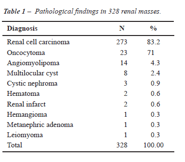
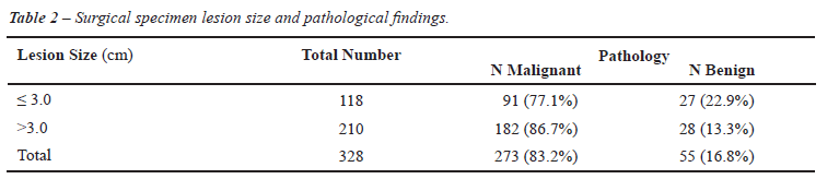
In this study, the diameter of the evaluated lesions ranged from 0.2 cm to 24.0 cm, with a median of 4.0 cm. According to Table-1, among the 328 renal lesions found in 305 patients, 273 (83.2%) were renal cell carcinomas. In the other cases, 43 (13.2%) were benign lesions and 12 (3.6%) were non-neoplasic lesions.
When the incidence of benign lesions was
evaluated as a function of the renal mass diameter, a frequency of 13.3%
and 22.9% was observed, respectively, in the masses with diameters equal
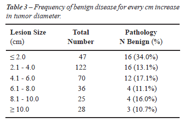
or greater and smaller than 3 cm (Tables 2 and 3) with a statistically
significant difference between the frequency of benign pathologies and
the lesion size (p = 0.026). These same numbers show that the chance of
finding a malignant lesion in nodes greater than 3 cm in diameter is 1.93
times greater (IC at 95%, 1.07-3.46) than in nodes with diameters of 3
cm.
COMMENTS
This
study shows that there is a true correlation between the diameter or solid
renal nodes and the incidence of benign lesions. To perform our analysis
we compared lesion smaller or larger than 3 cm and we choose this cut-off
value because it has been demonstrated that it correlates well with the
risks of metastatic disease in renal cell carcinoma. Metastatic disease
is usually found in tumors larger than 3 cm (12) which makes this threshold
a valuable clinical parameter for handling patients with solid renal lesions.
Benign lesions were seen in 13.3% of 210 (two hundred and ten) masses
less than 3 cm in diameter and in 22.9% of 118 nodes greater than 3 cm
in diameter. We believe these results are significant, as the present
study included a large number of patients, the surgical specimens were
all analyzed by a single pathologist, and the largest tumor diameter was
measured in the surgical specimen. Some previous studies published regarding
the same subject evaluated a smaller number of cases (13,14) and the measurement
of the tumor size was made by imaging studies that show a certain variability
due to heterogeneous cuts and random radiologist readings (3,15,16).
The greatest frequency of benign lesions
in renal masses smaller than 3 cm found in our study agrees with other
studies previously published (4,6,7,15). According to those studies, between
12.5% and 46.3% of solid renal nodes less than 3 cm in diameter are benign,
and this frequency decreases, from 0% to 19.9%, when the solid mass is
more than 3 cm in diameter (7,15). A single study, published by Snyder
et al., has shown the absence of correlation between the size of renal
nodes and the incidence of benign lesions. However, this study excluded
greater diameter masses, a fact that probably reduced the number of malignant
lesions in this sub-group when the patients were treated surgically (16).
One of the main limitations of the present
study is that only tumor size was taken in account to predict the pathological
outcome of the solid renal masses. It has been already shown that other
relevant variables such as other radiological features, clinical presentation,
age, gender or tumor growth velocity can help in the differential diagnosis
of renal tumors (17,18). On the other hand, renal tumor size correlates
with the risks of metastasis and with patient survival. Furthermore measuring
tumor diameter is a simple way that helps physician to plan patient care
when a more detailed analysis of imaging studies are not available or
are incomplete.
Fifty-five of the 328 lesions (16.8%) evaluated
in our study represented benign disorders, with a prevalence of oncocytomas
(7.1%) and angiomyolipomas (4.3%). This distribution is similar to that
seen in other studies (15,16), showing that although there is an absence
of aggressiveness, most of the benign renal lesions found in practice
are neoplasic in nature, with a possibility of generating difficulties
in interpretation when percutaneous biopsies are diagnosed in patients
with solid renal lesions. In fact, in one of the contemporary series of
percutaneous biopsies to study renal masses, 21% of the cases did not
have the etiology defined on the first attempt, generating problems in
terms of an effective therapeutic intervention (8).
According to the data of the present study,
it is important for specialists to consider the possibility of benign
lesions when identifying solid or mixed renal nodes. The incidence of
benign lesions is significantly greater in solid renal masses with diameters
of less than 3 cm and this must be taken into account when advising patients
about the relevance of the disease and about treatment options. In lesions
with diameters less than 3 cm, the frequency of benign masses is almost
twice as common as that found in greater masses. This finding can be relevant
when we are treating patients with multiple bilateral small solid nodules
or patients unsuited for surgery. In these settings, the use of percutaneous
lesion biopsies has a great chance to show a benign lesion and this finding
will obviate the need for an unnecessary surgical intervention (9). Furthermore,
the adoption of surveillance in a patient with a solid renal mass can
be done with less concern if the lesion has less than 3 cm in diameter.
Considering that such tumors have no or negligible growth when followed
for years (19), based on tumor size and tumor growth physicians can safely
monitor their patients treated with active surveillance.
ACKNOWLEDGEMENT
Adriana Sañudo performed the statistical analyses.
CONFLICT OF INTEREST
None declared.
REFERENCES
- Jemal A, Siegel R, Ward E, Hao Y, Xu J, Murray T, et al.: Cancer statistics, 2008. CA Cancer J Clin. 2008; 58: 71-96.
- Chow WH, Devesa SS, Warren JL, Fraumeni JF Jr: Rising incidence of renal cell cancer in the United States. JAMA. 1999; 281: 1628-31.
- Pahernik S, Ziegler S, Roos F, Melchior SW, Thüroff JW: Small renal tumors: correlation of clinical and pathological features with tumor size. J Urol. 2007; 178: 414-7; discussion 416-7.
- Glassman D, Chawla SN, Waldman I, Johannes J, Byrne DS, Trabulsi EJ, et al.: Correlation of pathology with tumor size of renal masses. Can J Urol. 2007; 14: 3616-20.
- Dall’Oglio M, Srougi M, Ortiz V, Nesrallah L, Gonçalves PD, Leite KM, et al.: Incidental and symptomatic kidney cancer: pathological features and survival. Rev Assoc Med Bras. 2004; 50: 27-31.
- Schachter LR, Cookson MS, Chang SS, Smith JA Jr, Dietrich MS, Jayaram G, et al.: Second prize: frequency of benign renal cortical tumors and histologic subtypes based on size in a contemporary series: what to tell our patients. J Endourol. 2007; 21: 819-23.
- Frank I, Blute ML, Cheville JC, Lohse CM, Weaver AL, Zincke H: Solid renal tumors: an analysis of pathological features related to tumor size. J Urol. 2003; 170: 2217-20.
- Lebret T, Poulain JE, Molinie V, Herve JM, Denoux Y, Guth A, et al.: Percutaneous core biopsy for renal masses: indications, accuracy and results. J Urol. 2007; 178: 1184-8; discussion 1188.
- Volpe A, Kachura JR, Geddie WR, Evans AJ, Gharajeh A, Saravanan A, et al.: Techniques, safety and accuracy of sampling of renal tumors by fine needle aspiration and core biopsy. J Urol. 2007; 178: 379-86.
- Dechet CB, Zincke H, Sebo TJ, King BF, LeRoy AJ, Farrow GM, et al.: Prospective analysis of computerized tomography and needle biopsy with permanent sectioning to determine the nature of solid renal masses in adults. J Urol. 2003; 169: 71-4.
- Störkel S, Eble JN, Adlakha K, Amin M, Blute ML, Bostwick DG, et al.: Classification of renal cell carcinoma: Workgroup No. 1. Union Internationale Contre le Cancer (UICC) and the American Joint Committee on Cancer (AJCC). Cancer. 1997; 80: 987-9.
- Kunkle DA, Crispen PL, Li T, Uzzo RG: Tumor size predicts synchronous metastatic renal cell carcinoma: implications for surveillance of small renal masses. J Urol. 2007; 177: 1692-6; discussion 1697.
- Kutikov A, Fossett LK, Ramchandani P, Tomaszewski JE, Siegelman ES, Banner MP, et al.: Incidence of benign pathologic findings at partial nephrectomy for solitary renal mass presumed to be renal cell carcinoma on preoperative imaging. Urology. 2006; 68: 737-40.
- Harada K, Sakai I, Ishimura T, Inoue TA, Hara I, Miyake H: Clinical symptoms in localized renal cell carcinoma reflect its invasive potential: comparative study between incidentally detected and symptomatic diseases. Urol Oncol. 2006; 24: 201-6.
- Schlomer B, Figenshau RS, Yan Y, Venkatesh R, Bhayani SB: Pathological features of renal neoplasms classified by size and symptomatology. J Urol. 2006; 176: 1317-20; discussion 1320.
- Snyder ME, Bach A, Kattan MW, Raj GV, Reuter VE, Russo P: Incidence of benign lesions for clinically localized renal masses smaller than 7 cm in radiological diameter: influence of sex. J Urol. 2006; 176: 2391-5; discussion 2395-6.
- Lane BR, Kattan MW: Prognostic models and algorithms in renal cell carcinoma. Urol Clin North Am. 2008; 35: 613-25.
- Volpe A: The role of surveillance in the management of small renal masses. ScientificWorldJournal. 2007; 7: 860-8.
- Volpe A, Panzarella T, Rendon RA, Haider MA, Kondylis FI, Jewett MA.: The natural history of incidentally detected small renal masses. Cancer. 2004; 100: 738-45.
____________________
Accepted after revision:
March 2, 2009
_______________________
Correspondence address:
Dr. Miguel Srougi
Rua Barata Ribeiro, 414, 7º andar
São Paulo, SP, 01308-000, Brazil
E-mail: srougi@uol.com.br