BULBAR
URETHROPLASTY USING THE DORSAL APPROACH: CURRENT TECHNIQUES
(
Download pdf )
GUIDO BARBAGLI, ENZO PALMINTERI, GIORGIO GUAZZONI, ANDRE CAVALCANTI
Center for Urethral and Genitalia Reconstructive Surgery, Arezzo, Italy, and Ospedale ed Università “Vita e Salute – San Raffaele”, Milan, Italy
ABSTRACT
Introduction:
The use of flaps or grafts is mandatory in patients with longer and complex
strictures. In 1995-96 we described a new dorsal onlay graft urethroplasty.
Over time, our original technique was better defined and changed. Now
this procedure (also named Barbagli technique) has been greeted with a
fair amount of enthusiasm in Europe and in the United States.
Surgical Technique: The patient is placed
in normal lithotomy position, and a midline perineo-scrotal incision is
made. The bulbar urethra is then free from the bulbo-cavernous muscles,
and is dissected from the corpora cavernosa. The urethra is completely
mobilized from the corpora cavernosa, it is rotated 180 degrees, and is
incised along its dorsal surface. The graft (preputial skin or buccal
mucosa) or the flap is fixed and quilted to the tunica albuginea of the
corporal bodies. The right mucosal margin of the opened urethra is sutured
to the right side of the patch-graft. The urethra is rotated back into
its original position. The left urethral margin is sutured to the left
side of the patch graft and to the corporal bodies, and the grafted area
is entirely covered by the urethral plate. The bulbo-cavernous muscles
are approximated over the grafted area. A 16F silicone Foley catheter
is left in place.
Comments: Dorsal onlay graft urethroplasty
is a versatile procedure that may be combined with various substitute
materials like preputial skin, buccal mucosa grafts or pedicled flaps.
Key
words: urethra; urethral stricture; reconstructive surgical procedures;
tissue transplantation
Int Braz J Urol. 2003; 29: 155-61
INTRODUCTION
A
wide array of techniques is used in reconstructive surgery for bulbar
urethral stricture diseases, and modifications are continuously added
to them. Stricture excision and anastomotic repair is appropriate only
for short and untreated lesions of traumatic origin, following a blunt
perineal trauma. End-to-end urethroplasty for bulbar adult-urethral stricture
has greater than 95% durable cure rates and low complication rates (1).
The use of flaps or grafts is mandatory in patients with longer and complex
strictures.
In 1995-96 we described a new dorsal onlay
graft urethroplasty (2-4): an external longitudinal urethrotomy is created
in the dorsal urethral surface, the graft (skin or buccal mucosa) is sutured
and quilted over the corpora cavernosa, and the urethra is sutured over
the graft (5). Over time, our original technique was better defined and
new changes were added to it (6). Now, this procedure (also named Barbagli
technique) has been greeted with a fair amount of enthusiasm in Europe
and in the United States (7-12).
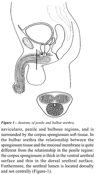
Anatomical
Remarks
The male urethra can be divided into 2 different
portions: the posterior urethra, which includes the membranous and the
prostatic regions, and the anterior urethra. The anterior urethra includes
navicularis, penile and bulbous regions, and is surrounded by the corpus
spongiosum soft tissue. In the bulbar urethra the relationship between
the spongiosum tissue and the mucosal membrane is quite different from
the relationship in the penile region: the corpus spongiosum is thick
in the ventral urethral surface and thin in the dorsal urethral surface.
Furthermore, the urethral lumen is located dorsally and not centrally
(Figure-1).
SURGICAL
TECHNIQUE
Preparation
of the Bulbar Urethra
The patient is placed in normal lithotomy
position, and a midline perineo-scrotal incision is made. The bulbo-cavernous
muscles are separated in the midline and, in patients with proximal bulbar
urethral stricture, the central tendon of the perineum is dissected. The
bulbar urethra is then free from the bulbo-cavernous muscles, and it is
dissected from the corpora cavernosa (Figure-2, A). The urethra is completely
mobilized from the corpora cavernosa, it is rotated 180 degrees, and is
incised along its dorsal surface (Figure-2, B). The stricture is opened
along its whole length (Figure-2, C).
Preparation
and Suture of the Graft
(Skin or Buccal Mucosa)
In patients with a shorter than 4 cm stricture,
an ovoid strip of ventral penile skin is outlined for harvesting. In patients
with a longer than 4 cm stricture, a double circumferential subcoronal
incision is made for harvesting a longer preputial skin strip. When local
epithelial foreskin is unavailable or when the patient does not agree
with harvesting from the prepuce, the buccal mucosa is preferred to other
various types of extra-genital free grafts because of its qualities. We
choose the inner cheek over the lip as a donor site, because the width
of the lip limits the size of the graft. Moreover, the buccal mucosa is
thicker and more resistant in the cheek than the buccal mucosa from the
lip. Buccal mucosa harvesting increases operative time by 1 hour. Thus,
a 2-team approach should be used. The perineal team exposes and calibrates
the strictured tract, while another team simultaneously harvests the graft
from the mouth. This procedure also increases sterilization of the surgical
field. Reduced operative time offers remarkable advantages and may prevent
troublesome complications due to prolonged lithotomy position.
The fenestrated ovoid preputial skin or
buccal mucosa graft is spread-fixed and quilted to the overlying tunica
albuginea of the corporal bodies (Figure-2D). The right mucosal margin
of the opened urethra is sutured to the right side of the patch graft,
spaying open the strictured tract to the new roof, which is the spread,
fixed graft (Figure-2E). The urethra is rotated back into its original
position (Figure-2E). The left urethral margin is sutured to the left
side of the patch graft and to the corporal bodies, and the grafted area
is entirely covered by the urethral plate (Figure-2F). The bulbo-cavernous
muscles are approximated over the grafted area. A small suction drain
is placed, and an indwelling 16F silicone Foley catheter is left in place.
Suprapubic cystostomy is unnecessary.
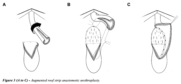
In patients with a stricture that requires
a complete removal of the scar, the urethra is completely transected.
The distal urethra is mobilised from the underlying corpora cavernosa.
The proximal mucosal edge is spatuled and spayed over the corpora cavernosa,
and the mobilised distal urethra is widely opened along its dorsal surface
(Figure-3, A). The skin or buccal mucosa graft is spread-fixed and quilted
to the underlying corpora, and its lower margin is sutured to the proximal
mucosal edge of the urethra (Figure-3, B). The left mucosal margin of
the opened distal urethra is sutured to the left side of the graft (Figure-3,
C). The urethra is rotated back over the grafted area, sutured to the
proximal mucosal edge and to the right corpora cavernosa. The bulbo-cavernous
muscles are sutured over the bulbar urethra, and the perineal closure
is made as described.
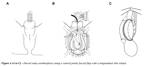
Preparation and Suture of the Flap
In patients with a stricture that
recurred after urethroplasty, or in patients who have serious ischemic
urethral disease or damage, it may be useful the use of a pedicled flap
owing to improved graft take and neovascularization. A midline perineal
incision is made, and a longitudinal ventral penile skin island is outlined
in the penile shaft (Figure-4, A). The bulbar urethra is circumferentially
mobilized from the corpora cavernosa and rotated 180 degrees (Figure-4,
B). The longitudinal penile skin island carried on the ventral dartos
fascial pedicle (Figure-4, B). The fascial pedicle is extensively dissected
and mobilized to allow the transposition of the skin island into the perineum,
using a finger-made tunnel (Figure-4, B). The bulbar urethra is opened
along its dorsal surface; the skin island is sutured to the corpora cavernosa
(Figure-4, C). The urethra is rotated back over the island flap, and the
grafted area is covered by the urethral plate.
Postoperative
Course
On the day after the surgery the drain is
removed, and the patient is discharged from hospital. Three weeks later,
the bladder is filled with contrast medium, the Foley catheter is removed
and a voiding cystourethrography is obtained. Uroflowmetry and urine culture
is repeated every 4 months during the first year and yearly thereafter.
Radiological studies are repeated when uroflowmetry is less than 14 mL
per second. Clinical outcome was considered a failure in any case postoperative
instrumentation was needed, including dilatation.
Intraoperative,
Perioperative and Postoperative Complications
In patients who have undergone repeated
internal urethrotomies it may be difficult or impossible to separate the
urethra from the corpora cavernosa, and the tunica albuginea may be opened
or injured. In this case it is important to realise that there is damage
on the corpora cavernosa and the opening must be repaired immediately.
If it is difficult to free the urethra from the corpora, a lateral or
ventral opening can be made into the urethral lumen. In this case, it
might be better to use buccal mucosa instead of preputial skin as a graft.
In patients who have undergone an augmentation
urethroplasty for a longer than 6 cm stricture, the presence of an urethral
fistula or an extravasation of the contrast medium can be observed during
the first radiological investigation. In this case a 14F Foley silicone
catheter should be left in place, and a new radiological study should
be performed 2 weeks later.
Long-term Results and Attrition Rate of Dorsal Onlay Graft Urethroplasty
Table-1 summarizes the results from 5 different
series of a total of 176 patients with a follow-up of 19 to 43 months
(8-10,13,14). The overall success rate ranged from 85% to 97% (8-10,13,14).
Any patient who required postoperative instrumentation - including periodic
dilation – was considered a treatment failure.
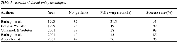
In 1998, we reported our results in 37 patients
who had undergone dorsal onlay procedures with prepuce (n = 31) or buccal
mucosa (n = 6) (13). With a mean follow-up of 21.5 months, the procedure
was classified as successful in 34 (92%) patients (13). In 2001, with
a mean follow-up pf 21.5 to 48 months in 40 patients with preputial grafts,
the success rate declined to 85% (14). The latter series did not include
patients who had a buccal mucosa graft because the follow-up period was
insufficient in these cases (14).
COMMENTS
Surgical
repair of bulbar urethral strictures is based on anastomotic repair for
short lesions, while free grafts (preputial skin or buccal mucosa) or
pedicled flaps are suggested for longer and complex strictures.
Current techniques adopt graft or flap apposition
on the ventral surface of the urethra, but this graft often lacks the
mechanical support of a fixed bed, which allows it to fold on itself,
reducing the opportunity of neovascularization, and decreasing the calibre
of the reconstructed urethra (8). Moreover, sacculation at the graft side
may occur, causing post voiding dribbling and ejaculatory failure. Sequestration
of semen and residual infected urine inside the pseudodiverticulum may
further compromise the state of the adjacent urethra and facilitate recurrent
stricture disease (8,15-19).
Recently, buccal mucosa grafts have been
used instead of preputial skin in a wide series of patients who were treated
with ventral onlay patch urethroplasty. The incidence of mechanical weakening
of the buccal mucosa graft is actually unknown. Buccal mucosa is thicker
and has a higher density of elastic fibres than preputial skin; it is
probably more resistant to mechanical weakening over time. A long-term
follow-up of these cases is mandatory.
The dorsal approach to strictures of the
bulbar urethra is anatomically simpler than the ventral one, requiring
less extensive opening of the spongy tissue since the urethral lumen is
located dorsally in this region (Figure-1) (5). Using this approach, significant
bleeding from the corpus spongiosum is avoided and the mechanical weakening
of the graft is unlikely. A serious complication of free graft urethroplasty
is the necrosis of the patch, caused by vascularization failure from its
bed. When this occurs in ventrally placed grafts, an urethro-perineal
fistula of considerable size is inevitable; this effect did not occur
in patients treated with dorsal graft apposition (3). The dorsal placement
of the graft provides a better opportunity for roof-strip epithelial regeneration,
according to the principles described by different authors (20-24). The
reported experience with dorsal onlay graft urethroplasty shows that graft
take is also adequate in the corporal bodies, although the opened corpus
spongiosum may contribute to vascularization on both sides of the graft
(2-14).
The Barbagli procedure has many advantages
(Table-2). Dorsal onlay graft urethroplasty is a versatile procedure which
may be combined with various substitute materials like preputial skin,
buccal mucosa grafts or pedicled flaps. Other substitute materials like
human urethral mucosa from corpses or collagen matrix will be possible
in the future.

Long-term results of a wide series of patients
showed a final success rate of 92% to 97%. Any substitution urethroplasty
deteriorates over time. In our series of patients the success rate of
dorsal onlay graft urethroplasty decreased from 92% to 85% with an extended
follow-up from 21.5 to 43 months.
With regard to substitute material concerns
(buccal mucosa versus preputial skin), a long-term follow-up is mandatory
to establish whether buccal mucosa is superior to foreskin as an urethral
substitute material.
At present we currently use both according
to the patient’s preference, to the status of the genital tissues
or to stricture characteristics.
REFERENCES
- Santucci RA, Mario LA, McAninch JW: Anastomotic urethroplasty for bulbar urethral stricture: analysis of 168 patients. J Urol. 2002; 167: 1715-9.
- Barbagli G, Menghetti I, Azzaro F: A new urethroplasty for bulbar urethral strictures. Acta Urol Ital. 1995; 9: 313-7.
- Barbagli G, Selli C, di Cello V, Mottola A: A one-stage dorsal free graft urethroplasty for bulbar urethral strictures. Br J Urol. 1996; 78: 929-32.
- Barbagli G, Selli C, Tosto A, Palminteri E: Dorsal free graft urethroplasty. J Urol. 1996; 155: 123-6.
- Iselin CE, Webster GD: Dorsal onlay graft urethroplasty for repais of bulbar urethral stricture. J Urol. 1999; 161: 815-8.
- Palminteri E, Lazzeri M, Guazzoni G, Turini D, Barbagli G: New 2-stage buccal mucosa graft urethroplasty. J Urol. 2002; 167: 130-2.
- Jordan GH: Anterior urethral reconstruction: concepts and concerns. Cont Urol. 1998; 10: 80-96.
- Iselin C, Webster GD: Dorsal onlay graft urethroplasty for repair of bulbar urethral strictures. J Urol. 1999; 161: 815-8.
- Guralnick ML, Webster GD: The augmented anastomotic urethroplasty: indications and outcome in 29 patients. J Urol. 2001; 165: 1496-501.
- Andrich DE, Leach CJ, Mundy AR: The Barbagli procedure gives the best results for patch urethroplasty of the bulbar urethra. BJU Int. 2001; 88: 385-9.
- Andrich DE, Mundy AR: Surgery of urethral stricture disease. Cont Urol. 2001; 13: 32-44.
- Barbagli G, Palminteri E, Lazzeri M: Dorsal onlay techniques for urethroplasty. Urol Clin North Am. 2002; 29: 389-95.
- Barbagli G, Palminteri E, Rizzo M: Dorsal onlay graft urethroplasty using penile skin or buccal mucosa in adult bulbo-urethral strictures. J Urol. 1998; 160: 1307-9.
- Barbagli G, Palminteri E, Lazzeri M, Guazzoni G, Turini D: Long-term outcome of urethroplasty after failed urethrotomy versus primary repair. J Urol. 2001; 165: 1918-9.
- Barbagli G, Selli C, Tosto A: reoperative surgery for recurrent strictures of the penile and bulbous urethra. J Urol. 1996; 156: 76-7.
- Pansadoro V, Emiliozzi P: Wich urethroplasty for wich results? Curr Opin Urol. 2002; 12: 223-7.
- Morey AF, Duckett CP, McAninch JW: Failed anterior uretrhoplasty: guidelines for reconstruction. J Urol. 1997; 158: 1383-7.
- Mundy AR: Results and complications of urethroplasty and its future. Br J Urol. 1993; 71: 322-5.
- Webster GD, Robertson CN: The vascularized skin island urethroplasty: its role and results in urethral stricture management. J Urol. 1985; 133: 31-3.
- Browne D: An operation for hypospadias. Proc Roy Soc Med. 1949; 42: 466-8.
- Weaver RG, Schulte JW: Experimental and clinical studies of urethral regeneration. Surg Gynec Obst. 1962; 115: 729-36.
- Weaver RG, Schulte JW: Clinical aspects of urethral regeneration. J Urol. 1965; 93: 247-54.
- Moore CA: One-stage repair of stricture of bulbous urethra. J Urol. 1963; 90: 203-7.
- Monseur J: L’elargissement de l’urètre au moyen du plan sou-urètral. J Urol.[Paris] 1980; 6: 439-42.
_____________________
Received: August 8, 2002
Accepted after revision: November 2, 2002
_______________________
Correspondence address:
Dr. André Guilherme Cavalcanti
Av. N.S. de Copacabana, 1066 / 1109
Rio de Janeiro, RJ, 22060-010, Brazil
Fax: + 55 21 2521-0893
E-mail: andre70211@hotmail.com