ASSESSMENT
OF SERUM LEVEL OF PROSTATE-SPECIFIC ANTIGEN ADJUSTED FOR THE TRANSITION
ZONE VOLUME IN EARLY DETECTION OF PROSTATE CANCER
(
Download pdf )
MARCOS D. FERREIRA, WALTER J. KOFF
Division of Urology, General Hospital of Porto Alegre, Federal University of Rio Grande do Sul, UFRGS, Porto Alegre, Rio Grande do Sul, Brazil
ABSTRACT
Objective:
To determine the clinical usefulness of prostate-specific antigen (PSA)
density in the transition zone (PSADTZ) for increasing the specificity
in early detection of prostate cancer (PCa) and reducing unnecessary biopsies
in males with PSA between 4.0 and 10 ng/mL.
Materials and Methods: This cross-sectional
study obtained PSADTZ measurements in 68 patients with PSA between 4.0
and 10 ng/mL. All patients underwent transrectal ultrasonography (TRUS)
with biopsies. PSADTZ was estimated by dividing the PSA value by the volume
of the transition zone (TZ) obtained. We compared performance measurements
for these parameters with those from the PSA itself, PSA density (PSAD)
and free PSA/total PSA ratio (F/T PSA). The ability of the method in increasing
PSA specificity was demonstrated and compared in univariate and multivariate
analyses, and by Receiver Operating Characteristic Curves (ROC).
Results: Of the 68 patients under study,
17 (25%) were diagnosed with PCa. The TZ volume (p = 0.001) and PSADTZ
(p = 0.001) variables presented means that exhibited statistically significant
differences. When compared with the area under the curve (AUC), ROC curves
obtained by this method revealed that PSADTZ was the strongest predictor
for PCa when considering the cut-off point provided by the curve; that
is, 0.35 ng/mL/cc. When PSADTZ was employed, the detection failure would
be close to 20%, and less than 45% of cases would undergo unnecessary
biopsies. On the other hand, when F/T PSA was used, the loss would reach
almost 40%; however less than 30% would undergo unnecessary biopsies.
Nevertheless, PSADTZ had the only AUC presenting p < 0.05 in significance
when compared with 50%, and was consequently discriminative.
Conclusions: PSADTZ increased PSA specificity
in early detection of PCa in males with PSA between 4.0 and 10 ng/mL.
However, it was shown to have lower predictive value and lower accuracy
than the percentage of free PSA since it presents a higher negative predictive
value than all other parameters assessed, and it can be considered clinically
useful for reducing unnecessary indications for biopsy.
Key
words: prostatic neoplasms; diagnosis; prostate-specific antigen;
neoplasm staging
Int Braz J Urol. 2005; 31: 137-46
INTRODUCTION
Prostate
cancer (PCa) is considered the most frequent neoplasia in men and the
second cause of death by cancer in males. Over the past 12 years, the
incidence of PCa has increased over 50% and deaths from the disease risen
40% (1).
The isolation of the prostate-specific antigen
(PSA) in 1979 led to a dramatic change in the diagnosis and management
of PCa (2). PSA is the most sensitive serum marker in males with prostate
disease. Though periurethral glands are known to release PSA into the
urine, it is usually accepted that, at least for practical purposes, PSA
is produced exclusively by prostate epithelial cells (3).
Therefore, PSA has proven its usefulness
as a serum marker for PCa. However, PSA sensitivity and specificity are
not yet enough to turn it into the ideal screening test for PCa, since
increased levels can also be seen in prostatitis and benign prostate hyperplasia
(BPH) (4). The majority of PSA that is released by the prostate into the
serum comes from the transition zone and BPH results almost exclusively
from the transition zone hyperplasia. Kalish et al. (5) introduced the
concept of serum PSA adjusted for the transition zone volume (called PSADTZ)
and some studies, like his own, have suggested higher accuracy of this
method for early detection of PCa. From all the referenced studies, we
can extract a clear impression that their conclusions are not definitive.
The introduction of this new concept opens a wide range of new diagnostic
possibilities; however, though its usefulness has been statistically demonstrated,
it must still be confirmed and validated through other studies that are
equally well controlled and methodologically well conducted (reproducible).
In this study, we intend to assess the potential
role of PSADTZ in the early diagnosis of PCa in our setting through measurement
of early detection from a screening study.
MATERIALS AND METHODS
This
research has been performed with a cross-sectional design in a screening
study. The study factor was PSADTZ and other tests for early diagnosis
of PCa, and the end-point was the pathological identification of BPH or
PCa.
The sample consisted of 68 patients undergoing
screening for early diagnosis of PCa and with indication of transrectal
ultrasound (TRUS) with biopsy (altered PSA and/or digital rectal examination).
In this research, population, in relation to PSA, only individuals with
total PSA values between 4.0 and 10 ng/mL were included.
The exclusion criteria were a total PSA
lower than 4.0 ng/mL or higher than 10 ng/mL, patient refusal to participate
in any assessment procedure, a history of PCa, prostatitis, prostate intraepithelial
neoplasia (PIN), urinary tract infection, bladder catheterization or urinary
retention and previous prostate surgery.
Variables under study were total prostate
volume, transition zone volume, total PSA, free PSA, free-to-total PSA
ratio, age, PSADTZ, result of biopsy and digital rectal examination.
All patients from the screening study were
assessed for lower urinary tract symptoms by completing the international
prostate symptom score, then had their blood collected for determining
PSA levels and underwent digital rectal examination.
TRUS with prostate biopsy was indicated
for patients who presented changes in their digital rectal examination,
which suggested the presence of PCa, and for those with total PSA higher
than 4.0 ng/mL. Also, regardless of any adjustments for age, their free
PSA was dosed in serum before the TRUS. The serum PSA level was determined
by radioimmunoassay method performed on serum collected before digital
rectal examination and analyzed with the Immulite-PSA kit (CA, USA).
A digital rectal examination was classified
as suspected or non-suspected for neoplasia.
The TRUS measured the total prostate volume
and the specific volume of the transition zone (TZ) through a bi-planar
2-probe transductor with 7.5 MHz in the sagittal probe and 6.5 MHz in
the transversal probe, coupled to a model AI 5200 S Envision Acoustic
Imaging device (Dornier Co.). Prostate was measured in its transversal
and sagittal planes using the formula for estimating the ellipsoid volume
(width x length x height x 0.52). Once the measures were obtained, sextant
puncture biopsies were systematically performed using a 16-gauge needle
mounted on a Pro-Mag 2.2 automatic pistol.
Biopsy results were generically classified
as PCa or BPH and/or prostatitis.
The value of PSADTZ was estimated by dividing
the serum PSA level value (between 4.0 and 10 ng/mL) by the value obtained
in an echographic measurement of the TZ volume and expressed as ng/mL/cc.
As a post-diagnosis approach, individuals
who were identified as having PCa were referred to the staging protocol
and their therapeutic follow-up was based on the protocol’s findings.
Statistical
Analysis
Receiver Operating Characteristic (ROC)
curves were used to graphically demonstrate the sensitivity and specificity
of the assessed tests (PSA parameters), over a variety of “cut-off”
points. The area under the curve (AUC) was calculated using PEPI version
3.0 (Computer Programs for Epidemiologists by J.H. Abramson and P.M. Gahlinger).
Different AUCs were compared as described
by Hanley & McNeil, using the correlation coefficient corrected for
AUC as a test for comparing proportions, while the McNemar test uses data
that have been previously categorized by their cut-off points.
Wilcoxon-Mann-Whitney U test (WMW) was used
for non-parametric analysis of intergroup comparison of data from patients
with and without PCa, considering the analysis of means and medians. The
relationship between variables was analyzed using Spearman’s correlation
coefficient.
On the other hand, the linear correlation
coefficient (Pearson Product-Moment) was calculated in order to assess
the association intensity between total gland volume, TZ volume and PSA.
Finally, we used the multivariate logistic
regression analysis model in the “stepwise” system for assessing
PSA parameters in relation to its ability to predict PCa by applying systematic
selection procedures based on data from the general and negative digital
rectal examination groups. The statistical significance level established
for all described tests was p < 0.05.
RESULTS
Of
the 68 assessed patients, 17 were diagnosed as having PCa, and BPH was
detected in 51. The digital rectal examination revealed changes suggestive
of malignant neoplasia in 15 patients (22.1%) where the presence of PCa
was confirmed in only 4 (26.7%). Additionally, among patients with normal
(unsuspected) digital rectal examination, histology compatible with malignancy
was subsequently found in 13 (24.5%) cases. These findings indicate that,
in this study group, due to its low sensitivity (23%), the digital rectal
examination was not good as a method for early diagnosis of PCa, despite
its good specificity (78%) and low positive (29%) and negative (24%) predictive
value.
When considering their asymmetrical distribution,
mean values for parameters in both groups, cancer and benign disease,
did not show statistically significant differences for the variables age,
PSA, total gland volume and PSAD. On the other hand, the variables TZ
volume (p = 0.001) and PSADTZ (p = 0.001) presented means that revealed
statistically significant differences in both groups. For estimating the
means, the p value considered the Levene’s test for equality of
variances when applying the t test for independent samples (Table-1).

In relation to the median analysis, the
only variables presenting statistical differences were TZ volume (p =
0.0002) and PSADTZ (p = 0.0012) (Table-2).

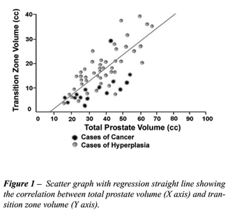
Bivariate analysis of correlations between
total prostate volume and TZ volume using the scatter plot presented a
regression straight line (Figure-1) as demonstrated by the plot’s
points, suggesting a strong correlation between them (r = 0.825, p = 0.0001).
On the other hand, when analyzing PSA and other volumes, the correlation
coefficient was very low both at the crossing of PSA and total volume
(r = 0.05, p = 0.664) and at the crossing of PSA and TZ volume (r = 0.01,
p = 0.928).
Data on each parameter shown in the Tables
demonstrating performance measurements were obtained from the best cut-off
value for each parameter (which, in turn, was determined by the ROC curve),
considering both the general and the negative digital rectal examination
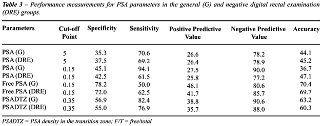
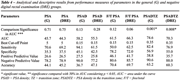
groups (Table-3). In relation to absolute values in the negative digital
rectal examination group, the AUC of PSADTZ presented higher absolute
values than did F/T PSA, in addition to being more significant (considering
the significance compared with 50%). Its specificity, positive predictive
value (PPV) and accuracy are surpassed by the performance f F/T PSA; however,
its sensitivity and its negative predictive value (NPV) are clearly superior.
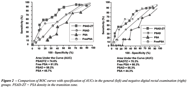
When compared, ROC curves obtained for each
parameter from their specific cut-off values revealed that PSADTZ was
the strongest predictor of PCa when considering the cut-off point in the
upper left area of the curve – that is 0.35 ng/mL/cc (Figure-2).
It means that when considering the best cut-off point for PSAD (0.15 ng/mL/cc),
in the group with negative digital rectal examination approximately 40%
of PCa cases would be missed (since sensitivity was approximately 60%)
and about 60% would undergo unnecessary biopsies (since specificity was
approximately 40%). Using the PSADTZ parameter, the missing rate would
be close to 20% and less than 45% of patients would undergo unnecessary
biopsies.
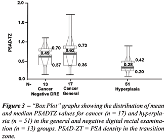
The “box plot” percentile graphs
demonstrated central trend values for PSADTZ, whose median was 0.62 ng/mL/cc
for PCa and 0.28 ng/mL/cc for BPH. The cut-off value for PSADTZ was 0.36
ng/mL/cc, which corresponded to values between the 75th percentile for
cancer (75% of cases of PCa were above this value) and the 25th percentile
for benign disease (75% of cases of BPH were below this value). When considering
the values obtained by the same analysis, we verified a statistically
significant difference between PSADTZ values for both groups (p = 0.0012),
Figure-3.
Considering the ROC curves for each parameter
in the parameter analysis, PSA presented a smaller area (AUC = 45.7%)
and low specificity (35%), while PSADTZ showed a larger area (AUC = 74.6%)
and higher specificity (56.9%) and was the only parameter to present significance
compared with 50% at significant levels; that is, it was discriminative.
At its best cut-off point (0.35 ng/mL/cc) whose sensitivity reached 82.4%,
there would be a more accentuated reduction in the number of unnecessary
biopsies (over 55%) in comparison with other diagnostic tests (PSA parameters),
Table-4.
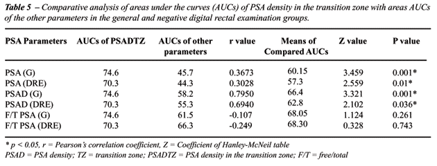
In the comparative analysis of the respective
AUCs, using the Hanley-McNeil method, statistically significant differences
were observed between the AUC of the PSADTZ parameter and those of the
PSA and PSAD parameters (Table-5). The area under the curve of the parameter
F/T PSA, however, was not statistically different from the PSADTZ, thus
suggesting a similar overall performance for both tests in increasing
PSA specificity.
Results obtained with the multiple logistic
regression analysis model for prediction of PCa, which applied the “stepwise”
selection procedure based on the 4 variables, excluded non-significant
variables – that is PSA and PSAD – demonstrating that PSADTZ
and free-to-total PSA ratio were the strongest and most significant predictors
of PCa. When considering values obtained at the best cut-off points in
the original ROC curves, the odds ratio was 9.07 (with p = 0.032) for
PSADTZ and 7.45 (with p = 0.027) for free-to-total ratio. When the assessment
was performed using cut-off points with highest specificity of the ROC
curve, odds ratio was 13.71 (p = 0.005) and 6.33 (p = 0.069) respectively,
showing statistical superiority of PSADTZ in predicting PCa (Table-6).

COMMENTS
Studies
have indicated that 2/3 of patients undergoing biopsy based exclusively
on the finding of an intermediary PSA value show no histological evidence
of PCa (6). In this respect, several new concepts have been introduced
regarding PSA, all of them intended to optimize the clinical usefulness
of PSA by increasing its sensitivity and specificity and, thus, trying
to reduce the numbers of unnecessary biopsies in men with benign disease.
These new concepts include PSA density, PSA velocity, PSA adjusted for
age, and determination of molecular forms of PSA (free versus conjugated
to proteins) (7,8).
For this purpose, the concept of PSADTZ
has been recently suggested as a valuable approach. When comparing the
results from the assessment of mean and median values for the parameters
obtained in our study with those demonstrated by the majority of reviewed
authors, we found an important similarity in the significant differences
obtained in the 2 subgroups (PCa and BPH) for the variables TZ volume
and PSADTZ (6-8).
On bivariate analysis of correlations, results
observed in the literature have presented correlation coefficients between
the variables total volume and TZ volume that, when compared, confirm
those obtained in our study (7,9-11). Comparative analyses of the areas
under ROC curves of several parameters demonstrate that higher indexes
are observed by the PSADTZ parameter, both in the total group and in the
segment’s negative digital rectal examination, volume < 30cc
and volume > 30 cc. These results are comparable in the total group
to those found by other authors (6,7,9-13). However, in a deviation from
the study by Maeda et al. (7) who detected a higher index in the group
with negative digital rectal examination, the present project did not
attain such an outcome. On the other hand, Lin et al. (15), Akduman et
al. (16), Gohji et al. (17) and Rietbergen et al. (18) did not find significant
statistical differences between the AUCs of PSAD and PSADTZ, even if they
used different sampling criteria and statistical methods in relation to
our study.
When using the method described by Hanley
and McNeil for comparison between the AUC parameters estimated both for
the total group and for the group with negative digital rectal examination,
we observed that they showed significant differences, especially when
comparing PSADTZ with the PSA and PSAD parameters. Theses differences
have been demonstrated by other studies as well, thus revealing the high
predictive power of this diagnostic test (6,7,10,12).
On the other hand, when PSADTZ was compared
with the F/T PSA parameter, no statistically significant difference was
found in either group (p = 0.261 and p = 0.743, respectively). The same
result has been obtained by the majority of previous studies observing
the absolute value of AUC for PSADTZ (6,7,9,11,13,14). Searching for a
cut-off limit for all parameters that maintained the sensitivity for cancer
detection around minimal limits of 90 to 95%, values were defined that,
in addition to being close to those evidenced in the literature (except
for F/T PSA), presented very similar corresponding specificity as well
with no exceptions. At 0.3 ng/mL/cc, PSADTZ reached 47% of specificity,
the same one obtained by the cut-off value of 0.25 in the study by Djavan
et al. (6).
Finally, by applying the “stepwise”
selection procedure which has been performed by a few authors, our results
were in agreement with those demonstrated by Djavan et al. (6,12) in 2
of their studies (n = 559 and n = 1051 respectively) where the same exclusions
for the PSA and PSAD parameters were performed due to their low predictive
value in both studies, thus revealing the F/T PSA and PSADTZ parameters
as the strongest and highly predictive for PCa. This result was also similar
to the one presented by Moon et al. (9).
Among limitations observed in our analysis
is the sample “n” itself, which, despite being regarded as
reduced and consequently presenting a wide confidence interval, was surprisingly
able to provide statistical results that were comparable to larger series.
Analyses related to the predictive ability for extraprostatic disease
were not performed as well, despite being the object of study in some
of the reviewed references (19).
The need for cost-effectiveness studies
is evident as the main components of this diagnostic test involve the
performance of 2 procedures, which, in turn, have their specific costs
– even if we can infer a decreased cost when considering savings
due to fewer indications of TRUS with biopsies, its own associated costs,
and risks that are certainly more relevant. Another fact to be considered,
which is still unedited, is the proposal for using the method presented
here in the routine for performance of transrectal biopsies in patients
with indication who, in case of a benign diagnosis even in the presence
of suggestive changes (PSA and/or digital recital examination) could benefit
from another parameter for increasing the specificity of PSA when altered
values are maintained. When considering the possibility in the near future
that these patients undergo a new invasive procedure that is not exempt
from risks, we must remember the recent studies published by Okegawa et
al. (20) and Singh et al. (21), showing that PSADTZ, as well as advanced
age and presence of high grade PIN, is one of the strongest predictors
for PCa in subsequent biopsies following the initial biopsy.
CONCLUSION
The PSA parameter known as PSADTZ has shown itself to possess a statistical power for increasing the specificity, positive predictive value, negative predictive value and accuracy, which, in addition to being similar to performance measures verified in the comparative analysis with F/T PSA, were superior to the latter in some analyses. The best cut-off point established by our study was 0.35 ng/mL/cc.
REFERENCES
- Parker SL, Tong T, Bolden S, Wingo PA: Cancer statistics, 1996. CA Cancer J Clin. 1996; 46: 5-27.
- Schellhammer PF, Wright GL: Biomolecular and clinical characteristics of PSA and other candidate prostate tumor markers. Urol Clin North Am. 1993; 4: 597-606.
- Noldus J, Stamey TA: Limitations of serum prostate specific antigen in predicting peripheral and transition zone cancer volumes as measured by correlation coefficients. J Urol. 1996; 155: 232-7.
- Partin AW, Oesterling JE: The clinical usefulness of prostate specific antigen: update 1994. J Urol. 1994; 152: 1358-64.
- Kalish J, Cooner WH, Graham SD Jr.: Serum PSA adjusted for volume of transition zone (PSAT) is more accurate than PSA adjusted for total gland volume (PSAD) in detecting adenocarcinoma of the prostate. Urology. 1994; 43: 601-7.
- Djavan B, Zlotta AR, Byttebier G, Shariat S, Omar M, Schulman CC, et al.: Prostate specific antigen density of the transition zone for early detection of prostate cancer. J Urol. 1998; 160: 411-8; discussion 418-9.
- Maeda H, Arai Y, Ishitoya S, Okubo K, Aoki Y, Okada T: Prostate specific antigen adjusted for the transition zone volume as an indicator of prostate cancer. J Urol. 1997; 158: 2193-6.
- Catalona WJ, Smith DS, Wolfert RL, Wang TJ, Rittenhouse HG, Ratliff TL, et al.: Evaluation of percentage of free serum prostate-specific antigen to improve specificity of prostate cancer screening. JAMA. 1995; 274: 1214-20.
- Moon DG, Cheon J, Kim JJ, Yoon DK, Koh SK: Prostate-specific antigen adjusted for the transition zone volume versus free-to-total prostate-specific antigen ratio in predicting prostate cancer. Int J Urol. 1999; 6: 455-62.
- Kurita Y, Terada H, Masuda H, Suzuki K, Fujita K: Prostate specific antigen (PSA) value adjusted for transition zone volume and free PSA (gamma-seminoprotein) /PSA ratio in the diagnosis of prostate cancer in patients with intermediate PSA levels. Br J Urol. 1998; 82: 224-30.
- Michielsen DP, De Boe VR, Braeckman JG, Keuppens FI: Specificity and accuracy of TRUS-measured PSA-density and transition zone-PSA in the diagnosis of prostate cancer. Eur J Ultrasound. 1998; 8: 125-8.
- Djavan B, Zlotta A, Remzi M, Ghawidel K, Basharkhah A, Schulman CC, et al.: Optimal predictors of prostate cancer on repeat prostate biopsy: a prospective study of 1,051 men. J Urol. 2000; 163: 1144-1148; discussion 1148-49.
- Kikuchi E, Nakashima J, Ishibashi M, Ohigashi T, Asakura H, Tachibana M, et al.: Prostate specific antigen adjusted for transition zone volume: the most powerful method for detecting prostate carcinoma. Cancer. 2000; 8: 842-9.
- Roumeguere T, Zlotta AR, Djavan BR, Marberger M, Schulman CC: PSA level of the transitional zone: a new marker especially reliable for the detection of prostatic cancer. Acta Urol Belg. 1997; 65: 5-9.
- Lin DW, Gold MH, Ransom S, Ellis WJ, Brawer MK: Transition zone prostate specific antigen density: lack of use in prediction of prostatic carcinoma. J Urol. 1998; 160: 77-81; discussion 81-2.
- Akduman B, Alkibay T, Tuncel A, Bozkirli I: The value of percent free prostate specific antigen, prostate specific antigen density of the whole prostate and of the transition zone in Turkish men. Can J Urol. 2000; 7: 1104-9.
- Gohji K, Nomi M, Egawa S, Morisue K, Takenaka A, Okamoto M, et al.: Detection of prostate carcinoma using prostate specific antigen, density and the density of the transition zone in Japanese men with intermediate serum prostate specific antigen concentrations. Cancer. 1997; 79: 1969-76.
- Rietbergen JB, Kranse R, Hoedemaeker RF, Kruger AE, Bangma CH, Kirkels WJ, et al.: Comparison of prostate-specific antigen corrected for total prostate volume and transition zone volume in a population-based screening study. Urology. 1998; 52: 237-46.
- Horiguchi A, Nakashima J, Horiguchi Y, Nakagawa K, Oya M, Ohigashi T, et al.: Prediction of extraprostatic cancer by prostate specific antigen density, endorectal MRI, and biopsy Gleason score in clinically localized prostate cancer. Prostate. 2003; 56: 23-9.
- Okegawa T, Kinjo M, Ohta M, Miura I, Horie S, Nutahara K, et al.: Predictors of prostate cancer on repeat prostatic biopsy in men with serum total prostate-specific antigen between 4.1 and 10 ng/mL. Int J Urol. 2003; 10: 201-6.
- Singh H, Canto EI, Shariat SF, Kadmon D, Miles BJ, Wheeler TM, et al.: Predictors of prostate cancer after initial negative systematic 12 core biopsy. J Urol. 2004; 171: 1850-4.
__________________________
Received:
September 20, 2004
Accepted after revision: March 17, 2005
_______________________
Correspondence address:
Dr. Marcos Dias Ferreira
Rua Ladislau Neto, 474 / casa 01, Bairro Ipanema
Porto Alegre, RS, 91760-070, Brazil
Fax: + 55 51 3249-9653
In this study, the authors used ultrasound to determine the volume of the transition zone where BPH would be expected to occur and where cancer is less likely to occur. When the PSA is factored by the volume of the transition zone, they found that the PSA density in transition zone was more predictive of prostate cancer than either serum total PSA or PSA density as routinely determined in patients with a PSA of 4 to 10 ng %. The percentage of free PSA and transition zone PSA density enhance the specificity of PSA when determining which patients should undergo repeat biopsy. Overall, 25% of the patients were diagnosed with prostate cancer. The high cancer rate can be explained by the fact that patients included in this study were referred for early diagnosis and not for screening purposes. Accuracy of transition zone volume measurement is ultrasonographer-dependent, which may influence the reproducibility of the procedure. Of some concern may be the fact that the predictive power of the PSA transition zone is significantly affected by prostate size in prostates weighing less than 30g in which the transition zone is not markedly enlarged and is therefore more difficult to measure. The usefulness of the PSA transition zone density is significantly diminished and less accurate than free-to-total PSA (1). Finally, unlike PSA parameters, PSA density in the transition zone requires the use of transrectal ultrasound, escalating the costs of the test.
REFERENCE
1. Djavan B, Zlotta A, Kratzik C, Remzi M, Seitz C, Schulman CC, et al.: PSA, PSA density, PSA density of transition zone, free/total PSA ratio, and PSA velocity for early detection of prostate cancer in men with serum PSA 2.5 to 4.0 ng/mL. Urology. 1999; 54: 517-22.
Dr. Valdemar Ortiz
Division of Urology
Federal University of Sao Paulo, UNIFESP
São Paulo, SP, Brazil