RENAL
CELL CARCINOMA PRESENTING AS A CERVICAL MASS
(
Download pdf )
ANTONIO C. POMPEO, HIDEKI KANASHIRO, MATHEUS N. SILVA
Department
of Urology, General Hospital, School of Medicine, University of Sao Paulo,
USP,
Sao Paulo, SP, Brazil
ABSTRACT
The
authors report a case of a 60-year-old woman presenting with a renal cell
carcinoma in which the first sign leading to its diagnosis was a cervical
metastasis, an uncommon site of distant disease in renal neoplasms.
The patient had an 18-month history of a
progressively enlarging cervical mass at the anterior aspect of the neck.
After laboratory and radiological evaluation, the cervical mass was excised,
and the microscopic and immunohistochemical patterns suggested the possibility
of a metastatic renal cell carcinoma. Computerized tomography of the abdomen
showed a solid, 4 cm left renal mass. A radical left nephrectomy was performed,
and the histology confirmed the suspected diagnosis. The patient received
immunotherapy, and in a follow-up period of 9 months, there was no evidence
of recurrent disease. It seems that head and neck metastasis of renal
cell carcinoma should preferentially be treated with surgical excision
because of the associated morbidity and quality-of-life issues.
Key
words: kidney neoplasms; renal cell, carcinoma; neoplasms metastasis;
head and neck
Int Braz J Urol. 2005; 31: 151-2
INTRODUCTION
Renal cell carcinoma represents approximately 3% of all adult malignancy. The most common site of metastases is bone and lung, but it has been documented to metastasize to every organ and site in the body (1). Renal cell carcinoma is the third most common infraclavicular neoplasm to metastasize to head and neck following lung and breast carcinoma. The authors report a case of a patient who had a cervical mass as the first sign leading to diagnosis of renal cell carcinoma.
CASE REPORT
A
60-year-old woman was referred to the head and neck surgery service of
our hospital with an 18-month history of a progressively enlarging cervical
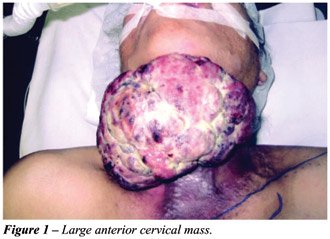
mass at the anterior aspect of the neck. Physical examination disclosed
a friable, ulcerated, 20 cm anterior cervical mass (Figure-1). A plain
radiography of the thorax was normal.
Computerized tomography of the neck showed
a solid mass in the superficial space of the neck, invading pre-laryngeal
muscles in the visceral space. Incisional biopsy was inconclusive.
A wedge resection of the cervical mass was
performed, and a microsurgical antero-lateral thigh flap closed the wound.
Microscopic examination was suggestive of metastatic carcinoma. The immunohistochemical
analysis revealed a possible renal cell carcinoma.
The patient was then referred for urological
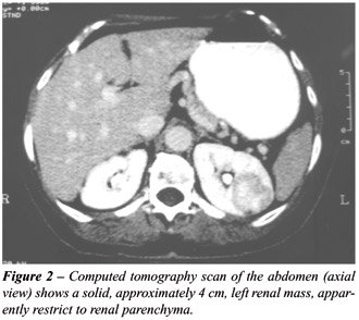
evaluation. She denied any urinary symptoms. Computed tomography of the
abdomen showed a solid, 4 cm left renal mass, which seemed restrict to
renal parenchyma (Figure-2). On physical examination, there was no abdominal
or flank mass. A radical left nephrectomy was then performed, and the
patient received immunotherapy there after. The patient has been under
continual medical observation, and there was no evidence of recurrent
disease in a follow-up period of 9 months.
COMMENTS
Metastases
commonly occur in renal cell carcinoma, about 40% of patients presenting
with metastatic disease. The most frequent sites are lung, regional lymph
nodes, bone, and liver (2). Approximately 15% of patients with renal cell
carcinoma have extracranial head and neck metastases (2). In 7.5% of patient
with renal cell carcinoma, the head and neck metastasis is the presenting
complaint. However, only 1% of patients with renal cell carcinoma have
metastases confined only to the head and neck, and solitary cervical metastatic
mass, as in our patient, is rare.
Usually, the role of surgery in metastatic
renal cell carcinoma is for diagnosis and debulking of disease. Excision
of solitary metastatic lesion of renal cell carcinoma following nephrectomy
results in a 41% survival at 2 year and 13% survival at 5 years. Pritchyk
et al. (3) consider head and neck metastasis should be viewed differently
because the lesion can lead to airway compromise, severe bleeding and
severe disfigurement. Based on the presented case, we agree with them
that depending on the site of presentation, local resection may improve
quality of life and can provide a chance for cure in the head and neck.
The experience described herein confirms
that bizarre sites of metastases from renal cell carcinoma should be kept
in mind by clinicians and surgeons. Moreover, renal cell carcinoma should
be considered in the differential diagnosis of any growing lesion in the
head and neck.
REFERENCES
- Savas MC, Celik, I, Benekli M, Gullu IH, Tekuzman G: Renal cell carcinoma presenting as a solitary cervical node metastasis compressing the brachial plexus. Nephron. 1998; 79: 107-8.
- Boles R, Cerny J: Head and neck metastases from renal cell carcinomas. Mich Med. 1971; 70: 616-8.
- Pritchyk KM, Schiff BA, Newkirk KA, Krowiak E, Deeb ZE: Metastatic renal cell carcinoma to the head and neck. Laryngoscope. 2002; 112: 1598-602.
____________________
Received: July 28, 2004
Accepted after revision: September 14, 2004
_______________________
Correspondence address:
Dr. Matheus Neves Ribeiro da Silva
Rua Dr Ovidio Pires de Campos 171 / 313
São Paulo, SP, 05403 010, Brazil
E-mail: ribeiromed@hotmail.com