LAPAROSCOPIC
PARTIAL NEPHRECTOMY FOR CANCER: TECHNIQUES AND OUTCOMES
(
Download pdf )
MAURICIO RUBINSTEIN, JOSE R. COLOMBO JR, ANTONIO FINELLI, INDERBIR S. GILL
Section of Laparoscopic and Robotic Surgery, Glickman Urological Institute, Cleveland Clinic Foundation, Cleveland, Ohio, USA
ABSTRACT
Open partial nephrectomy is the gold standard nephron-sparing treatment for small renal tumors. Technical aspects of laparoscopic partial nephrectomy have evolved considerably, and the technique is approaching established status at our institution. Over the past 4 years, the senior author has performed more than 400 laparoscopic partial nephrectomies at the Cleveland Clinic. Herein we present our current technique and review contemporary outcome data.
Key
words: kidney neoplasms; laparoscopy; surgery, conservative;
partial nephrectomy
Int Braz J Urol. 2005; 31: 100-4
INTRODUCTION
Laparoscopic partial nephrectomy (LPN) was first performed more than 10 years ago (1,2). Since the widespread use of contemporary imaging techniques has resulted in an increased detection of incidental small renal tumors, many centers have published their experiences with LPN (3-9). Initially, small exophytic tumors were selected for LPN. With increasing experience, larger, infiltrating tumors have been submitted to laparoscopic partial nephrectomy (4,5,10). Securing renal parenchymal hemostasis and sutured water-tight caliceal repair after tumor excision is paramount. In an attempt to minimize these technical problems, several new techniques and technologies have recently been explored. Herein we describe our current technique and review the outcomes of laparoscopic partial nephrectomy.
TECHNIQUE
Our
laparoscopic technique attempts to duplicate established oncological and
reconstructive principles inherent to open partial nephrectomy (10). Selection
of the laparoscopic approach depends on tumor location. Posterior or posterolateral
tumors are approached retroperitoneoscopically, while anterior, antero-lateral,
or lateral tumors are approached transperitoneally. Precise preoperative
imaging using three-dimensional computed tomography (CT) with volume-rendered
video reconstruction, and real-time intraoperative ultrasonography of
the tumor provide the surgeon with detailed information that facilitates
the laparoscopic procedure.
All patients undergo cystoscopic placement
of a 5F open ended ureteral catheter that is positioned in the renal pelvis.
A 60 cc syringe filled with dilute indigo carmine dye is attached to the
ureteral catheter. Retrograde injection via this catheter is used to confirm
collecting system entry and water-tight closure.
Transperitoneal
Approach
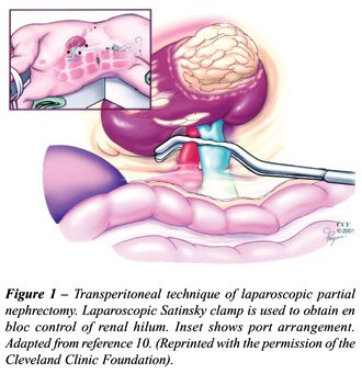
Typically, the patient is secured to the
table in a 45 to 60-degree lateral position, and a 4 or 5-port transperitoneal
approach is employed (Figure-1). The ureter and gonadal vein are identified
and retracted laterally. Dissection is carried cephalad along the psoas
muscle and the renal hilum is dissected en bloc. Gerota’s fascia
is dissected off the kidney, preserving the perirenal fat in contact with
the tumor. A laparoscopic Satinsky clamp is then positioned for hilar
clamping (Figure-1). Attention must be taken not to disrupt any lumbar
vessels in the hilum when applying the clamp. Occasionally, small, superficial,
completely exophytic tumors may be managed without hilar clamping (11).
Intraoperatively, a laparoscopic flexible
ultrasound color Doppler probe is introduced through a 10/12 mm port and
positioned in direct contact with the surface of the kidney. Information
regarding tumor size, depth of intraparenchymal extension and distance
from the collecting system is obtained. The renal capsule is scored circumferentially
with J-hook electrocautery under sonographic guidance maintaining an approximate
0.5 cm margin of normal renal parenchyma around the tumor. One to two
prepared Surgicel (Johnson & Johnson, New Brunswick, New Jersey) bolsters
and a needled suture (#1 Vicryl sutures on a CT-X needle) are introduced
into the abdomen through a 12 mm port and positioned lower down in the
paracolic gutter. Mannitol (12.5 to 25 mg) and furosemide (10 to 20 mg)
are given intravenously. If the warm ischemia time is anticipated to last
longer than 30 minutes renal hypothermia is achieved (12).
The severity of renal ischemic injury is
directly proportional to the duration of ischemia (13). Clinically, an
accepted practice during nephron-sparing surgery has been to limit warm
ischemia to £ 30 min. Regional hypothermia is often utilized when
prolonged times are anticipated. Various methods have been studied to
address this issue (12,14,15). At the Cleveland Clinic, regional hypothermia
is employed with ice slush only when prolonged ischemic times are anticipated.
In addition, adequate hydration and mannitol are administered to optimize
renal perfusion and urine output.
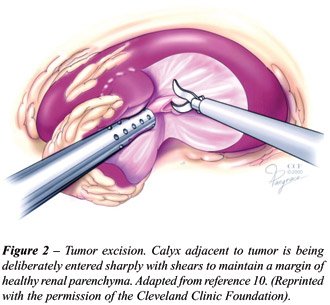
The hilum is then clamped and the tumor
excised with cold scissors. If achievement of an adequate margin requires
entry into the collecting system, the calyx or renal pelvis is divided
sharply without electrocautery. (Figure-2). An excisional biopsy of the
base is sent for frozen section analysis. The collecting system is closed
with a running 2-0 Vicryl on CT-1 needle. Injection of dilute indigo carmine
via the preplaced ureteral catheter is performed to confirm watertight
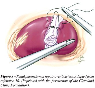
closure of the collecting system. Renal parenchymal repair is completed
using simple #1-Vicryl sutures on a CT-X needle. Briefly, interrupted
sutures are placed over the Surgicel bolster (Figure-3), a Hem-o-Lok clip
(Weck Closure System, Research Triangle Park, NC) is secured on the suture
to prevent it from pulling through, and FloSeal (Baxter, Mountain View,
CA) is applied to the cut surface beneath the bolster. The suture is then
tied, maintaining adequate compression. One or more sutures are placed
depending on the extent of resection. The Satinsky clamp is released and
complete hemostasis and renal revascularization is confirmed. The excised
tumor is placed in an impermeable sac and extracted through a minimally
extended lower abdominal port site incision. A Jackson-Pratt drain is
placed in patients where calyceal entry has occurred and laparoscopic
exit is performed.
Retroperitoneal
Approach
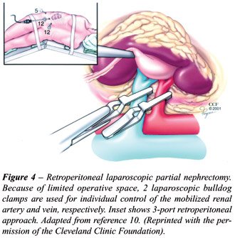
With the retroperitoneal approach, following
balloon dilation and placement of 3 (12 mm) ports, the renal artery and
vein are dissected to facilitate application of laparoscopic bulldog clamps
to each vessel (Figure-4). Recently, we have clamped the hilum en bloc
using a Satinsky clamp positioned through a separate (12 mm) trocar. Similar
to the transperitoneal approach, the tumor is excised, and renal parenchymal
repair and hemostasis are achieved, with caliceal suturing as necessary.
The bulldog clamp is removed from the renal vein initially, and then,
from the renal artery. Drain placement and exit are performed.
RESULTS
AND COMMENTS
We
have approached more than 350 LPN at our institution. A cohort of 100
patients undergoing laparoscopic partial nephrectomy was compared to a
group of 100 patients undergoing open nephron sparing surgery for a sporadic
single renal tumor of 7 cm or less at our institution (16). Since our
laparoscopic technique was based on our established open surgical principles,
the 2 approaches were similar in regards all the steps of partial nephrectomy.
The median tumor size was 2.8 cm in the laparoscopic group compared to
3.3 cm in the open group (p = 0.005). When comparing the laparoscopic
to open groups, the median surgical time was 3 vs. 3.9 h (p < 0.001);
estimated blood loss was 125 vs. 250 mL (p < 0.001); and the mean warm
ischemic time was 28 vs. 18 min (p < 0.001). The laparoscopic group
required less postoperative analgesia and experienced a shorter hospital
stay and period of convalescence (p < 0.001 for all 3 comparisons).
Although there were more intraoperative complications in the laparoscopic
group (5% vs. 0), the frequency of postoperative complications was similar
(9% vs. 14%; p = 0.27). There were 3 positive surgical margins in the
laparoscopic group and none in the open group. One of the patients had
an oncocytoma and the other 2 had renal cell carcinoma. At 2- and 3-year
follow-up, both patients have remained free of disease.
At our institution, Desai et al. (17) recently
evaluated the impact of warm ischemic renal hilar occlusion on renal function
in 179 patients after laparoscopic partial nephrectomy. Mean duration
of warm ischemia for the entire group was 31 minutes (range 4-55 min).
The study revealed no significant change in serum creatinine when dividing
patients according to duration of warm ischemia, age and or baseline serum
creatinine. In patients with a solitary kidney (N = 15), there was a transient
rise in serum creatinine in the immediate postoperative period; however,
the percent rise in serum creatinine from baseline (mean 1.3%) at latest
follow-up (mean 4.8 months) approximated the subjective amount of renal
parenchyma excised (mean 29%). Preexisting azotemia and advanced age increased
the risk of postoperative kidney dysfunction if warm ischemia time was
greater than 30 minutes.
Guillonneau et al. (18) retrospectively
performed a comparison of laparoscopic partial nephrectomy with (N = 12)
and without (N = 16) renal hilar clamping. Tumor size was larger in the
group with renal hilar clamping (2.5 vs. 1.9 cm.). The group without renal
hilar clamping was associated with a significantly greater blood loss
(708 mL vs. 270 mL, p = 0.014), and longer operative time (179 minutes
vs. 121 minutes, p = 0.004) as compared to the group with renal hilar
control. There was no significant difference in postoperative serum creatinine
(1.26 mg/dL vs. 1.45 mg/dL, p = 0.075) between the groups. They concluded
that renal hilum clamping during tumor resection and renorrhaphy seems
to be associated with less blood loss and shorter laparoscopic operative
times.
In another study (19) we evaluated the outcome
of laparoscopic heminephrectomy (defined as excision of ³ 30% renal
parenchyma) in 41 patients with renal tumor and compared outcome data
to a contemporary cohort undergoing laparoscopic partial nephrectomy (excision
of < 30% renal parenchyma). The laparoscopic heminephrectomy group
had larger tumors (4.0 cm vs. 2.4 cm, p < 0.001) with greater intraparenchymal
extension (2.3 cm vs. 1.4 cm, p < 0.001). Additionally, laparoscopic
heminephrectomy was associated with a longer warm ischemia time (38.7
min. vs. 34.2 min., p = 0.01). There were no significant differences between
the 2 groups as regards blood loss (210 mL vs. 172 ml, p = 0.32), intraoperative
complications (2.4% vs. 2.4%, p = 1.0), postoperative complications (7.3%
vs. 7.3%, p = 1.0), and late complication rate (9.8% vs. 7.3%, p = 0.72).
In an effort to improve hemostasis, the
use of ancillary agents has been studied. Our group (20) retrospectively
compared outcome data in 131 patients undergoing laparoscopic partial
nephrectomy with (N = 63) or without the use of FloSeal (N = 68). Both
groups were comparable as regards to tumor size, number of central tumors,
performance of pelviocaliceal suture-repair, operative time, duration
of warm ischemia, blood loss, and hospital stay. The group using FloSeal
had significantly less complications (16% vs. 37%, p = 0.008), and tended
towards a lower incidence of hemorrhagic complications (3% vs. 12%, p
= 0.08).
CONCLUSION
LPN is an emerging, efficacious treatment option for select patients. We are expanding our indications to include tumors that are larger, deeply infiltrating and present in less technically favorable locations. However, LPN is still a challenging operation that must be performed by surgeons with experience in advanced urologic laparoscopic procedures.
REFERENCES
- McDougall EM, Clayman RV, Chandhoke PS, Kerbl K, Stone AM, Wick MR, et al.: Laparoscopic partial nephrectomy in the pig model. J Urol. 1993; 149: 1633-6.
- Winfield HN, Donovan JF, Godet AS, Clayman RV: Laparoscopic partial nephrectomy: initial case report for benign disease. J Endourol. 1993; 7: 521-6.
- Janetschek G, Jeschke K, Peschel R, Strohmeyer D, Henning K, Bartsch G: Laparoscopic surgery for stage T1 renal cell carcinoma: radical nephrectomy and wedge resection. Eur Urol. 2000; 38: 131-8.
- Gill IS, Matin SF, Desai MM, Kaouk JH, Steinberg A, Mascha E, et al.: Comparative analysis of laparoscopic versus open partial nephrectomy for renal tumors in 200 patients. J Urol. 2003; 170: 64-8.
- Kim FJ, Rha KH, Hernandez F, Jarrett TW, Pinto PA, Kavoussi LR: Laparoscopic radical versus partial nephrectomy: assessment of complications. J Urol. 2003; 170: 408-11.
- Simon SD, Ferrigni RG, Novicki DE, Lamm DL, Swanson SS, Andrews PE: Mayo Clinic Scottsdale experience with laparoscopic nephron sparing surgery for renal tumors. J Urol. 2003; 169: 2059-62.
- Hoznek A, Salomon L, Antiphon P, Radier C, Hafiani M, Chopin DK, et al.: Partial nephrectomy with retroperitoneal laparoscopy. J Urol. 1999; 162: 1922-6.
- McDougall EM, Elbahnasy AM, Clayman RV: Laparoscopic wedge resection and partial nephrectomy - the Washington University experience and review of the literature. JSLS. 1998; 2: 15-23.
- Rassweiler JJ, Abbou C, Janetschek G, Jeschke K: Laparoscopic partial nephrectomy. The European experience. Urol Clin North Am. 2000; 27: 721-36.
- Gill IS, Desai MM, Kaouk JH, Meraney AM, Murphy DP, Sung GT, et al.: Laparoscopic partial nephrectomy for renal tumor: duplicating open surgical techniques. J Urol. 2002; 167: 469-7; discussion 475-6.
- Licht MR, Novick AC, Goormastic M: Nephron sparing surgery in incidental versus suspected renal cell carcinoma. J Urol. 1994; 152: 39-42.
- Gill IS, Abreu SC, Desai MM, Steinberg AP, Ramani AP, Ng C, et al.: Laparoscopic ice slush renal hypothermia for partial nephrectomy: the initial experience. J Urol. 2003; 170: 52-6.
- Novick AC: Renal hypothermia: in vivo and ex vivo. Urol Clin North Am. 1983; 10: 637-44.
- Janetschek G, Abdelmaksoud A, Bagheri F, Al-Zahrani H, Leeb K, Gschwendtner M: Laparoscopic partial nephrectomy in cold ischemia: renal artery perfusion. J Urol. 2004; 171: 68-71.
- Landman J, Venkatesh R, Lee D, Vanlangendonck R, Morissey K, Andriole GL, et al.: Renal hypothermia achieved by retrograde endoscopic cold saline perfusion: technique and initial clinical application. Urology. 2003; 61: 1023-5.
- Gill IS, Matin SF, Desai MM, Kaouk JH, Steinberg A, Mascha E, et al.: Comparative analysis of laparoscopic versus open partial nephrectomy for renal tumors in 200 patients. J Urol. 2003; 170: 64-8.
- Desai MM, Gill IS, Ramani AP, et al.: Impact of warm ischemia on renal function after laparoscopic partial nephrectomy. J Urol. (Submitted).
- Guillonneau B, Bermudez H, Gholami S, El Fettouh H, Gupta R, Adorno Rosa J, et al.: Laparoscopic partial nephrectomy for renal tumor: single center experience comparing clamping and no clamping techniques of the renal vasculature. J Urol. 2003; 169: 483-6.
- Finelli A, Gill IS, Desai MM, Tam YH, Moinzadeh A, Singh D, Kaouk JH: Laparoscopic heminephrectomy for tumor. Urology (in press).
- Gill IS, Ramani AP, Spaliviero M, Xu M, Kaouk JH, Desai MM: Improved hemostasis during laparoscopic partial nephrectomy using gelatin matrix thrombin sealant. J Urol. (submitted).
_________________________
Received: November 19, 2004
Accepted: December 22, 2004
_______________________
Correspondence address:
Dr. Mauricio Rubinstein
Av N. Sra de Copacabana, 1066 / 1109
Rio de Janeiro, RJ, 22060-010, Brazil
Fax: +55 21 2247-7796
E-mail: mrubins@attglobal.net