RENAL
LYMPHOMA. ATYPICAL PRESENTATION OF A RENAL TUMOR
(
Download pdf )
FRANCUALDO BARRETO, MARCOS F. DALL’OGLIO, MIGUEL SROUGI
Department of Urology, Federal University of Sao Paulo (UNIFESP), Paulista School of Medicine, Sao Paulo, Brazil
ABSTRACT
Primary renal lymphoma is a rare lesion that represents less than 1% of the kidney’s lesions. The authors discuss the case of a 67-year-old woman with a renal mass identified 7 years after treatment of a non-Hodgkin’s lymphoma, and analyze clinical and prognostic aspects of renal lymphomas. Radiological findings in this case showed an uncommon presentation of the renal lymphomatous lesion which served as a warning that tumors might appear during follow-up as atypical and uncommon lesions.
Key
words: kidney neoplasms; nephrectomy; lymphoma
Int Braz J Urol. 2006; 32: 190-2
INTRODUCTION
A
primary renal lymphoma is a rare lesion that represents less than 1% of
the lesions in this organ (1). Renal lymphoma has an insidious clinical
presentation that occurs late in the course of the disease (2,3). It can
present in many ways, however the most common are primary tumors presenting
single or multiple nodules, or that involve the kidney, either in a hematogenic
dissemination form or through a contiguous retroperineal disease (2).
The authors discuss the case of a 67-year-old
patient presenting a solitary renal mass 7 years after the chemotherapy
and radiotherapy treatment of a non-Hodgkin’s lymphoma.
CASE REPORT
A
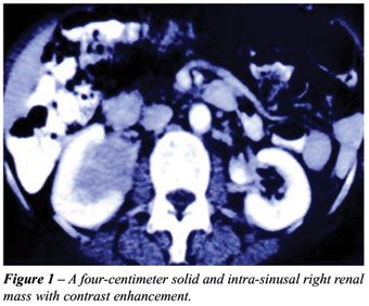
67-year-old female asymptomatic patient with no palpable lymph nodes presented
a right renal mass (Figure-1) in an abdomen computerized tomography (CT)
in a routine check up for a non-Hodgkin’s lymphoma that had been
diagnosed and treated with chemotherapy and radiotherapy 7 years earlier.
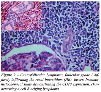
The patient underwent a radical right nephrectomy
and the macroscopic exam revealed an expansion of fat caused by a homogeneous,
yellowish tumor infiltration. The microscopic exam revealed a centrofollicular
lymphoma, follicular grade I, diffusely infiltrating the renal interstitium.
The immunohistochemical analysis demonstrated a CD 20 positivity, characterizing
a lymphoma originating in lymphocytes B (Figure-2). This finding led to
adjuvant radiotherapy and the patient is now with a 16-month follow-up.
COMMENTS
Among
cases of renal lymphoma, between 37% and 47% occur due to dissemination
of an advanced systemic disease (2), while 0.1% are due to primary involvement
of the kidney (1). The term, primary renal lymphoma, can be used either
when the initial manifestation involves the kidney or the lesion is limited
to it (3).
Renal lymphoma usually is of small cells
(Burkitt´s), or less frequently of lymphocytes B (1). Prognosis
of these cases is not well established, but is typically defined using
the criteria of the Working Formulation, Ann Arbor Stage and International
Prognostic Index (3). The disease has the same prevalence in both sexes
and is predominant in patients with a mean age of 66 years (3). The symptoms
observed during the later stages are lumbalgia, hematuria and fever (3).
However, in the series studied by Dimopoulos et al., all 6 patients presented
symptoms related to the involvement of the urinary tract and the absence
of peripheral palpable lymph nodes (3). Even with chemotherapy, only 2
patients had complete remission of the disease. The patients with unfavorable
prognostic factors as defined by the International Prognostic Index presented
poor results with chemotherapy treatment (3), which is based on cyclofosfamide,
doxorubicin, vincristine and prednisone.
Lesions can be solitary masses (10-20%)
or multiple masses (60%). They are generally bilateral and present extension
by contiguity (25%-30%), diffuse infiltration (20%) or perirenal involvement
10% (2). Radiological findings frequently indicate renal involvement with
multiple nodules (60%) and help in clarifying the diagnosis when considered
along with previous family history. Renal lymphoma generally is presented
as a bilateral nodular infiltration with a diffuse kidney increase and
infiltration of the renal parenchyma by a diffuse invasion of the retroperitoneum
(3). Solitary unilateral renal mass, perirenal mass with distortion of
the renal architecture in the CT scan and absence of lymph node enlargements
are more suggestive of renal cell carcinomas (3), however the presence
of solid masses can require a biopsy to rule out other pathologies (2).
Radiological findings in this case showed
an uncommon presentation of the renal lymphomatous lesion which served
as a warning that tumors might appear during follow-up as atypical and
uncommon lesions.
CONFLICT OF INTEREST
None declared.
REFERENCES
- Stallone G, Infante B, Manno C, Campobasso N, Pannarale G, Schena FP: Primary renal lymphoma does exist: case report and review of the literature. J Nephrol. 2000; 13: 367-72.
- Urban BA, Fishman EK: Renal lymphoma: CT patterns with emphasis on helical CT. Radiographics. 2000; 20: 197-212.
- Dimopoulos MA, Moulopoulos LA, Costantinides C, Deliveliotis C, Pantazopoulos D, Dimopoulos C: Primary renal lymphoma: a clinical and radiological study. J Urol. 1996; 155: 1865-7.
____________________
Accepted
after revision:
August 31, 2005
_______________________
Correspondence address:
Dr. Marcos F Dall’Oglio
Rua Manoel da Nóbrega, 853 / 22
São Paulo, SP, 04001-084, Brazil
Fax: + 55 11 3885-0658
E-mail: marcosdallogliouro@terra.com.br