INCIDENTAL
PROSTATIC ADENOCARCINOMA IN PATIENTS WITH PSA LESS THAN 4 NG/ML UNDERGOING
RADICAL CYSTOPROSTATECTOMY FOR BLADDER CANCER IN IRANIAN MEN
(
Download pdf )
S.Y. HOSSEINI, A.K. DANESH, M. PARVIN, A. BASIRI, T. JAVADZADEH, M.R. SAFARINEJAD, A. NAHABEDIAN
Urology and Nephrology Research Center, Shaheed Modarress and Shaheed Labbafinejad Hospitals, Shaheed Beheshti University of Medical Sciences, Tehran, Iran
ABSTRACT
Objective:
To assess the incidence of prostate adenocarcinoma in patients undergoing
radical cystoprostatectomy due to bladder cancer in Iranian men.
Materials and Methods: Fifty cystoprostatectomy
specimens removed due to bladder malignancy (2004-2005) at two referral
centers (Shaheed Modarress and Shaheed Labbafinejad Hospitals, Tehran,
Iran) were examined for the coincidental finding of prostate cancer (PCa).
At the time of surgery the patient’s serum PSA was less than 4 ng/mL
and there were no suspicious lesions by digital rectal examination. Pathologic
grade, stage, morphometric volume, number of tumor foci and association
with areas of high grade prostatic intraepithelial neoplasia (HGPIN) were
assessed by light microscopy. All specimens were totally embedded and
whole-mounted. Clinically significant cancers were defined as tumors with
≥ 0.5 mL volume, Gleason pattern 4 or 5, pT3, positive surgical
margin, and multifocality > 3.
Results: Incidentally detected cancer was
found in 7 (14%) of cystoprostatectomy specimens. HGPIN was present in
1 (14.3%) of the cystoprostatectomies with incidentally detected prostate
cancer. None of cystoprostatectomies without prostate cancer had HGPIN.
Four (57%) of the detected cancers were significant.
Conclusion: We conclude that incidentally
detected prostate cancer in Iran is lower than the rates reported in other
countries. Further studies are warranted for better declaration of variability
of prostate cancer between different ethnic groups.
Key
words: Incidental cancer, prostate cancer, bladder cancer, cystoprostatectomy
Int Braz J Urol. 2007; 33: 167-75
INTRODUCTION
The
distribution of cancer varies significantly from country to country all
over the world. The latest estimates of global cancer incidence show that
prostate cancer has become the third most common cancer in men, with half
a million new cases every year, almost 10% of all cancers in men (1-3).
The lifetime risk of clinically detected prostate cancer is 9.5%, and
the probability of dying from prostate cancer is 3%. The frequency of
incidentally detected cancer is approximately 42% in men older than 50
years of age; the frequency of autopsy-detected cancer is similar or higher
(4). In no other malignancy, there is such a vast reservoir of undetected
cases that may never be clinically significant or cause death (4). Prostate
cancer incidence is characterized by a very large geographical variability.
Asian countries present much lower rates of occurrence of the disease
when compared to North American, North and Western European countries,
with Southern European and South American countries displaying an intermediate
incidence rate (5). The incidence of clinical prostate cancer in Black
men is greater than in any other ethnic group. Japanese and Chinese men
are less likely to develop prostate cancer (6). The incidence of prostate
cancer is considerably low in Orientals. Such differences seem to be linked
to ethnic characteristics.
Because Iranian men are ethnically and racially
different from most of Asian countries’ men (e.g. Japanese, Chinese,
and Arabic men) the prevalence of prostate cancer should be different.
We conducted a prospective study in Iranian men undergoing cystoprostatectomy
to study this issue.
MATERIALS AND METHODS
Between
2004 and September 2005, fifty men with bladder cancer underwent radical
cystoprostatectomy at Shaheed Modarress and Shaheed Labbafinejad Hospitals,
Tehran Iran. Mean age of patients was 62.5 ± 10.56 years, with
age limits ranging between 44 and 82 years. The inclusion criteria comprised
a serum PSA level < 4 ng/mL and normal digital rectal examination.
Patients with a history of radiotherapy, chemotherapy, previous prostate
surgery and any medical therapy for benign prostatic hyperplasia (BPH)
were also excluded from the study. The product from the radical cystoprostatectomies
was fixed in 10% formalin solution and processed according to the usual
standards for fixation and inclusion routinely employed in pathology services.
After receiving the specimens, they were measured and weighed, the outer
surface of the specimen was inked and they were opened totally and fixed
in buffered formalin for 24 hours. After fixation, the prostate including
prostatic urethra was sectioned in quadrants. Sections from transitional
and peripheral zones of the prostate and from apical, middle and basal
regions in both lobes were included, resulting, in average, in 6 blocks
per case. The margin of prostatic urethra was represented separately.
The blocks were sectioned in slices with 3- to 5-micrometers in thickness
and the resultant histological slides were stained by hematoxylin-eosin.
If adenocarcinoma was discovered, then tumor location and Gleason score
was determined and involvement of the margins or seminal vesicle extension
was evaluated. If there was HGPIN, it was also mentioned. Cancer location
and extent were determined and mapped in each section. The presence of
tumor cells at the inked margin of resection (defined as the presence
of ink on neoplastic cells) was considered to present a positive surgical
margin. A positive surgical margin in an area where no capsule was identified
was referred to as pT2+ and was thought to indicate where the plane of
dissection entered the prostatic capsule or otherwise where no capsule
was present, i.e. apex and anteriorly (7). A single pathologist reviewed
all tumors for tumor stage (1997 AJCC TNM classification) (8), grade (Gleason
scoring system) (9), and surgical margin status. Cancer volume was calculated
from histological tissue sections using the grid method (10). All disease-containing
areas were outlined in each prostatectomy specimens section. Tumor area
was measured using a 1 mm grid, and aggregate tumor volume was estimated
by multiplying the sum of tumor areas in consecutive sections by the section
thickness. To calculate the tumor volume it was multiplied by a factor
of 1.25 to correct the shrinkage that occurs during fixation. The volume
of the single largest cancer focus was the incident tumor for investigation.
The number of PCa foci within the prostate, the presence and volume of
prostate HGPIN, and the proximity of HGPIN to PCa were also gauged for
each specimen. Criteria for defining HGPIN included (1) intraluminal proliferation
of the secretory cells in the prostate duct-acinar system, forming pseudostratified
layers, (2) large nuclei of relatively uniform size, an increased chromatin
content, which may be irregularly distributed, and (3) multiple prominent
nucleoli (11,12). High-grade PIN was classified as “low volume”
if there were three or fewer separate foci/acini of high-grade PIN, and
as “high volume” if there were more than three foci/acini
of high-grade PIN on different sections.
Extraprostatic extension was defined as
seminal vesicle involvement, malignant cells outside the prostatic capsule,
or lymph node metastases. Seminal vesicle invasion was diagnosed when
tumor penetrated the muscular coat of the seminal vesicles. Prostate cancers
with one of the following characteristics were regarded as clinically
significant: an estimated tumor volume ≥ 0.50 mL, contains a component
of Gleason histologic pattern 4 or 5, exhibits extraprostatic extension
(pT3), has a positive surgical margin, or is recognized in more than three
separate areas of the prostate (multifocal).
Clinical features were summarized with mean
and ranges or as percentages. Linear regression to complete a bivariate
fit of the number of cancer foci by the number of HGPIN foci was done
using the computer statistical package SPSS/10.0 (SPSS, Chicago, IL).
RESULTS
The
mean age was 62.5 ± 10.56 years (range 44-82 years). This was 57.35
± 9.75 and 63.19 ± 10.5 years for patients with and without
prostate cancer, respectively. Of fifty patients, 7 (14%) had the incidental
finding of PCa within the radical cystoprostatectomy specimen. The mean
serum PSA level was 1.89 ± 1.32 and 1.33 ± 1.095 ng/mL in
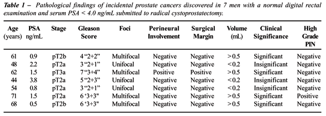
patients with and without prostate cancer, respectively. Table-1 details
the patient characteristics and associated pathologic findings. The majority
(57%) was pT2a and 28.6% pT2b, with lower frequency in other pT categories
(14.3% pT3a, 0% pT3b, and 0% pT4). In 14.3%, 28.6 and 14.3% of cases,
Gleason scores were 5, 6, and 7 respectively. The most prevalent (28.6
%) Gleason histological pattern was 3+3=6. One patient (14.3%) demonstrated
a focus of Gleason pattern 4 carcinoma. All patients were pN0 for prostate
cancer and one (14.3%) had positive surgical margin. High-grade PIN was
present in 14.3% of incidentally detected prostate cancer. None of the
cystoprostatectomies without prostate cancer had HGPIN. A single patient
had a 0.15 mL volume, Gleason score 7 cancer with clear extraprostatic
extension (pT3a) at the prostatic base. More than half of the patients
(4/7) had ≥ 3 separate foci of PCa identified. The largest tumor
volume exceeded 0.5 mL in 4 patients (57%).
As defined, clinically significant cancers
were present in 57% of the studied patients having a mean age of 61 ±
6.5 years (range 49-72). The remainder 43% had insignificant prostate
cancer.
COMMENTS
The
incidence of prostate cancer varies considerably across populations. The
highest reported incidence of prostate cancer in the world is in Jamaica.
The average age adjusted incidence of prostate cancer in Kingston, Jamaica
is 304/100,000 men (13). In Asian countries, particularly in china, the
incidence of PCa is low. According to Deng et al. in 1995 the incidence
rate of PCa in Shanghai was only 2.4/100,000 men (14). It was reported
in Los Angeles County, United States that the incidence rate was highest
in African Americans (116/100,000 person-years) and lowest among Asians
(Japanese, 39/100,000 person-years) and Chinese (28/100,000 person-years)
(15). The significance of environmental factors in the development of
prostate cancer is apparent from studies of migrants. Japanese men have
a low incidence of prostate cancer in Japan. These rates are only one-fifteenth
of those of white men in the United States, and they quadruple among first
and second generation Japanese migrants to the United States (16). It
remains to be determined whether similar environmental influences are
responsible for the high incidence of prostate cancer in United States
black men.
The frequency of incidentally detected cancer
is approximately 42% in men older than 50 years of age; the frequency
of autopsy-detected cancer is similar or higher (4). We found that 14%
of cystoprostatectomy specimens in patients with bladder cancer also contained
incidental prostate cancer. This result was much lower than overall mean
frequency of incidentally detected prostate cancer in other series of
cystoprostatectomy cases (range, 23%-68%) (4,17-23) and also much lower
than the age-adjusted frequency of autopsy-detected prostate cancer (mean
frequency, 40%; range, 36%-46% (24-27). Incidental prostate cancer in
our cystoprostatectomy cases was usually stage pT2a or pT2b (57% and 28.6%,
respectively). Of incidentally detected prostate cancer, 57% were low
grade (Gleason scores 3, 4, and 5) and 43% were high grade (Gleason scores
6 and 7). In a study from Brazil, 28.3% of patients had prostate carcinoma
in cystoprostatectomy specimens (28). Though the discrepancies between
studies could be related to the method of pathologic evaluation employed,
all indicate the presence of a significantly high incidence of prostate
cancer.
High-grade PIN was present in association
with 70% of cases of incidentally detected prostate cancer and in 54%
of cystoprostatectomies without prostate cancer (29,30). Interestingly
in our series, only 14.3% of cystoprostatectomies with prostate cancer
had HGPIN and none of the specimens without cancer had HGPIN. Arakawa
et al. (29) found that PIN was present in association with 78% of cases
of incidentally detected prostate cancer. Qian et al. (31) found PIN in
87% of radical prostatectomy specimens with localized cancer.
This percentage was higher in Iran, and
it was lower than the percentages reported in the other Asian countries.
Our finding may reflect a recent decrease in the incidence of prostate
cancer in Iran. The incidence of prostate cancer varies considerably.
Prostate cancer shows significant racial variation (32-34). The incidence
and mortality rates for American black men are almost twice those for
American white men (35). This increased incidence in black men cannot
be ascribed to differences in socioeconomic status (36). Cancer registries
are available in many countries but it is important to note that the degree
of accuracy may vary. Possible explanations for low rates of cancer in
some countries may be due to under-reporting (37).
The clinical incidence of adenocarcinoma
of the prostate is 75.3 per 100,000 men (38). The life time risk of American
men is calculated to be between 8 and 9.5% with a 2.9% risk of dying of
prostate cancer (39). Mortality rates for black American men are the highest
reported in the world until now. Even correcting for clinical stage at
diagnosis, the mortality from prostate cancer in black men is 2 times
higher than in white men (35). Prichett et al. (40) reviewed a 3-year
experience and found 45 adenocarcinomas of the prostate in 165 male cystectomy
patients with bladder cancers (27%). Patients with bladder neoplasia can
present prostate neoplasia with a relative risk up to 19 times higher
than what would be expected (41). However, incidental prostate tumors
present characteristics that are similar to latent tumors found in autopsy
series, some have a proven potential of progressive disease (42).
The objective of this work is to verify
the incidence of incidental prostate adenocarcinoma in patients who underwent
radical cystoprostatectomy for bladder urothelial carcinoma.
Our study was limited by the moderate number
of cases studied and potential bias in patient selection for surgery at
our medical centers. We attempted to minimize biases by using totally
embedded specimens and using a consecutive series of cases.
CONCLUSION
The present results indicate that the percentage of incidentally detected prostate cancer in cystoprostatectomies specimens in Iran is much lower than reported rates in the world until now. We therefore assumed regional differences in prostate cancer incidence rates to be related to environmental and racial factors. Still more epidemiologic research is essential to further understand the distribution as well as the prevalence and incidence of prostate cancer in certain ethnic groups.
CONFLICT OF INTEREST
None declared.
REFERENCES
- Parkin DM, Laara E, Muir CS: Estimates of the worldwide frequency of sixteen major cancers in 1980. Int J Cancer. 1988; 41: 184-97.
- Parkin DM, Pisani P, Ferlay J: Estimates of the worldwide incidence of 25 major cancers in 1990. Int J Cancer. 1999; 80: 827-41.
- Parkin DM, Bray FI, Devesa SS: Cancer burden in the year 2000. The global picture. Eur J Cancer. 2001; 37 Suppl 8: S4-66.
- Stamey TA, Freiha FS, McNeal JE, Redwine EA, Whittemore AS, Schmid HP: Localized prostate cancer. Relationship of tumor volume to clinical significance for treatment of prostate cancer. Cancer. 1993; 71: 933-8.
- [No authors listed]: Cancer incidence in five continents. Volume VIII. IARC Sci Publ. 2002; 155: 1-781.
- Gohji K, Nomi M, Egawa S, Morisue K, Takenaka A, Okamoto M, et al.: Detection of prostate carcinoma using prostate specific antigen, its density, and the density of the transition zone in Japanese men with intermediate serum prostate specific antigen concentrations. Cancer. 1997; 79: 1969-76.
- Stamey TA, Villers AA, McNeal JE, Link PC, Freiha FS: Positive surgical margins at radical prostatectomy: importance of the apical dissection. J Urol. 1990; 143: 1166-72; discussion 1172-3.
- Fleming ID: AJCC cancer staging manual/American Joint Committee on Cancer. Fifth edition. Philadelphia, Lippincott-Raven. 1997.
- Gleason DF, Mellinger GT: Prediction of prognosis for prostatic adenocarcinoma by combined histological grading and clinical staging. J Urol. 1974; 111: 58-64.
- Humphrey PA, Vollmer RT: Intraglandular tumor extent and prognosis in prostatic carcinoma: application of a grid method to prostatectomy specimens. Hum Pathol. 1990; 21: 799-804.
- Montironi R, Mazzucchelli R, Algaba F, Lopez-Beltran A: Morphological identification of the patterns of prostatic intraepithelial neoplasia and their importance. J Clin Pathol. 2000; 53: 655-65.
- Montironi R, Mazzucchelli R, Scarpelli M: Precancerous lesions and conditions of the prostate: from morphological and biological characterization to chemoprevention. Ann N Y Acad Sci. 2002; 963: 169-84.
- Glover FE Jr, Coffey DS, Douglas LL, Cadogan M, Russell H, Tulloch T, et al.: The epidemiology of prostate cancer in Jamaica. J Urol. 1998; 159: 1984-6; discussion 1986-7.
- Deng J, Gao JT, Xie T: Clinical epidemiological study on prostate cancer. Tumor 1995; 15: 340-5.
- Irvine RA, Yu MC, Ross RK, Coetzee GA: The CAG and GGC microsatellites of the androgen receptor gene are in linkage disequilibrium in men with prostate cancer. Cancer Res. 1995; 55: 1937-40.
- Karr JP: Prostate cancer in the United States and Japan. Adv Exp Med Biol. 1992; 324: 17-28.
- Abbas F, Biyabani SR, Pervez S: Incidental prostate cancer: the importance of complete prostatic removal at cystoprostatectomy for bladder cancer. Urol Int. 2000; 64: 52-4.
- Abbas F, Hochberg D, Civantos F, Soloway M: Incidental prostatic adenocarcinoma in patients undergoing radical cystoprostatectomy for bladder cancer. Eur Urol. 1996; 30: 322-6.
- Babaian RJ, Troncoso P, Ayala A: Transurethral-resection zone prostate cancer detected at cystoprostatectomy. A detailed histologic analysis and clinical implications. Cancer. 1991; 67: 1418-22.
- Cindolo L, Benincasa G, Autorino R, Domizio S, De Rosa G, Testa G, et al.: Prevalence of silent prostatic adenocarcinoma in 165 patients undergone cystoprostatectomy: a retrospective study. Oncol Rep. 2001; 8: 269-71.
- Kabalin JN, McNeal JE, Price HM, Freiha FS, Stamey TA: Unsuspected adenocarcinoma of the prostate in patients undergoing cystoprostatectomy for other causes: incidence, histology and morphometric observations. J Urol. 1989; 141: 1091-4; discussion 1093-4.
- Konski A, Rubin P, DiSantangnese PA, Mayer E, Keys H, Cockett A, et al.: Simultaneous presentation of adenocarcinoma of prostate and transitional cell carcinoma of bladder. Urology. 1991; 37: 202-6.
- Sanchez-Chapado M, Olmedilla G, Cabeza M, Donat E, Ruiz A: Prevalence of prostate cancer and prostatic intraepithelial neoplasia in Caucasian Mediterranean males: an autopsy study. Prostate. 2003; 54: 238-47.
- Sakr WA, Grignon DJ, Crissman JD, Heilbrun LK, Cassin BJ, Pontes JJ, et al.: High grade prostatic intraepithelial neoplasia (HGPIN) and prostatic adenocarcinoma between the ages of 20-69: an autopsy study of 249 cases. In Vivo. 1994; 8: 439-43.
- Sakr WA, Haas GP, Cassin BF, Pontes JE, Crissman JD: The frequency of carcinoma and intraepithelial neoplasia of the prostate in young male patients. J Urol. 1993; 150: 379-85.
- Scott R Jr, Mutchnik DL, Laskowski TZ, Schmalhorst WR: Carcinoma of the prostate in elderly men: incidence, growth characteristics and clinical significance. J Urol. 1969; 101: 602-7.
- Silvestri F, Bussani R, Pavletic N, Bassan F: Neoplastic and borderline lesions of the prostate: autopsy study and epidemiological data. Pathol Res Pract. 1995; 191: 908-16.
- Romero FR, de Castro MG, Andriolo Junior A, de Meneses AH, Fernandes RC, Perez MD: Coexistence of prostate neoplasia in patients undergoing radical cystoprostatectomy due to vesical neoplasia. Int Braz J Urol. 2004; 30: 296-301.
- Arakawa A, Soh S, Wheeler TM: High-grade prostatic intraepithelial neoplasia in cystoprostatectomy specimens. J Urol Pathol. 1997; 7:1-8.
- Prange W, Erbersdobler A, Hammerer P, Graefen M, Hautmann SH, Hautmann RE, et al.: High-grade prostatic intraepithelial neoplasia in cystoprostatectomy specimens. Eur Urol. 2001; 39 Suppl 4: 30-1.
- Qian J, Wollan P, Bostwick DG: The extent and multicentricity of high-grade prostatic intraepithelial neoplasia in clinically localized prostatic adenocarcinoma. Hum Pathol. 1997; 28: 143-8.
- Whittemore AS, Wu AH, Kolonel LN, John EM, Gallagher RP, Howe GR, et al.: Family history and prostate cancer risk in black, white, and Asian men in the United States and Canada. Am J Epidemiol. 1995; 141: 732-40.
- Severson RK, Schenk M, Gurney JG, Weiss LK, Demers RY: Increasing incidence of adenocarcinomas and carcinoid tumors of the small intestine in adults. Cancer Epidemiol Biomarkers Prev. 1996; 5: 81-4.
- Gilliland FD, Becker TM, Key CR, Samet JM: Contrasting trends of prostate cancer incidence and mortality in New Mexico’s Hispanics, non-Hispanic whites, American Indians, and blacks. Cancer. 1994; 73: 2192-9.
- Ernster VL, Winkelstein W Jr, Selvin S, Brown SM, Sacks ST, Austin DF, et al.: Race, socioeconomic status, and prostatic cancer. Cancer Treat Rep. 1977; 61: 187-91.
- Walker B, Figgs LW, Zahm SH: Differences in cancer incidence, mortality, and survival between African Americans and whites. Environ Health Perspect. 1995; 103 Suppl 8: 275-81.
- Muir CS, Nectoux j:Nectoux international patterns of cancer. In: Cancer Epidemiology and Prevention. Scottenfield D. & Fraumen JF (Eds.). Philadelphia, Saunders. 1982.
- Devesa SS, Silverman DT, Young JL Jr, Pollack ES, Brown CC, Horm JW, et al.: Cancer incidence and mortality trends among whites in the United States, 1947-84. J Natl Cancer Inst. 1987; 79: 701-70.
- Seidman H, Mushinski MH, Gelb SK, Silverberg E: Probabilities of eventually developing or dying of cancer—United States, 1985. CA Cancer J Clin. 1985; 35: 36-56.
- Pritchett TR, Moreno J, Warner NE, Lieskovsky G, Nichols PW, Cook BA, et al.: Unsuspected prostatic adenocarcinoma in patients who have undergone radical cystoprostatectomy for transitional cell carcinoma of the bladder. J Urol. 1988; 139: 1214-6.
- Chun TY: Coincidence of bladder and prostate cancer. J Urol. 1997; 157: 65-7.
- Moutzouris G, Barbatis C, Plastiras D, Mertziotis N, Katsifotis C, Presvelos V, et al.: Incidence and histological findings of unsuspected prostatic adenocarcinoma in radical cystoprostatectomy for transitional cell carcinoma of the bladder. Scand J Urol Nephrol. 1999; 33: 27-30.
____________________
Accepted
after revision:
January 5, 2007
_______________________
Correspondence address:
Dr. M.R. Safarinejad
P.O. Box 19395-1849
Tehran, Iran
Fax: + 90 212 635-1918
E-mail: safarinejad@unrc.ir
Prostate
cancer is unique among the potentially lethal human malignancies in the
wide discrepancy between the high prevalence of histological (incidentally
found) cancer and the much lower prevalence of the clinical disease. In
50 year-old men and with an expectancy of life more than 25 years, the
risk for prostatic carcinoma is estimated to be 42% for histological (incidentally
found) cancer, 9.5% for clinical cancer, and 2.9% for fatal cancer (1).
These
epidemiological findings suggest the existence of latent or clinically
unimportant cancers that should be distinguished from those that are clinically
important by the larger volume, higher grade, and greater invasiveness.
Unfortunately, when dealing with small volume cancers, there is no marker
to predict whether a tumor will behave as latent or progress to clinical
disease.
In
spite of striking differences in the frequency of clinical carcinoma (in
Asian countries being the lowest), the frequency of histological (incidentally
found) carcinoma is fairly similar around the world. According to the
theory of multistep events in carcinogenesis, molecular events (initiation)
resulting in histological prostate carcinoma probably occur equally around
the world. For evolvement to clinical carcinoma, further events related
to race, food, environmental pollution, etc (promoting factors) must be
implicated (2).
Incidentally
found carcinoma can be studied in two ways: in autopsies and in cystoprostatectomies.
The frequency of incidentally found cancer in both ways varies considerably
and the main cause is the method of examination of the prostate. Baron
& Angrist (3) compared the frequency of histological (incidentally
found) cancer in autopsies conducted by two methods: examining routine
fragments and step-sectioning the prostate. Using the first method the
frequency was 9.9% and using the latter 46%.
Bean
et al. (4) found a frequency of 6.6% in routine processing and 27.2% in
step-sectioning. The same applies to the examination of cystoprostatectomy
specimens. The number of fragments processed is critical for properly
evaluating the frequency of a lesion. In a series of 265 consecutive radical
prostatectomies in our Institution with step-sectioning of the surgical
specimen, the mean number of blocks examined (excluding blocks from the
seminal vesicles, vas deferens and cone amputated base and apex of the
prostate) was 31 with a minimum of 10 and a maximum of 56.
REFERENCES
- Scardino PT, Weaver R, Hudson MA: Early detection of prostate cancer. Hum Pathol. 1992; 23: 211-22.
- Dhom G: Epidemiologic aspects of latent and clinically manifest carcinoma of the prostate. J Cancer Res Clin Oncol. 1983; 106: 210-8.
- Baron E, Angrist A: Incidence of occult adenocarcinoma of the prostate after fifty years of age. Arch Pathol. 1941; 32: 787-93.
- Bean MA, Yatani R, Liu PI, Fukazawa K, Ashley FW, Fujita S: Prostatic carcinoma at autopsy in Hiroshima and Nagaski Japanese. Cancer. 1973; 32: 498-506.
Dr. Athanase Billis
Full-Professor of Pathology
State University of Campinas, Unicamp
Campinas, Sao Paulo, Brazil
E-mail: athanase@fcm.unicamp.br
EDITORIAL COMMENT
In
recent years, incidentally detected prostate carcinoma (PCa) in patients
undergoing radical cystoprostatectomy has become a concern for practicing
urologists because of the suggestion of prostate sparing cystoprostatectomy
by several authors (1). To our opinion, the major questions that merit
comment about this issue are the following. 1) Should all patients undergoing
radical cystoprostatectomy be screened for the coexistence of PCa? 2)
Does PCa coexist with bladder cancer? 3) Is prostate sparing cystoprostatectomy
established in terms of oncological principles? The current opinions of
the authors about these questions are as follows.
1)
Screening of patients who are candidates for radical cystoprostatectomy
with serum PSA determination and digital rectal examination (DRE) have
a risk of overdiagnosis for prostate cancer. Overdiagnosis is the diagnosis
of cancers that for whatever reason do not threaten the health condition
or the life of a given patient, which is the rationale for PSA determination
to the patients with an estimate life expectancy over 10 years. For this
reason, it is logical for not to evaluate the patients with bladder cancer
requiring cystoprostatectomy in terms of PCa. However, we recently reported
that despite the vast majority of the patients had organ confined PCa
(90.5%) after surgery, 9.5% of the patients had capsular extension and
4.75% were lymph node positive (2). Moreover, only 57.1% of the patients
survived after a mean follow-up of 24.3 months. Similarly, Hosseini et
al., in the present paper, reported that 14.3% of the patients had capsular
extension and 14.3% had positive surgical margin. In patients without
organ-confined disease, the extent of PCa may threaten the life of patient
instead of bladder cancer, which is especially important for patients
with clinically low stage (Ta, T1) cancer. For this reason, we believe
that all patients undergoing radical surgery for bladder cancer should
receive DRE and PSA testing. In addition, in the case of palpable prostatic
abnormalities or elevated PSA levels, more accurate clinical staging (with
transrectal ultrasound biopsy or sophisticated imaging modalities) should
be attempted, especially in patients with clinically low stage disease.
2)
Around 20% of prostate cancers detected during incidental autopsies are
clinically significant PCa by the tumor volume criteria (> 0.5 mL).
Hautmann et al. (3) reported that 44% of the patients had clinically significant
PCa, which is significantly higher than the estimated percentage in autopsy
studies. Meanwhile, this incidence is reported as 14% by Hosseini et al.,
in the present paper. It is hard to interpret the difference between this
study and the former study and autopsy series. However, it may be attributed
to the lower incidence of prostate cancer in particular countries. Furthermore,
despite the lower rates of PCa, the rate of incidental prostate carcinoma
is as high as western countries. This observation advocates that the data
of the previous surveys suggesting a carcinogenic correlation between
bladder and prostate cancer such that the incidence of PCa is 9 to 20.5
times greater following cystoprostatectomy (4-6).
3)
Recently, oncological justification of the prostate sparing cystoprostatectomy
was critically evaluated by Hautmann & Stein (7). Briefly, the authors
noted that distant failure rate of patients with sexuality sparing surgery
is at least twice as high as expected for superficial or organ-confined
transitional cell carcinoma. Moreover, they addressed a 6% risk of leaving
PCa in any residual tissue. For this reason, until long term data is available
from the patients receiving prostate sparing cystoprostatectomy, we continue
to perform a complete removal of the prostate during surgery in our clinical
practice. On the other hand, it should be mentioned that prostate sparing
cystoprostatectomy is an attractable option for a young patient with superficial
bladder cancer. However, before performing this surgery, urologists should
discuss the risks of this “experimental” surgery with the
patient.
REFERENCES
- Meinhardt W, Horenblas S: Sexuality preserving cystectomy and neobladder (SPCN): Functional results of a neaobladder anastomosed to the prostate. Eur Urol. 2003; 43: 646-50.
- Sanli O, Acar O, Celtik M, Oktar T, Kilicarslan I, Ozcan F, et al.: Should prostate cancer status be determined in patients undergoing radical cystoprostatectomy. Urol Int. 2006; 77: 307-10.
- Hautmann SH, Conrad S, Henke RP, Erbersdobler A, Simon J, Straub M, et al.: Detection rate of histological insignificant prostate cancer with systematic sextant biopsies and fine needle aspiration cytology. J Urol. 2000; 163; 1734-38.
- Kurokawa K, Ito K, Yamamato, Takechi H, Miyamoto S, Suzuki K, et al.: Comparative study on the prevalence of clinically detectable prostate cancer in patients with and without bladder cancer. Urology. 2004; 63: 268-72.
- Kinoshita Y, Singh A, Rovito PM Jr, Wang CY, Haas GP: Double primary cancers of prostate and bladder:a literature review. Clin Postate Cancer. 2004; 3: 83-6.
- Singh A, Kinoshita Y, Rovito PM Jr, Landas S, Silberstein J, Nsouli I, et al.: Higher than expected association of clinical prostate and bladder cancers. J Urol. 2005; 173: 1526-9.
- Hautmann RE, Stein JP: Neobladder with prostate capsule and seminal-sparing cystectomy for bladder cancer: A step in the wrong direction. Urol Clin North Am. 2005; 32: 177-85.
Dr. Oner Sanli
Dr. Tarik Esen
Department of Urology
Istanbul University
Istanbul, Turkey
E-mail: sanlio@istanbul.edu.tr