LAPAROSCOPIC
RADICAL PROSTATECTOMY: OMITTING A PELVIC DRAIN
(
Download pdf )
DAVID CANES, MICHAEL S. COHEN, INGOLF A. TUERK
Lahey Clinic Medical Center, Burlington, Massachusetts, USA
ABSTRACT
Purpose:
Our goal was to assess outcomes of a selective drain placement strategy
during laparoscopic radical prostatectomy (LRP) with a running urethrovesical
anastomosis (RUVA) using cystographic imaging in all patients.
Materials and Methods: A retrospective chart
review was performed for all patients undergoing LRP between January 2003
and December 2004. The anastomosis was performed using a modified van
Velthoven technique. A drain was placed at the discretion of the senior
surgeon when a urinary leak was demonstrated with bladder irrigation,
clinical suspicion for a urinary leak was high, or a complex bladder neck
reconstruction was performed. Routine postoperative cystograms were obtained.
Results: 208 patients underwent LRP with
a RUVA. Data including cystogram was available for 206 patients. The overall
rate of cystographic urine leak was 5.8%. A drain was placed in 51 patients.
Of these, 8 (15.6%) had a postoperative leak on cystogram. Of the 157
undrained patients, urine leak was radiographically visible in 4 (2.5%).
The higher leak rate in the drained vs. undrained cohort was statistically
significant (p = 0.002). Twenty-four patients underwent pelvic lymph node
dissection (8 drained, 16 undrained). Three undrained patients developed
lymphoceles, which presented clinically on average 3 weeks postoperatively.
There were no urinomas or hematomas in either group.
Conclusions: Routine placement of a pelvic
drain after LRP with a RUVA is not necessary, unless the anastomotic integrity
is suboptimal intraoperatively. Experienced clinical judgment is essential
and accurate in identifying patients at risk for postoperative leakage.
When suspicion is low, omitting a drain does not increase morbidity.
Key
words: prostatectomy; urinoma; laparoscopy; complications; drainage;
surgical anastomosis
Int Braz J Urol. 2008; 34: 151-8
INTRODUCTION
Acceptance
of the laparoscopic radical prostatectomy (LRP) since its inception, and
later its robotic counterpart, has been motivated by a drive to minimize
perioperative morbidity. Room for improvement still exists, with the hope
that minor technical adjustments will further decrease morbidity. The
following editorial remark accompanies a 1996 article addressing the morbidity
of drains following radical retropubic prostatectomy (RRP): “anything
that reduces patient discomfort deserves consideration” (1).
Savoie et al. first suggested a pelvic drain
might be omitted following an open RRP in an analysis of 116 consecutive
cases (2). These same authors updated this concept with 552 patients,
arriving at the same conclusion (3). In a recent comprehensive review
of published LRP literature, drain placement was not addressed (4), since
published series seldom report this detail. In many centers, pelvic drainage
remains a routine part of open and minimally invasive prostatectomy. We
hypothesized that improved optical magnification and a running urethrovesical
anastomosis (RUVA) may obviate routine pelvic drainage. We assessed the
relationship between pelvic drainage and postoperative complications in
a consecutive series of 268 LRP in which routine postoperative cystography
was performed in all patients, regardless of drain status.
MATERIALS AND METHODS
Laparoscopic
radical retropubic prostatectomy (LRP) was performed on 268 patients at
the Lahey Clinic Medical Center between January 2003 and December 2004.
Two hundred and eight patients were identified in whom an RUVA was performed.
Complete preoperative, operative, and post-operative patient information
was obtained from a combination of a prospective database maintained by
the Department of Urology Clinical Research Assistants and from a retrospective
chart review. The age, co-morbidities, prostate specific antigen (PSA),
Gleason score, clinical stage, estimated blood loss, blood transfusions,
pelvic lymph node dissection (PLND), pathological stage, pathological
Gleason score, prostate size, intravenous narcotic use, length of stay,
and complications were recorded. The body mass index (BMI) was calculated
from the preoperative height and weight documented in the anesthesia report.
The operative time was calculated from incision start time to procedure
end time as recorded in the operative nursing report. Narcotic use was
calculated to be the sum of intravenous narcotics recorded by the nursing
staff and administered via patient controlled analgesic or on an as needed
basis. Different narcotics medications were converted to morphine equivalents
for comparison.
All patients underwent either a transperitoneal
or extraperitoneal LRP as described previously (5,6) by a single surgeon
(IT). When nerve sparing was indicated and technically feasible, this
was performed using a harmonic scalpel (Ethicon Endo-Surgery). Lymphadenectomy
included the external iliac and obturator lymph nodes. The anastomosis
was performed with two 2-0 monocryl sutures each with a polydioxanone
absorbable suture clip Lapra-TyTM on one end. The first suture
was placed at the 5:30 position, and 2 - 3 running stitches were made
in the counterclockwise direction. The second suture was placed at the
6:30 position, and 2 - 3 running stitches made in the clockwise direction.
Therefore, prior to cinching the sutures, at least 4 to 6 running stitches
were placed. Therefore, the initial tension is distributed over 4 - 6
stitches instead of 1. Then the sutures were continued in a running manner
in their appropriate direction until they meet at the anterior aspect
of the anastomosis and tied together with an intracorporeal knot.
Anastomotic integrity was tested by distending
the bladder with approximately 200 mL of saline, prior to inflating the
Foley balloon. A Jackson-Pratt closed suction or Penrose drain was placed
at the discretion of the senior surgeon (IT) when a leak was visualized
at the anastomosis or a complex bladder neck reconstruction was performed.
Indications for drain placement were obtained from the operative report.
When omitted from the operative report, the indication was recorded as
unspecified. The drain was placed in close proximity to the anastomosis.
If drainage was less than 50 cc per 8-hour shift, the drain was removed.
A routine cystogram was performed within the first 7 postoperative days
in 90% of patients. The remaining patients had a cystogram prior to postoperative
day (POD) 14 due to scheduling difficulties.
Patients were seen postoperatively at 1
week, 5 weeks, 3 months, 6 months, 9 months, and 1 year in follow-up.
The Foley catheter was removed on POD 7 if the cystogram showed no evidence
of leak, defined as any amount of contrast extravasation. Patients were
asked a series of questions to screen for symptomatic intra-abdominal
collections, and complete abdominal examination was performed. Directed
radiographic imaging was performed when warranted by clinical symptoms.
The primary endpoint was the incidence of early postoperative complications:
urine leak, urinoma, lymphocele, and hematoma.
Fisher’s exact test was used to analyze
the association between categorical data: (1) urine leak and drain placement,
(1) urine leak and surgical approach, and (3) drain placement and performance
of PLND. Two-tailed p values were reported. Unpaired t-test was used to
compare mean values between the drained and undrained groups for the following
parameters: age, PSA, biopsy Gleason, pathologic Gleason, BMI, prostate
size, operative time, estimated blood loss, morphine equivalents used,
and length of stay. The chi-square test was used to compare the distribution
of clinical and pathologic stage between both groups.
RESULTS
A
total of 208 patients underwent LRP with a RUVA between January 2003 and
December 2004. The drained and undrained groups did not differ with respect
to age, preoperative PSA, biopsy Gleason sum, pathologic Gleason sum,
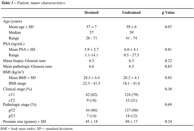
BMI, clinical stage, pathologic stage, or prostate size (Table-1).
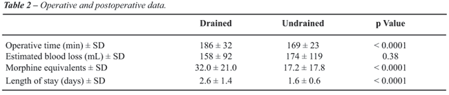
The operative and post-operative data demonstrated
a statistically significant difference between the drain and undrained
groups in regards to operative time, post-operative narcotic use, and
length of stay (Table-2). However, there was no statistical difference
in estimated blood loss between groups. When a drain was placed, operative
time was longer by an average of 17 minutes (95% CI 9-25, p < 0.0001).
Postoperative narcotic use and average length of stay were significantly
greater when comparing the drained and undrained groups, respectively.
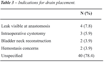
In the majority of cases, the indication for drain placement was not specified
in the operative record (Table-3). Reasons stated for drain placement
included the following: visible leak during testing of the anastomosis,
inadvertent cystotomy during bladder neck dissection, extensive bladder
neck reconstruction, and concerns for hemostasis (Table-3).
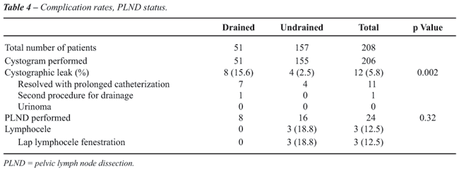
The incidence of urinary extravasation on
post-operative cystogram is outlined in Table-4. Drains were placed in
51 patients (25%), and omitted in 157 (75%). Cystograms were available
for 206 patients (99%). Mean duration of drainage was 48 hours. Overall,
12 patients had radiographic evidence of a urinary leak (5.8%). The patients
with a drain had a statistically higher incidence of a urinary leak. Presence
of radiographic urine leak was not significantly associated with surgical
approach (p = 0.23), either transperitoneal (n = 32, 4 leaks) or extraperitoneal
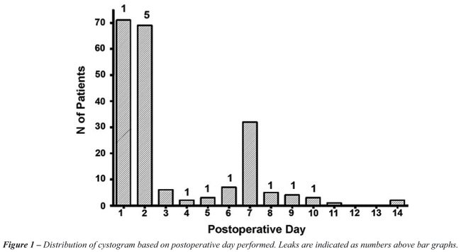
(n = 172, 8 leaks). As shown in Figure-1, 90% of patients had postoperative
cystograms within the first 7 postoperative days; the remainder were performed
the following week. As expected, earlier cystograms demonstrate the majority
of leaks, with 50% seen on POD 1 or 2. All leaks resolved on follow-up
imaging after prolonged drainage. No patient developed a urinoma in this
series.
Of the 208 patients, non-nerve sparing,
unilateral nerve sparing, and bilateral nerve sparing procedures were
performed in 21 (10.1%), 55 (26.4%), and 126 (60.6%) patients. Nerve sparing
data was missing in 6 (2.9%) patients. There were no postoperative bleeding
complications, including hemorrhage or hematoma. No patient required intraoperative
or postoperative blood transfusion, or reoperation for bleeding. One patient
required a secondary procedure for anastomotic urine leak. This patient
had mild unilateral hydronephrosis and a clinical leak with increasing
drain output. He returned to the OR on POD 7 for ureteral stent and suprapubic
catheter placement. Of note, had cystograms not been performed in any
patient, this is the only patient whose leak was apparent from increased
pelvic drain output. The remaining 11 patients with a radiographic leak
had no increased drainage.
Three patients underwent laparoscopic lymphocele
fenestration. Their lymphoceles (12.5% of patients undergoing PLND) presented
on average 3 weeks postoperatively with symptoms of low-grade fever, urinary
retention with bladder spasms, or lower extremity edema. Of the patients
undergoing PLND in whom drains were omitted, the approach was extraperitoneal
in 14/16 (87.5%). Drain placement was not significantly associated with
the performance of PLND (p = 0.32).
COMMENTS
The
Miami group, who were the first to suggest that routine pelvic drainage
after open RRP was unnecessary (2,3) placed drains with a similar selective
strategy. Since their report, the Roswell Park group has also supported
the safety of drain omission (7). These reports relied on global comparisons
of complication rates, without imaging studies. The present study is the
first, to our knowledge, in which routine postoperative cystograms were
used to assess the true radiographic leak rate underlying this clinically
driven algorithm, adding to a growing body of literature to support selective
drain omission. Using this selective algorithm, drains were placed in
25% of patients, approximately the same frequency as prior reports (2,7).
The overall cystographic leak rate defined as any radiographic extravasation
in this series is 5.8% and objective imaging was available for 99% of
patients. Interestingly, only one of the ten patients with radiographic
evidence of a leak had a clinical leak.
What is the correlation between clinical
and radiographic impressions of water tightness? Ischia and Lidsay, in
a study of 68 patients undergoing open prostatectomy, found a strong correlation
between intraoperative assessment with saline instillation, and subsequent
low leak rate on cystogram. Out of 68 consecutive patients, 53 had no
intraoperative leakage, and of these only two (3.7%) had leaks on day
7 cystograms (8). Our data are similar, in that unsuspected leaks in the
undrained group occurred in only 4 patients (2.5%).
Our overall cystographic leak rate compares
favorably with published series. Cystography data has generally been analyzed
to assess the feasibility of early catheter removal, and to correlate
leak rates with the occurrence of anastomotic strictures. Studies often
have inconsistent reporting (clinical vs. radiographic leak rate) and
discrepant testing intervals. Leibovitch et al. found 5.7% of patients
with significant contrast extravasation in a consecutive series of 245
patients undergoing open RRP. However, minimal or contained extravasation
was observed in an additional 11.4%, for a total leak rate of 17.1% (9).
Cystography was performed late (19.2 days postoperatively) compared to
the current study.
Contrast extravasation in the first 5-8
days postoperatively has historically been reported to range from 67-78%
(9). Similar statistics have fueled conventional wisdom that early extravasation
was common following open RRP. Dalton et al. (10) reported a leak rate
of 34.5% in a series of 55 patients studied with cystograms starting on
day 7. Ramsden et al. reported a 31% leak rate in 275 consecutive open
RRP cases where cystography was performed between postoperative days 8
and 10 (11). Contemporary numbers are much lower. Guazzoni et al. reported
a 12% leak rate on postoperative day 5 cystograms in patients undergoing
LRP with an interrupted anastomosis (12). In another review of 619 open
RRP with cystograms at day 10 a leak rate of 4.6% was reported (13), which
is similar to our findings.
Even when timing of cystography and anastomotic
technique are similar, leak rates may differ. Nadu et al. reported the
only other series of LRP with a RUVA in which cystography was routinely
performed (14). A cystographically apparent leak was present in 17/113
patients (15.1%), even though most parameters mirror our series. The RUVA
was performed with a single 3-0 polyglactin suture. Cystograms were performed
between postoperative days 2-4. No urinomas developed, and drain status
was not reported. What accounts for the higher leak rate? Patients were
asked to Valsalva during cystography, which may transmit greater pressures
to the anastomosis, whereas patients are imaged while voiding without
Valsalva at our institution.
Hoznek et al. were the first to describe
a RUVA, which has significantly decreased operative time and efficiency
during this portion of the procedure (15). The difference in early integrity
between running and interrupted techniques is not known. Theoretically,
suture tension may be distributed more evenly over the circumference of
the anastomosis. Authors have assumed, based on lack of symptomatic urine
leak, that the anastomosis is watertight (16). Our cohort includes the
learning curve for the RUVA, as well as objective imaging. Therefore,
a low leak rate of 5.8% lends further evidence to this clinical observation.
Although a selective drain placement strategy may be appropriate when
a laparoscopic interrupted anastomotic technique is employed, our study
did not include patients with this technique, nor was it designed to compare
different anastomotic techniques. We also noted that although the senior
surgeon had performed several hundred LRP prior to this time period, this
cohort contains his learning curve for the RUVA. We have previously reported
that a low leak rate may be a good surrogate endpoint for advanced acquisition
of technical skill (17).
Intuitively, a senior surgeon’s judgment
of anastomotic integrity should be accurate, and objective imaging substantiates
this impression. When a drain was placed, the leak rate was significantly
higher. When the anastomosis was watertight intraoperatively and a drain
omitted, the leak rate was indeed significantly lower, but not zero. Four
patients in the undrained group did have leaks, none of which developed
into urinomas. The longer operative time in the drained group was statistically
significant. However, we did not conclude that placing a drain led to
a statistically longer operative time. In the majority of cases the reason
for drain placement was not specified. A narrow pelvis or otherwise small
working space, fibrotic bladder neck tissue, presence of a median lobe,
or a difficult posterior dissection from previous prostatitis may all
contribute a sense of complexity to performing a LRP. These factors are
less quantifiable, and translate into prolonged operative time. Since
indications for placement were neither prospective nor randomized, selection
bias of the senior surgeon is inherent in this study. That this bias has
statistically significant clinical utility, however, is an important finding.
Traditionally, drains are placed to allow
the egress of urine, blood, and lymphatic fluid. Clinical suspicion was
sufficient to omit a drain without increasing the chance of urinoma. Symptomatic
lymphoceles were also acceptably uncommon (3/24 PLND). Since PLND was
performed in only 24 patients, we are unable to draw firm conclusions
regarding drainage following PLND. However, our data suggests that lymphoceles
should generally not be used as a justification for drain placement. Lymphoceles
in this series became symptomatic 3 weeks postoperatively. Lymphoceles
therefore accumulate long after the pelvic drain has been removed. Fried
et al. observed a similar time course, where two symptomatic lymphoceles
occurred at 4 and 9 weeks postoperatively (18). Pepper et al. reported
a series of 260 open RRP with PLND in which 9 patients developed lymphoceles
(3.5%) 12-120 days postoperatively (19). The mean time at diagnosis was
not provided.
In general, the lymphocele rate after open
PLND is between 4.7-14.8% (19). The wide range depends on the surgical
technique used, and whether clinical or radiographic diagnostic triggers
are employed. Freid et al. reported clinically detected lymphoceles in
1% of 111 patients, although 7/23 (30.4%) who subsequently underwent CT
imaging for adjuvant radiation had lymphoceles (18). The approach, whether
transperitoneal or extraperitoneal, also contributes. With the former,
lymphatic fluid is absorbed, compared to an extraperitoneal approach where
the retropubic space is an enclosed area where any lymphatic drainage
can readily form a lymphocele. However, our data suggests that drainage
is not mandatory even after PLND and an extraperitoneal approach. Of the
16 patients undergoing PLND without postoperative drainage, 87.5% were
approached extraperitoneally and only 3 developed symptomatic lymphocele.
A larger study with power to address this question is needed before definitive
recommendations can be made.
The morbidity of the drain itself is not
a primary endpoint of this study. The drained group utilized more narcotic
medication than the undrained group. We cannot conclude that the increased
pain was attributable to the drain. Without directed questionnaires and
pain score assessment, the contribution of drains to increased narcotic
use is speculative. Evidence for drain related pain was reported by Niesel
et al., who found that roughly one out of every four patients experience
pain after RRP attributable only to the drain site and not the incision
(1). The longer length of stay in the drained group is also likely multifactorial.
The single patient with a clinical urine leak had an 11 day hospital stay,
which may have contributed to the increased mean length of stay in the
drained group.
In addition to the retrospective, nonrandomized
nature of this study, a potential criticism is the role of the cystogram
itself in subsequent decision making. Here, 90% of leaks were only apparent
radiographically, and prolonged catheterization and repeat imaging were
performed. Can the cystogram itself be omitted? At the present time, after
the results of the present study and with increased experience, we have
ceased performing routine cystography. Using this selective drain algorithm
we have found no increased incidence of complications.
CONCLUSIONS
Routine pelvic drainage has traditionally accompanied radical prostatectomy. Our results suggest a pelvic drain can be omitted in patients undergoing an LRP with a RUVA if the anastomosis is watertight intraoperatively. Incidence of clinically detected urine leak, urinoma, hematoma, and lymphocele is not increased with this selective strategy.
CONFLICT OF INTEREST
None declared.
REFERENCES
- Niesel T, Partin AW, Walsh PC: Anatomic approach for placement of surgical drains after radical retropubic prostatectomy: long-term effects on postoperative pain. Urology. 1996; 48: 91-4.
- Savoie M, Soloway MS, Kim SS, Manoharan M: A pelvic drain may be avoided after radical retropubic prostatectomy. J Urol. 2003; 170: 112-4.
- Araki M, Manoharan M, Vyas S, Nieder AM, Soloway MS: A pelvic drain can often be avoided after radical retropubic prostatectomy--an update in 552 cases. Eur Urol. 2006; 50: 1241-7; discussion 1246-7.
- Trabulsi EJ, Guillonneau B: Laparoscopic radical prostatectomy. J Urol. 2005; 173: 1072-9.
- Rhee HK, Triaca V, Sorcini A, Tuerk IA: Transperitoneal laparoscopic radical prostatectomy: descending technique. J Endourol. 2004; 18: 601-4; discussion 604.
- Stolzenburg JU, Do M, Rabenalt R, Pfeiffer H, Horn L, Truss MC, et al.: Endoscopic extraperitoneal radical prostatectomy: initial experience after 70 procedures. J Urol. 2003; 169: 2066-71.
- Sharma S, Kim HL, Mohler JL: Routine pelvic drainage not required after open or robotic radical prostatectomy. Urology. 2007; 69: 330-3.
- Ischia JJ, Lindsay S: Is a cystogram necessary after radical prostatectomy? ANZ J Surg. 2005; 75: 825-7.
- Leibovitch I, Rowland RG, Little JS Jr, Foster RS, Bihrle R, Donohue JP: Cystography after radical retropubic prostatectomy: clinical implications of abnormal findings. Urology. 1995; 46: 78-80.
- Dalton DP, Schaeffer AJ, Garnett JE, Grayhack JT: Radiographic assessment of the vesicourethral anastomosis directing early decatheterization following nerve-sparing radical retropubic prostatectomy. J Urol. 1989; 141: 79-81.
- Ramsden AR, Chodak GW: Can leakage at the vesico-urethral anastomosis be predicted after radical retropubic prostatectomy? BJU Int. 2004; 93: 503-6.
- Guazzoni G, Cestari A, Naspro R, Riva M, Centemero A, Zanoni M, et al.: Intra- and peri-operative outcomes comparing radical retropubic and laparoscopic radical prostatectomy: results from a prospective, randomised, single-surgeon study. Eur Urol. 2006; 50: 98-104.
- Varkarakis J, Wirtenberger W, Pinggera GM, Berger A, Harabayashi T, Bartsch G, et al.: Evaluation of urinary extravasation and results after continence-preserving radical retropubic prostatectomy. BJU Int. 2004; 94: 991-5.
- Nadu A, Salomon L, Hoznek A, Olsson LE, Saint F, de La Taille A, et al.: Early removal of the catheter after laparoscopic radical prostatectomy. J Urol. 2001; 166: 1662-4.
- Hoznek A, Salomon L, Rabii R, Ben Slama MR, Cicco A, Antiphon P, et al.: Vesicourethral anastomosis during laparoscopic radical prostatectomy: the running suture method. J Endourol. 2000; 14: 749-53.
- Van Velthoven RF, Ahlering TE, Peltier A, Skarecky DW, Clayman RV: Technique for laparoscopic running urethrovesical anastomosis: the single knot method. Urology. 2003; 61: 699-702.
- Cohen MS, Canes D, Tuerk IA: Secondary learning curve for the laparoscopic radical prostatectomy. J Urol. 2006; 175: (Suppl. 4): 367.
- Freid RM, Siegel D, Smith AD, Weiss GH: Lymphoceles after laparoscopic pelvic node dissection. Urology. 1998; 51 (5A Suppl): 131-4.
- Pepper RJ, Pati J, Kaisary AV: The incidence and treatment of lymphoceles after radical retropubic prostatectomy. BJU Int. 2005; 95: 772-5.
____________________
Accepted after revision:
January 9, 2008
_______________________
Correspondence address:
Dr. David Canes
Lahey Clinic Medical Center
41 Mall Road
Burlington, MA, 01805, USA
Fax: + 1 781 744-8427
E-mail: david@canes.net