HISTOPATHOLOGICAL
CHARACTERIZATION OF A SYNGENEIC ORTHOTOPIC MURINE BLADDER CANCER MODEL
(
Download
pdf )
DAHER C. CHADE, PRISCILA M. ANDRADE, RICARDO C. BORRA, KATIA R. LEITE, ENRICO ANDRADE, FABIOLA E. VILLANOVA, MIGUEL SROUGI
Laboratory of Medical Investigation (DCC, PMA, KRL, EA, FEV, MS), Division of Urology, University of Sao Paulo Medical School, and Bioodontology (RCB), Ibirapuera University, Sao Paulo, Brazil
ABSTRACT
Purpose:
We developed and characterized by histopathology and immunohistochemistry
a syngeneic murine bladder tumor model derived from the MB49 tumor cell
line.
Materials and Methods: Bladder tumor implantation
was achieved by intravesical instillation of 5 x 105 MB49 tumor
cells in C57BL/6 mice. A chemical lesion of the bladder was performed
in order to promote intravesical tumor implantation. The bladder wall
lesion was accomplished by transurethral instillation of silver nitrate
(AgNO3). After 15 days, the animals were sacrificed, examined
macroscopically for intravesical tumor and bladder weight. Histology and
immunohistochemistry were performed using cytokeratin 7 (CK7), carcinoembrionic
antigen (Dako-CEA), p53 and c-erbB2 oncoprotein (Her2/neu).
Results: Twenty-nine out of 30 animals (96.7%)
developed intravesical tumors in a 15-day period. Macroscopically, the
mean bladder weight was 0.196g (0.069-0.538g), 10 to 15 times the normal
bladder weight. The immunohistochemical analysis showed significant membrane
expression of CEA and CK7: a similar finding for human urothelial cancer.
We also characterized absence of expression of p53 and anti-Her2/neu in
the murine model.
Conclusions: High tumor take rates were
achieved by using the chemical induction of the bladder tumor. Although
electric cauterization is widely described in the literature for syngeneic
orthotopic animal models, the technique described in this study represents
an alternative for intravesical bladder tumor implantation. Moreover,
the histopathology and immunohistochemical analysis of the murine bladder
tumor model derived from the MB49 cell line showed a resemblance to human
infiltrating urothelial carcinoma, allowing clinical inference from experimental
immunotherapy testing.
Key
words: bladder cancer; intravesical instillation; tumor cell
line; mice/c57bl; experimental neoplasm
Int Braz J Urol. 2008; 34: 220-9
INTRODUCTION
Animal
models provide a system for understanding basic biological questions.
With animal models, adequate control of experimental design is possible
so that rigorous experiments can be performed to test various hypotheses
. In the case of testing therapeutic mechanisms, it is important to select
a model that is most analogous to the clinical setting so that observations
can be readily transferred to clinical studies for validation (1).
A murine bladder tumor model may offer some
of these characteristics, while having many controlled variables under
laboratory conditions (2). This permits inference from experimental data,
which can be helpful for clinical purposes. The murine bladder tumor models
may be a xenograft model (in immunodeficient mice) (3), a chemically induced
bladder cancer model (4), or a syngeneic animal model (5).
The use of immunodeficient mice in bladder
cancer research has allowed the implantation of human carcinomas in an
animal model. In addition, it has demonstrated the importance of the immune
system, in particular as regards T lymphocytes, in anti-tumor activity
(6).
The chemically induced models were achieved
by administering carcinogens such as N-[4-(5-nitro-2-furyl)-2-thiazolyl]
formamide (FANFT) in C3H/He mice (7) or 7,12-dimethylbenzanthracene in
mice strain C57BL/6 (8). Although adequate for immunotherapeutic testing,
a long period of time was required for carcinogen-induced bladder tumor
growth.
The syngeneic animal models were developed
with the objective of improving immunotherapeutic studies (9). It is characterized
by the transplantation of carcinogen-induced bladder cancer into syngeneic
immunocompetent mice (5,10). This murine bladder tumor model has been
considered appropriate for this purpose, as it permits the possibility
of mimicking intravesical conditions. Moreover, research can be improved
by testing local tumor response to drugs in an immunocompetent host (11).
Implantation of syngeneic tumor cells can be made subcutaneously (heterotopic
model) or by intravesical instillation (orthotopic model), in the anatomical
site.
In this study, our aim was to characterize
the syngeneic orthotopic murine bladder cancer model derived from the
MB49 tumor cell line by histopathology and immunohistochemistry. We focused
on the urothelial histogenesis of the murine tumor in order to demonstrate
its similarities to the human bladder tumor. Therefore, our findings may
support its use as an useful experimental bladder tumor model for drug
testing and new immunotherapeutic alternatives. In addition, we demonstrate
the feasibility of the implantation of the tumor cell line MB49 by the
chemical lesion of the bladder using silver nitrate, as described previously
by Luo et al. (12). Silver nitrate was chosen for this purpose due to
its controlled and stable characteristics. Similar effects may be achieved
using ethanol and poly-L-lysine, but this was not tested in this series.
The markers used for immunohistochemistry
testing were cytokeratin 7, carcinoembrionic antigen, p53 and c-erbB2
oncoprotein, all commonly used for evaluating human bladder tumor.
MATERIALS AND METHODS
Animals
- Eight- to 10-week-old female C57BL/6 mice, weighing 15-20g, were provided
by the Bioterism Center of the university and maintained at our animal
care facility for 1 week prior to use. The mice were housed five per cage
in a limited access area at a controlled room temperature, with food and
water ad libitum. The experiments were approved by the institution’s
Ethics Board Council.
Preparation of tumor cells - Syngeneic bladder
tumor cell line MB49 was kindly provided by Dr. Yi Lou (University of
Iowa, USA). The cells were maintained in vitro culture (DMEM, 10% FBS,
1% penicillin/streptomycin, at 37oC and 5% CO2).
Tumor cells were harvested by trypsinization and suspended in DMEM without
L-glutamine, FBS, and antibiotics. Viability was determined by trypan
blue exclusion and only tumor cell suspensions with more than 90% viable
cells were used for tumor implantation.
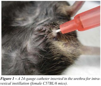
Intravesical tumor implantation - Mice were
anesthetized by the intraperitoneal administration of ketamine/xylazine
solution at a dose of 0.1 mL/10g body weight. Subsequently, a 24-gauge
Teflon i.v. catheter (NiproR) was inserted through the urethra
into the bladder using an inert lubricant (sterile contact gel) (Figure-1).
Then, in order to prepare the bladder for tumor implantation, a brief
acid exposure, followed by alkaline neutralization, promoted a chemical
lesion on the bladder wall, performed by intravesical instillation of
8µl 1M silver nitrate (AgNO3). This promoted an adequate
and controlled diffuse bladder wall lesion. After 10 seconds, the content
was washed out by transurethral infusion of phosphate-buffered saline.
The first catheter was removed and a new 24-gauge catheter was inserted
in the urethra for intravesical instillation of MB49 cells. A cell suspension
of MB49 tumor cells (5 x 10⁵ cells in 0.1 mL 50% normal mouse
serum) was instilled and retained for 2 hours by stitches.
The mice were evaluated on a daily basis
for viability and gross hematuria. After 15 days, the animals were sacrificed
by CO2 inhalation, examined macroscopically for intravesical
tumor and individually verified the bladder weight.
Histology and immunohistochemistry: After
gross examination, the bladders were fixed in buffered formalin 10%, routinely
processed and paraffin included and stained by hematoxylin and eosin .
Immunohistochemistry was performed to characterize the immunophenotype
and the antibodies used were cytokeratin 7 (CK7 OV-TL 12/30, 1:100), carcinoembrionic
antigen (Dako-CEA, II-7 1:200), p53 (DO7, 1:100) and c-erbB2 oncoprotein
(Her2/neu, 1:100), all produced by Dakocytomation, Glostrup, Denmark.
Three-micrometer sections from the paraffin block containing tumor were
placed on adhesive-coated slides. In a heat antigen retrieval process
the slides were placed in a citrate buffer (1mM, pH 6.0) and heated for
30 min. in the steamer. The slides were incubated overnight at 4ºC
with the above antibodies. Labelled Streptavidin Biotin (LSAB; Dako Cytomation,
CA,) at first biotinylated link universal for 35 min at room temperature,
then the slides were rinsed with Tris-buffer for 5 min., incubated for
a further 35 min. with streptavidin-HRP. The slides were rinsed in tap
water for 5 min. Color was developed by incubating the slides in 0.06%
diaminobenzidine in PBS for 15 minutes, and the slides were rinsed in
Tris-buffer and tap water, counterstained with Harris hematoxylin, dehydrated,
cover slipped, and reviewed under light microscope. Tissue sections of
a bladder urothelial carcinoma known to express p53 and Her2-neu, as well
as pulmonary adenocarcinoma positive for cytokeratin 7 and CEA were used
as positive controls. For each case a negative control was applied by
following all steps of IHC except for replacement of the primary antibody
by PBS.
RESULTS
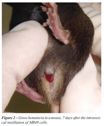
All
the animals survived the surgical intervention, no transmural bladder
injury or bladder perforation was observed. All animals, after 7 days,
presented evident gross hematuria (Figure-2), that persisted until the
sacrifice on day 15. There was no obstruction of the urinary flow, except
for the gross and intense hematuria. Considering all the animals (30)
that received intravesical instillation of MB49 cells, 29 (96.7%) developed
intravesical tumors after the 15-day period. At that time there was massive
growth of a solid tumor inside the bladder.
Macroscopically the mean bladder weight
was 0.196g (0.069-0.538g) in the tumor group, while a C57BL/6 mouse bladder
weighs between 0.010g to 0.015g approximately. The tumor was represented
by an usually solitary solid mass, growing inside the bladder, deeply
red, soft and extremely bloody. There were areas of necrosis and superficial
ulceration. Microscopically, there was a high grade urothelial solid carcinoma
composed of proliferation of large, cubic cells, arranged in solid nests,
with round, hypercromatic nuclei and one or more nucleoli. Scattered giant,
bizarre cells were seen in the tumor. The mitosis rate was high (50/HPF).
Superficial ulceration and foci of necrosis were identified. In all cases
the tumor was invasive through the bladder wall, reaching the muscularis
propria. No vascular invasion or perineural infiltration was seen. The
transition to normal urothelium was evident, and no in situ carcinoma
was identified (Figure-3).
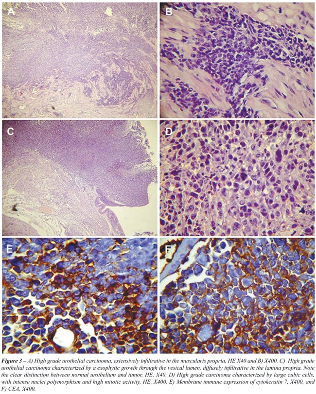
The immunohistochemical analysis showed
strong, membrane expression of CEA and CK7: a similar profile that we
commonly see for urothelial cancer that affects humans. Although p53 expression
is very common in urothelial cancer, due to p53 mutation, in the murine
model it does not seem to be a part of the carcinogenesis since we were
unable to detect p53 staining. Also, we used antibody anti-Her2/neu, an
oncogene super expressed in 30 to 60% of human urothelial carcinomas.
There was no Her2/neu expression in the cases we have evaluated. The molecular
evaluation of those two abnormalities among others should be the subject
of our next study.
COMMENTS
As
demonstrated above, the histological and immunohistochemical characteristics
described in the murine bladder tumor model derived from the MB49 cell
line mimics one of the main aspects of the human infiltrating urothelial
carcinoma, which is defined as a urothelial tumor that invades beyond
the lamina propria. By producing a pathologically similar cancer to the
human urothelial carcinoma, evidence from experimental research may become
closer to data from human testing.
Considering the immunoregulatory mechanisms
that participate in bladder tumor-host interaction (12), a wide field
of research is open for investigation. Consequently, adequate local and
systemic experimental environment model is required, allowing a significant
conclusion to be obtained for further studies.
A murine bladder tumor model may offer some
of these characteristics, while having many controlled variables under
laboratory conditions. These factors permit inference from experimental
data, which can be helpful for clinical purposes, specially considering
safety issues.
Despite the technical difficulty of orthotopic
tumor implantation, improvements have been made to avoid the variability
of tumor cell adhesion to the bladder wall (5). Soloway et al. simulated
a transurethral fulguration by cauterizing the murine bladder wall, creating
conditions for tumor cells implantation (10). The development of this
technical modification enhanced the model’s applicability by transforming
the orthotopic syngeneic bladder tumor model into a reliable tool for
investigating tumor growth mechanisms and intravesical drug testing. Furthermore,
the model closely resembles the clinical situation, making it very suitable
for bladder cancer research.
The use of immunohistochemistry for accurate
diagnosis of cancer has long been demonstrated (13). Determination of
cytokeratins, in this situation, may be helpful in tumors poorly differentiated
and for identifying the primary site of metastatic carcinomas (14). Moreover,
IHC provides information on tumor progression, giving p53 expression among
other markers, a significant prognostic value (15,16).
The murine bladder tumor model not only
creates adequate conditions for understanding tumor adhesion, proliferation
and invasiveness, but also allows the development of a cancer with many
similar histopathological characteristics to the human urothelial carcinoma,
making it a valuable tool for anti-tumor drug testing, based on immune,
viral or gene therapy(9,17,18).
CONCLUSIONS
The
tumor implantation procedures described herein provide a reproducible
experimental bladder cancer model. The orthotopic murine model has an
important role improving our knowledge of therapeutic mechanisms of superficial
bladder cancer in the proper anatomical site.
In conclusion, the histopathology and immunohistochemical
profile of the murine bladder tumor model derived from the MB49 cell line
resembles the human infiltrating urothelial carcinoma, allowing us to
make inferences about its behavior and response to different treatment
regimes.
ACKNOWLEDGEMENT
State of Sao Paulo Research Foundation (FAPESP) provided financial support.
CONFLICT OF INTEREST
None declared.
REFERENCES
- Böhle A, Brandau S: Immune mechanisms in bacillus Calmette-Guerin immunotherapy for superficial bladder cancer. J Urol. 2003; 170: 964-9.
- Ratliff TL: Role of animal models in understanding intravesical therapy with bacille Calmette-Guérin. Clin Infect Dis. 2000; 31 Suppl 3: S106-8.
- Hansson Y, Paulie S, Ben-Aïssa H, Rudberg U, Karlsson A, Perlmann P: Radioimmunolocalisation of bladder tumors xenotransplanted in nude mice. Anticancer Res. 1988; 8: 435-41.
- Soloway MS: Single and combination chemotherapy for primary murine bladder cancer. Cancer. 1975; 36: 333-40.
- Günther JH, Jurczok A, Wulf T, Brandau S, Deinert I, Jocham D, et al.: Optimizing syngeneic orthotopic murine bladder cancer (MB49). Cancer Res. 1999; 59: 2834-7.
- Abe T, Tada M, Shinohara N, Okada F, Itoh T, Hamada J, Harabayashi T, Chen Q, Moriuchi T, Nonomura K: Establishment and characterization of human urothelial cancer xenografts in severe combined immunodeficient mice. Int J Urol. 2006; 13: 47-57.
- Mickey DD, Mickey GH, Murphy WM, Niell HB, Soloway MS: In vitro characterization of four N-[4-(5-nitro-2-furyl)-2-thiazolyl] formamide (FANFT) induced mouse bladder tumors. J Urol. 1982; 127: 1233-7.
- Summerhayes IC, Franks LM: Effects of donor age on neoplastic transformation of adult mouse bladder epithelium in vitro. J Natl Cancer Inst. 1979; 62: 1017-23.
- Wang H, Satoh M, Abe H, Sunamura M, Moriya T, Ishidoya S, et al.: Oncolytic viral therapy by bladder instillation using an E1A, E1B double-restricted adenovirus in an orthotopic bladder cancer model. Urology. 2006; 68: 674-81.
- Soloway MS, Masters S: Urothelial susceptibility to tumor cell implantation: influence of cauterization. Cancer. 1980; 46: 1158-63.
- Fodor I, Timiryasova T, Denes B, Yoshida J, Ruckle H, Lilly M: Vaccinia virus mediated p53 gene therapy for bladder cancer in an orthotopic murine model. J Urol. 2005; 173: 604-9.
- Luo Y, Chen X, O’donnell MA: Use of prostate specific antigen to measure bladder tumor growth in a mouse orthotopic model. J Urol. 2004; 172: 2414-20.
- Debus E, Moll R, Franke WW, Weber K, Osborn M: Immunohistochemical distinction of human carcinomas by cytokeratin typing with monoclonal antibodies. Am J Pathol. 1984; 114: 121-30.
- Cid Mouteira P, Ortíz Rey JA, Antón Badiola I, San Miguel Fraile P, Alvarez Alvarez C, et al.: Coordinated expression of cytokeratin 7 and 20 in transitional carcinoma of the bladder: diagnostic usefulness. Actas Urol Esp. 2002; 26: 279-84.
- Schrier BP, Vriesema JL, Witjes JA, Kiemeney LA, Schalken JA: The predictive value of p53, p27(kip1), and alpha-catenin for progression in superficial bladder carcinoma. Eur Urol. 2006; 50: 76-82.
- McKenney JK, Amin MB: The role of immunohistochemistry in the diagnosis of urinary bladder neoplasms. Semin Diagn Pathol. 2005; 22: 69-87.
- Moltedo B, Faunes F, Haussmann D, De Ioannes P, De Ioannes AE, Puente J, et al.: Immunotherapeutic effect of Concholepas hemocyanin in the murine bladder cancer model: evidence for conserved antitumor properties among hemocyanins. J Urol. 2006; 176: 2690-5.
- Loskog AS, Fransson ME, Totterman TT: AdCD40L gene therapy counteracts T regulatory cells and cures aggressive tumors in an orthotopic bladder cancer model. Clin Cancer Res. 2005; 11: 8816-21.
____________________
Accepted after revision:
February 22, 2008
_______________________
Correspondence address:
Dr. Miguel Srougi
Rua Peixoto Gomide, 2055 / 81
São Paulo, SP, 01409-003, Brazil
Fax: + 55 11 3257-8002
E-mail: srougi@uol.com.br
EDITORIAL COMMENT
The
study presented by Chade et al. shows a nice modification of the previously
published orthotopic MB-49 bladder tumor model.
The main difference to the model optimization
published by Günther et al. (1999) is the initial bladder lesion
before tumor cell instillation. In the previous model by Günther
et al. mice were placed with their backs on the ground plate of the cautery
unit. To optimize contact, electrocardiogram electrode contact gel was
used. The soft-tipped end of a spring-wire guide of a 24-gauge central
venous catheter was inserted into the bladder via a Teflon catheter and
gently pushed forward until it reached the bladder wall. The guide wire
was attached to a cautery unit and a monopolar coagulation was applied
for 5 s at the lowest setting (5 W). After removal of the guide wire,
0.05 mL of the tumor cell suspension was instilled. Chade et al. induced
instead of physical alteration of the bladder wall a chemical lesion with
intravesical silver nitrate.
Furthermore, in the original description
catheters were after tumor cell instillation pinched off with a clamp,
kept locked with a Luer-Lock closing cone, and left in place until the
mice awakened. Using this method, a dwell time of approximately 3 h. was
given.
Here, a dwell time of 2 hours was performed
by temporary stitches (presumably of the urethra).
The effectivity of the described technique
is comparable to the previous model. The tumor take was almost 100% and
all animals developed gross hematuria. However, the number of animals
with pulmonary metastases, which was 20-70% before, was not mentioned
here.
Chade et al. examined the Her2/neu, p53,
CK7, and CEA expression in this model. Interestingly, p53 and Her2/neu
staining was negative. One has to be aware that this can also be due to
antibody-related problems since immunohistochemistry staining procedures
are frequently more difficult in mouse tissue than in human tissue. We
have previously examined Ki-67 (TEC-3) expression and found in the tumors
up to 70% positive cells (unpublished data).
In general, the molecular comparison between
human bladder cancers and mouse models is very interesting since the results
of therapeutic applications may be easier to interpret. As the authors
point out further molecular evaluations are planned. It would be highly
interesting to perform these evaluations also in an humanized immune incompetent
(SCID or Nude) mouse model. However, we were until now not able, to transfer
the intravesical model into SCID mice since most mice died after bladder
wall coagulation. Maybe the technical modification of Chade et al. would
lead to better results.
Dr.
Ingo Kausch
Department of Urology
University of Lubeck Medical School
Lubeck, Germany
E-mail: ingoKausch@uk-sh.de
Bladder
cancer is well suited for experimental therapies due to the isolated bladder
cavity in which therapies can be given locally. By transurethral noninvasive
surgery, bladder tumors can easily be monitored and biopsies taken for
further analyses. These features are beginning to attract a number of
drug developers within a variety of fields including chemo-, immuno- and
gene therapy. Bladder cancer is one of few cancers that have excellent
orthotopic murine experimental models that are closely mimicking the clinical
situation (1,2). Hence, in these orthotopic models, tumor biology can
be studied and tumor therapy can be given locally by instillation via
cauterization of urethra just as in the patients. In the current issue,
Chade et al. are describing a novel system to enhance tumor take in experimental
bladder cancer using silver nitrate (AgNO3) as well as giving
further insights into the biology of murine bladder tumors by performing
a histopathological evaluation.
The
most common murine bladder cancer cell lines are the mouse bladder-49
(MB49) and the mouse bladder tumor-2 (MBT2) cells (1-3). These two cell
lines can be used in syngeneic C57BL6 and C3H mice, respectively. The
cell lines are utilized to create subcutaneous, orthotopic or metastatic
tumors. In one of the first orthotopic models electrocautherization was
used for tumor take. The electric pulse created a burn wound to which
MB49 cells attached and formed tumors. In this model, the effect of Bacillus
Calmette-Guérin (BCG) therapy has been extensively evaluated and
immunological mechanisms found in this model have later been proven transferable
to human systems (1,4). However, the electrocautherization model has a
few drawbacks. The main issue is that it can be difficult to obtain the
technical equipment necessary. Further, this model does not give 100%
tumor take which increases the number of mice needed per treatment group
and may mask the true result in some treatment groups where differences
are slim. Many groups have tried to obtain similar or better tumor take
by chemical pretreatment of the bladder surface prior instillation of
tumor cells. Agents tested are for example ethanol and poly-L-lysine (PLL)
(2). Ethanol functions as an irritant and theoretically removes the mucin
layer in the bladder thereby facilitating tumor take. The latter, PLL,
is a polycathion that is thought to by its positive electrical charge
become a bridge between the negatively charged urothelium and the tumor
cells thereby aiding tumor attachment. PLL has so far been the only agent
that repeatedly gives tumor take in all mice. In this issue, Chade et
al has further improved the management of the orthotopic model via the
use of AgNO3 pretreatment of the bladders to irritate the bladder
wall prior tumor cell instillation. The tumor take is similar to that
of PLL but the model as such saves time since this agent only needs a
few seconds of incubation compared to PLL that needs to be incubated in
the bladder for 10-20 min. prior tumor instillation. It will be of interest
to evaluate effects of different therapies in this improved model.
The
MB49 tumor cells have many similarities to its human equivalent in terms
of antigens and immune escape mechanisms. The latter include infiltration
of T regulatory cells in the growing tumor, expression of TGF, attraction
of IL10-producing suppressor cells other than T regulatory cells etc (5-7).
This makes the MB49 model excellent for evaluation of novel immunotherapies.
The MB49 cells have expression of the male antigen HY and this antigen
has been used as a pseudo tumor antigen when the cells are used in female
mice (8). However, when antigen-directed approaches are evaluated, true
tumor antigens need to be targeted. Carcinoembryonic antigen (CEA) is
one of the first identified tumor antigens. It is expressed in about half
of all human tumors, especially in adenocarcinomas (9,10). The results
from Chade et al demonstrate that MB49 cells express CEA as do human bladder
cancer. This antigen is often used in tumor immunotherapy and the MB49
model can, hence, serve as a model system not only for bladder cancer
but for all CEA positive tumors.
The
murine experimental MB49 model gives new insights to tumor progression,
survival and immune escape in human bladder cancer. Currently, there are
several novel therapies such as immune and gene therapy that are proven
potent in the MB49 model and now translated into clinical Phase I and
II trials. It is important to further investigate murine experimental
models to simplify the techniques as well as to further enlighten biological
phenomena that may be translated into human cancer and get us closer to
better and more refined drugs.
REFERENCES
- Günther JH, Jurczok A, Wulf T, Brandau S, Deinert I, Jocham D, et al.: Optimizing syngeneic orthotopic murine bladder cancer (MB49). Cancer Res. 1999; 59: 2834-7.
- Loskog A, Ninalga C, Hedlund T, Alimohammadi M, Malmström PU, Tötterman TH: Optimization of the MB49 mouse bladder cancer model for adenoviral gene therapy. Lab Anim. 2005; 39: 384-93.
- Torti SV, Golden-Fleet M, Willingham MC, Ma R, Cline M, Sakimoto Y, et al.: Use of green fluorescent protein to measure tumor growth in an implanted bladder tumor model. J Urol. 2002; 167: 724-8.
- Suttmann H, Riemensberger J, Bentien G, Schmaltz D, Stöckle M, Jocham D, et al.: Neutrophil granulocytes are required for effective Bacillus Calmette-Guérin immunotherapy of bladder cancer and orchestrate local immune responses. Cancer Res. 2006; 66: 8250-7.
- Loskog A, Dzojic H, Vikman S, Ninalga C, Essand M, Korsgren O, et al.: Adenovirus CD40 ligand gene therapy counteracts immune escape mechanisms in the tumor Microenvironment. J Immunol. 2004; 172: 7200-5.
- Loskog AS, Fransson ME, Totterman TT: AdCD40L gene therapy counteracts T regulatory cells and cures aggressive tumors in an orthotopic bladder cancer model. Clin Cancer Res. 2005; 11: 8816-21.
- Loskog A, Ninalga C, Paul-Wetterberg G, de la Torre M, Malmström PU, Tötterman TH: Human bladder carcinoma is dominated by T-regulatory cells and Th1 inhibitory cytokines. J Urol. 2007; 177: 353-8.
- Melchionda F, McKirdy MK, Medeiros F, Fry TJ, Mackall CL: Escape from immune surveillance does not result in tolerance to tumor-associated antigens. J Immunother. 2004; 27: 329-38.
- Huang EH, Kaufman HL: CEA-based vaccines. Expert Rev Vaccines. 2002; 1: 49-63.
- Hörig H, Medina FA, Conkright WA, Kaufman HL: Strategies for cancer therapy using carcinoembryonic antigen vaccines. Expert Rev Mol Med. 2000; 2: 1-24.
Dr. Angelica Loskog, PhD
Clinical Immunology Division
Rudbeck Laboratory, Uppsala University
Uppsala, Sweden
Email: angelica.loskog@klinimm.uu.se
EDITORIAL COMMENT
An animal model that closely resembles human bladder cancer is needed for preclinical studies on the pathogenesis of bladder cancer and the development of therapeutic strategies for treating this disease. This paper describes a syngeneic murine bladder tumor model that is developed by intravesical implantation of MB49 cells, a commonly used murine bladder cancer cell line of the C57BL/6 origin. The authors have established the experimental conditions that result in a high incidence of orthotopic tumor in mice (96.7%; 29 out of 30 mice on day 15). By using a small volume of silver nitrate to traumatize the urothelium, the authors have demonstrated the feasibility of this method for intravesical bladder tumor implantation. The implanted animals developed a solid tumor inside the bladder that mimics human urothelial invasive carcinoma in histopathology. Immunohistochemical analysis showed the strong expression of cytokeratin 7 and carcinoembrionic antigen on the surface of MB49 tumor cells, which is similar to human urothelial cancer. These surface markers facilitate the identification of primary tumor when metastasis occurs. Although this model is feasible and provides a high rate of tumor intake, this model needs to be improved for its variability in tumor growth. Nevertheless, this model provides a useful means for the therapeutic studies of bladder cancer including immunotherapy, chemotherapy, and gene therapy.
Dr. Yi Luo
Department of Urology
University of Iowa
Roy J. and Lucille A. Carver College of Medicine
Iowa City, Iowa, USA
E-mail: yi-luo@uiowa.edu