POLYPROPYLENE
DISTAL URETHRAL SLING FOR TREATMENT OF FEMALE STRESS URINARY INCONTINENCE
(
Download pdf )
FERNANDO G. DE ALMEIDA, LARISSA V. RODRIGUEZ, SHLOMO RAZ
Department of Urology, University of California Los Angeles (UCLA), Los Angeles, California, USA
ABSTRACT
Introduction:
Sling procedures have been used successfully for the treatment of stress
urinary incontinence. Using similar surgical principles to the cadaveric
fascia sling, we describe the placement of a thinly woven polypropylene
(Prolene) mesh under the mid to distal urethra. We describe our technique
and report early outcomes.
Materials and Methods: A total of 263 consecutive
patients were evaluated. All patients had clinical evidence of stress
urinary incontinence. Patients underwent a preoperative evaluation with
video-urodynamic studies, symptom questionnaire, and cystoscopy. A 1 x
10-cm Prolene mesh was placed under the mid to distal urethra. At a minimum
follow-up of 1 year (12-24 months), patients were evaluated with a urogenital
symptom questionnaire, physical examination, and postvoid residual volume
determination.
Results: Twenty-six percent of the patients
had unsuccessful prior vaginal surgery. There were no major complications
such as permanent retention, erosion, infection or rejection to the mesh.
The mean operative time was 27 minutes. In 90% of the patients, the suprapubic
catheter was removed within 1 week. No patient experienced permanent retention.
One hundred and twenty-eight patients had at least 12 months of follow
up and were included for the outcome analysis. 96.4% of these patients
were cured or improved and only 3% developed urge-incontinence again.
Conclusions: We describe a new, simple,
quick, inexpensive, and effective method to correct stress urinary incontinence
by placing a Prolene mesh under the distal urethra.
Key words:
urinary incontinence; stress; female; surgical technique; sling; prolene
mesh
Braz J Urol, 28: 254-258, 2002
INTRODUCTION
Surgeries for the treatment of female urinary incontinence using “sling” procedures have been effective both for intrinsic sphincter deficiency and urethral hipermobility (1). There are many surgical techniques variations as well as variations in material forms and types used for urethral support. Among the options are the use of the vaginal wall, pubovaginal “sling” with the use of the autologous fascia, cadaveric fascia or synthetic materials placed at the vesical cervix level. Variations in the “sling” fixation method include pubis fixation (“bone anchors”), use of permanent or absorbable suprapubic sutures and, recently, “tension free vaginal tape” known as TVT. Nowadays, due to the morbidity and long postoperative after the autologous fascia harvesting, new materials have been developed and used. One example is the use of cadaveric fascia “sling” which presents good results. However, this material is relatively expensive and its long-term durability is still unknown. Many studies have suggested that the cadaveric fascia presents low durability and high recurrence rate (2-5). The reason is unknown, but the cadaveric fascia is seen absorbed or disintegrated in reinterventions. Using the same principle of distal urethra support used in TVT but with the use of the traditional technique of “sling” placement, we describe the placement of woven polypropylene mesh under the mid third to distal urethra, and report our results with a minimum follow up of one year.
PATIENTS AND METHODS
Two hundred and sixty-three women with stress urinary incontinence (SUI) were treated with the urethral sling technique with polypropylene mesh. Preoperative evaluation was performed through medical history, physical examination, urinary flow, post-voiding residue, video-urodynamics, cystoscopy and specific symptoms (UDI-6) (6) and quality of life questionnaires, besides voiding dysfunction and incontinence symptoms questionnaires. Postoperative evaluation was performed every three months with the same symptoms and quality of life symptoms questionnaire, physical examination, fluxometry and post-voiding urinary residue measurement. We used a strict criteria of cure defined as: cure reported by the patient, negative Marshall test, no use of absorbents, and absence of side effects. Failure was defined as negative patient report, with no or less than 50% improvement when compared to the preoperative. We have considered significant the improvement above 50% reported in the patients’ questionnaires.
SURGICAL TECHNIQUE
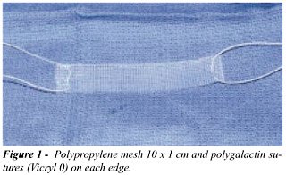
A
polypropylene mesh (Ethicon, New Jersey) is cut to form a 10X1 cm band
which will be used as a “sling”. A 0-polygalactine suture (Vicryl
0) is passed through each edge of the “sling” (Figure-1), which
is kept in povidine solution to avoid infection. The patient is placed
in forced lithotomy position and the inferior abdomen and perineal region
are prepared with povidine and sterile fields. The vaginal canal and the
urethra are exposed suturing the vaginal lips laterally and then placing
a weigh remover. A suprapubic cystostomy is performed using a Lowsley
and placing a Foley 16F probe, which will be used to evaluate the postvoiding
residue after the surgery. Another Foley 16F is placed through the urethra
to facilitate urethra and vesical cervix localization. An Allis clamp
is used to hold the vaginal wall close to the urethral meatus. Two parallel
incisions are done laterally on the anterior vaginal wall at distal urethra
level approximately 1 cm proximal to the urethral meatus (Figure-2). An
additional Allis clamp is placed distally to each incision to facilitate
exposure. The dissection is then performed laterally over the periurethral
fascia in the direction of the ipsilateral shoulder. Mayo scissors are
used to enter the retropubic space at distal urethra level (Figure-2).

A tunnel between the vaginal wall and the
urethra is created 1.5 cm from the urethral meatus to place the “sling”
(Figure-3). A 1-cm incision is done on the inferior region of the abdomen,
just above the pubic symphysis. Through this incision, a double needle
(Cook Urological, Spencer, Ind.) is guided by the surgeon’s index-finger,
placed through the lateral incision on the vagina entering through the
anus lifting muscle fascia inside the retropubic space (Figure-4). The
threads previously passed through the “sling” are suspended
using the double needle. A similar maneuver is performed on the opposite
side, after placing the “sling” on the suburethral region. A
cystoscopy is then performed to discard any vesical lesion.

To ensure that there is no tension on the
“sling”, an Allis clamp is placed in each of the vaginal incisions
holding the “sling” firmly in a horizontal position (Figure-5),
while the Vicryl 0 suprapubic sutures are tied by the assistant. This
way, the tension on the urethra is controlled avoiding postoperative urinary
retention. The retropubic space is irrigated with povidine solution. The
vaginal and suprapubic incisions are closed and a vaginal tampon soaked
in clindamycin ointment is placed.

POSTOPERATIVE CARE
The
vaginal tampon is removed two hours after the procedure in the recovery
room. The suprapubic catheter is closed and the patient is advised to
urinate every 3 hours measuring the postvoiding urinary residue. The patient
is discharged from hospital and the suprapubic catheter is removed when
the postvoiding urinary residue is below 50 mL.
RESULTS
Among
the 263 patients which were treated, 26% had been submitted to prior unsuccessful
vaginal surgeries. Forty-five per cent were treated only with the placement
of suburetheral “sling”, 14% received “sling” associated
with urethrolysis, 33% were concomitantly submitted to proctocele, 4%
enterocele and 11% vaginal hysterectomy. Mean operative time to place
the “sling” was 27 minutes. There were no intraoperative and
postoperative complications, such as permanent urinary retention, urethral
erosion, infections or need to repeat the surgery. Our routine is to keep
the Foley catheter in all patients for a minimum of 7 days to allow a
better regeneration. In 90% of the patients, the suprapubic catheter was
removed one week after the surgery. Two patients presented partial urinary
retention for 2-3 months, followed by normal voiding.
A total of 128 patients were followed during
a minimum of 1 year (12-24 months). From these, 96.5% were completely
cured or with significant improvement, without incontinence and without
the use of absorbents, or with an improvement of more than 50%. The mean
number of absorbents decreased from 2.7 to 0.9. Prior to the surgery,
16% of the patients reported some degree of urge incontinence. After the
surgery, only 3% reported its reincidence. In a scale from 0 to 6 (0 =
completely satisfied and 6 = very bad), the average score in the quality
of life index related to the incontinence after the surgery was 1.7, which
corresponds to a high satisfaction level. After the surgery, the average
urinary flow and postvoiding residue was 15 mL/s and 4 mL respectively.
There was no significant difference when these results were compared to
the preoperative.
DISCUSSIONS
Traditionally, surgeries using “sling” as urethral support for the treatment of urinary incontinence have been performed with the fascia of the abdominal rectum muscle or fascia lata. Recently, new materials have been used in this procedure to avoid the removal of autologous material, and to decrease morbidity and operative time. However, all the materials presented until now are expensive, being then difficult to make them accessible for the general public. Making a band from a polypropylene mesh easily found in any operating room allows the performance of a fast surgery, with low morbidity and reduced cost. Differently from the TVT, this mesh is placed in the retropubic space through a more reliable approach, is fixed with absorbable threads, does not require special instrumentation and presents an insignificant cost. After 283 surgeries without complications (infection, rejection and erosion) due to the use of synthetic materials, we believe that the use of the polypropylene “sling” is significantly safe.
CONCLUSION
Placement of a polypropylene “sling” for the treatment of female urinary incontinence is a safe, easy and fast procedure to be performed, presenting excellent results and low cost.
____________________________
Dr. Fernando G. de Almeida holds
a scholarship from CNPq, Brazil
REFERENCES
- Chaikin DC, Rosenthal J, Blaivas JG: Pubovaginal fascial sling for all types of stress urinary incontinence: long term analysis. J Urol, 160: 1312-1316, 1998.
- Carbone JM, Kavaler E, Hu JC, Raz S: Pubovaginal sling using cadaveric fascia and bone anchors: disappointing early results. J Urol, 165: 1605-1611, 2001.
- Chaikin DC, Blaivas JG: Weakened cadaveric fascial sling: an unexpected cause of failure. J Urol, 160: 2151, 1998.
- Admusen CL, Visco AG, Ruiz H, Webster GD: Outcome in 104 pubovaginal slings using freeze-dried allograft fascia lata from a single tissue bank. Urology, 56: 2-8, 2000.
- McGuire EF, Lytton B: Pubovaginal sling procedure for stress incontinence. J Urol, 119: 82-84, 1978.
- Uebersax JS, Wyman JF, Shumaker AS, McClish DK, Fantl JA: Short forms to assess life quality and symptom distress for urinary incontinence in women: The incontinence impact questionnaire and the urogenital distress inventory. Neurourol Urodynam, 14: 131-139, 1995.
______________________
Received:
March 13, 2002
Accepted: April 20, 2002
_______________________
Correspondence address:
Dr. Larissa V. Rodríguez
924 Westwood Blvd, Suite 520
Los Angeles, California 90024, USA
Fax: + + (1) (310) 794-0206
E-mail: lrodriguez@mednet.ucla.edu