GIANT
RETROPERITONEAL LIPOSARCOMA
(
Download pdf )
ROBERTO I. LOPES, ROBERTO N. LOPES, C.M. BARBOSA FILHO
Sírio Libanês Hospital, São Paulo, SP, Brazil
ABSTRACT
Introduction:
We report a case of a huge well-differentiated liposarcoma with dedifferentiated
areas contributing to the patient’s unfavorable outcome.
Case Report: A 77 year old woman presented
with a 8-month history of progressive abdominal enlargement accompanied
by diffuse abdominal discomfort. A firm mass was noted on the right quadrants
of her abdomen. Computed tomography revealed 2 large masses displacing
intra-abdominal organs. The masses were removed surgically and the histopathologic
examination of the specimens showed a well-differentiated liposarcoma
with dedifferentiated areas. Twenty six months after this surgery, the
patient underwent another surgery to remove a dedifferentiated liposarcoma
and died 2 years later.
Discussion: The outcome of a liposarcoma
is influenced by its histological subtype. Well-differentiated liposarcomas
are low-grade tumors that usually do not have an aggressive behavior,
but may recur locally. However, our patient had a huge tumor (> 20
cm) with dedifferentiated areas, which are associated with a poor prognosis.
Key words:
tumor; retroperitoneal neoplasm; sarcoma; liposarcoma
Braz J Urol, 28: 227-229, 2002
INTRODUCTION
Liposarcoma is a rare mesenchymal tumor that occurs more frequently in the soft tissues of the extremities between the fifth and seventh decades (1). However, other sites such as the retroperitoneum and the abdominal cavity may be affected (2). Retroperitoneal liposarcoma may achieve a considerable size before diagnosis, thanks to its clinical presentation characterized by symptoms which are usually late and nonspecific (1). We report a case of a huge well-differentiated liposarcoma with dedifferentiated areas contributing to the patient’s unfavorable outcome.
CASE REPORT
A
77-year-old woman presented with a 8-month history of progressive abdominal
enlargement accompanied by diffuse abdominal discomfort. During general
examination, a firm mass without tenderness was noted on the right upper
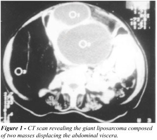
and lower quadrants of the abdomen. Computed tomography (CT) scan of the
abdomen revealed 2 large masses (Figure-1).
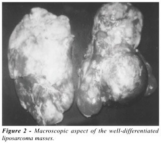
At laparotomy, a huge retroperitoneal tumor
consisting of 2 masses was completely removed (Figure-2). A multilobulated
mass involving the right kidney and the right adrenal gland weighed 3400
g and measured 37.5 x 11 x 8 cm. The histopathologic examination of the
surgical specimens showed a well-differentiated sclerosing liposarcoma.
There was no evidence of renal and adrenal tumor infiltration. Some areas
revealed dedifferentiation with a histological pattern compatible with
fibrous histiocytoma.
Twenty six months after this surgery, the
patient underwent another surgery to totally remove a dedifferentiated
liposarcoma, at L3-L4 level adherent to the inferior vena cava, characterized
by spindle cells arranged in fascicles in a schwannoma-like pattern. It
weighed 980 g and measured 18 x 13 x 8 cm. Immunohistochemistry study
was negative for S-100, CD68 and muscle-specific actin.
The patient died 2 years later by disease
progression. Caquexia was evident and although no metastases were observed,
another retroperitoneal mass was revealed at CT. At that point, the patient
refused any additional surgery.
DISCUSSION
Liposarcomas are classified according to
3 histological types: well-differentiated, myxoid, and pleomorphic (2).
Among all liposarcomas, the myxoid type is the most common, accounting
for 60% of cases, while well-differentiated liposarcoma occurs in 25%
and pleomorphic in 10% (2).
The outcome of liposarcomas is influenced
by their histological subtype (3). Pleomorphic liposarcomas are highly
aggressive with significant rates of recurrence and metastasis, while
well-differentiated liposarcomas are low-grade tumors that usually do
not have an aggressive behavior, but may recur locally (1,3).
Dedifferentiated
areas and large tumors (> 20 cm) have been associated with poor prognosis
(2,3). Dedifferentiation is defined as the presence of non-lipogenic high
grade areas within the well-differentiated liposarcoma (2). Dedifferentiation
is rare, occurring in 15% of the well-differentiated liposarcomas. In
most cases, it recurs in the primary tumor in most cases, but may also
develop later, usually after liposarcoma recurrence (2).
Divergent differentiation in schwannomas,
osteosarcomas, leiomyosarcomas and rhabdomyosarcomas is possible (2).
In our case, the histological presentation, at the moment of recurrence,
was a well-known phenomenon called dedifferentiation. Immunohistochemistry
study, using an antibody panel for identification of specific antigens
related to different sarcomas was performed to make the differential diagnosis.
All were negative excluding leiomyosarcoma, fibrous histiocytoma and schwannoma.
The other retroperitoneal mass, possibly another recurrence, will not
be discussed since its histological subtype was not diagnosed.
REFERENCES
- Susini T, Taddei G, Massi D, Massi G: Giant pelvic retroperitoneal liposarcoma. Obstet and Gynecol, 95: 1002-1004, 2000.
- Hasegawa T, Seki K, Hasegawa F, Matsuno Y, Shimoda T, Hirose T et al.: Dedifferentiated liposarcoma of retroperitoneum and mesentery: varied growth patterns and histological grades- a clinicopathologic study of 32 cases. Hum Pathol, 31: 717-727, 2000.
- Sato T, Nishimura G, Nonomura A, Miwa K: Intra-abdominal and retroperitoneal liposarcomas. Int Surg, 84: 163-167, 1999.
__________________________
Received: November 21, 2001
Accepted after revision: April 22, 2002
_______________________
Correspondence address:
Dr. Roberto Iglesias Lopes
Rua Baronesa de Itu, 721 / 121
São Paulo, SP, 01231-001, Brazil
Fax: + + (55) (11) 3666-8266
E-mail: robertoiglesias@terra.com.br