BILATERAL
HYDRONEPHROSIS CAUSED BY VAGINAL PROLAPSE
(
Download pdf )
HELIO BEGLIOMINI, BRUNO D. S. BEGLIOMINI
Humanae Vitae Medicine Institute, São Paulo, SP, Brazil
ABSTRACT
Introduction:
Even though it is uncommon, uterine prolapse can cause compression of
ureters and bilateral hydronephrosis, predisposing to arterial hypertension
and renal failure. Hydronephrosis consequent to cystocele and to vaginal
prolapse is even rarer.
Case Report: This paper reports on a 59
year-old patient, Caucasian, obese and hysterectomized who presented complete
vaginal prolapse with bilateral hydronephrosis and slight alteration in
serum urea and creatinine. Patient underwent correction of vaginal prolapse
by endoscopic suspension technique with improvement of hydronephrosis
and normalization of renal function. This work emphasizes the rarity of
such case and the requirement of surgical approach.
Key
words: vagina; vaginal prolapse; hydronephrosis
Int Braz J Urol. 2003; 29: 243-4
INTRODUCTION
Uterine prolapse can cause dilatation of upper urinary tract due to ureteral obstruction that, if left untreated, can impair renal function leading to anuria and arterial hypertension (1). Bilateral hydronephrosis due to cystocele and, especially, to vaginal prolapse, is very rare.
CASE REPORT
E.F.C.B.,
59 years old, Caucasian, widowed, was referred to the Urology Service
with vaginal prolapse and ultrasonography of urinary tract evidencing
bilateral grade II/III hydronephrosis.
As for her antecedents, she reported having
4 pregnancies in the past, with 2 normal deliveries, 1 cesarean and 1
miscarriage. She was hysterectomized by abdominal route 1 year before
due to uterine myoma, and on that occasion, a vesical suspension was also
performed. She did not present urinary incontinence.
On physical examination, she had a pyknic
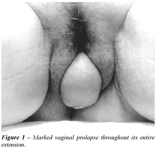
constitution, was obese and presented a good general state. Gynecologic
examination showed a marked vaginal prolapse throughout its entire extension
with excoriations, hyperemia and fissures on the posterior wall of vagina
(Figure-1). Laboratory tests showing alteration in urea 67.1 mg % (normal
< 40 mg %), creatinine 1.35 mg % (normal < 1.30 mg %) and glycemia
131 mg % (normal < 110 mg %). She did not present urinary infection.
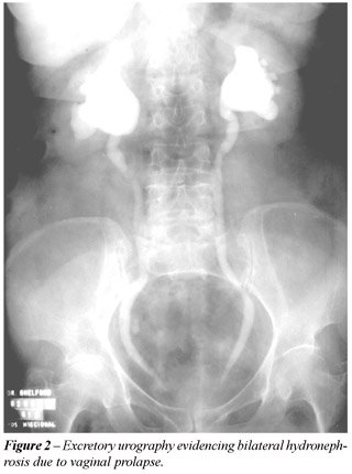
The excretory urography confirmed the presence of bilateral hydronephrosis
(Figure-2).
Patient underwent an endoscopic colposuspension
(3), with good post-operative results within 3 months of follow-up, and
improvement of hydronephrosis grade (grade I).
COMMENTS
It
is estimated that 4 to 7% patients with uterine prolapse have obstructive
uropathy. The mechanism most likely is direct compression of ureters (2).
In the uterine prolapse, there is herniation of bladder, uterus and ureters
through the pelvic floor and the ureters are compressed between the fundus
of uterus and the bladder, against the levator ani muscles. In this case,
since there was no uterus, we suspect that obstruction had occurred due
to ureteral compression against the pelvic musculature, as well as to
ureteral stretching itself, what makes peristaltic movements difficult.
Stress urinary incontinence usually is associated
to small cystoceles. Large cystoceles, associated or not with uterine
prolapse, predispose to obstructive voiding symptoms, chronic residual
urine and rarely to bilateral hydronephrosis with potential impairment
of renal function. In women presenting dilatation of upper urinary tract
one must always rule out, among other causes, uterine or vesical prolapse.
Surgical correction either by suprapubic
or vaginal approach, intends to resolve the obstructive urinary picture,
even though it is known that it can predispose to stress urinary incontinence.
When the uterus is present, hysterectomy and vaginal plastic surgery are
performed. When there are contraindications to surgery, the pressary can
be indicated in order to reduce the uterine prolapse (1).
In the case found in literature, it was
performed the fixation of the vaginal dome in sacral promontory complemented
with colpourethropexy in Cooper’s ligament (2). In the case reported
here, despite the patient being pyknic and obese, with 2 previous surgeries
in lower abdomen, the use of vaginal suspension with endoscopic control
has shown to be a simple and practical procedure.
REFERENCES
- Sudhakar AS, Reddi VG, Schein M, Gerst PH: Bilateral hydroureter and hydronephrosis causing renal failure due to a procidentia uteri: a case report. Int Surg. 2001; 86:173-5.
- Delaere K, Moonen W, Debruyne F, Jansen T: Hydronephrosis caused by cystocele. Treatment by colpopexy to sacral promontory. Urology. 1984; 24:364-5.
- Palma PCR, Rodrigues Netto N, Pinotti JA: Endoscopic suspension of vaginal vault prolapse. J Bras Urol. 1988; 14:41-2 [in Portuguese].
_____________________
Received: March 6, 2003
Accepted after revision: May 2, 2003
_______________________
Correspondence address:
Dr. Helio Begliomini
Rua Bias, 234
São Paulo, SP, 02371-020, Brazil