ROLE
OF INTRAVENOUS UROGRAPHY AND TRANSABDOMINAL ULTRASONOGRAPHY IN THE DIAGNOSIS
OF BLADDER CARCINOMA
(
Download pdf )
MUHAMMAD RAFIQUE, ABRAR A. JAVED
Instar Medical College, Multan, Pakistan
ABSTRACT
Introduction:
The present study was carried out to compare the efficacy of transabdominal
ultrasonography and intravenous urography in the diagnosis of bladder
carcinoma in those patients presenting painless hematuria.
Materials and Methods: Medical records of
100 patients who had both ultrasonography and intravenous urography were
studied. The reported findings of these investigations were correlated
with those of cystoscopy.
Results: Ultrasonography was significantly
more sensitive (96%) in the detection of bladder carcinoma compared to
urography (87%). By applying the test of equality of proportions, the
value of Z is 2.28, which is statistically significant (p < 0.01).
In addition, ultrasonography was more sensitive in clarifying the pathology
in upper renal tracts i.e. ureteric obstruction secondary to bladder carcinoma
when urography failed due to none or poor excretion of contrast.
Comments: We recommend the use of ultrasonography
as the initial radiological investigation for detection of bladder carcinomas
in patients presenting hematuria. Ultrasonography is safe, easily available,
cost effective and provides images of both upper and lower renal tract.
Patients diagnosed to be suffering from bladder carcinoma by ultrasonography
should be scheduled directly and promptly for cystoscopy and bladder tumor
resection.
Key
words: bladder; bladder neoplasms; ultrasonography; intravenous
urography
Int Braz J Urol. 2004; 30: 185-191
INTRODUCTION
Bladder
cancer is a disease of significant concern. In Europe (1) and USA (2)
it is the fourth most common cancer in men. In Pakistan, it is one of
the top ten common cancers in men and is the most common urological malignancy.
The majority of patients present painless hematuria, usually as the sole
presenting symptom (3). It has been the standard urological practice to
request an intravenous urogram as the initial radiological investigation
of patients with hematuria. Various authors have reported on the use of
transabdominal ultrasonography as the initial radiological investigation
for detection of bladder carcinomas in patients presenting hematuria (4-6).
Ultrasonography is safe and easily available and provides images of both
upper and lower renal tract. Confirmation of the bladder carcinoma requires
cystoscopy and histopathological diagnosis of the resected tumor tissue.
The present study was carried out in the
departments of Urology and Oncology of Nishtar Medical College Hospital,
Multan, to compare the efficacy of urography and ultrasonography in the
diagnosis of bladder carcinoma.
MATERIALS AND METHODS
In
this case controlled retrospective study medical records of 122 patients
who presented painless hematuria secondary to bladder carcinoma from January
2001 to June 2003 were evaluated. Only those patients who had both ultrasonography
of urinary tract and urography were included in the study. A hundred patients
satisfied this criterion. Those patients who had only one investigation
i.e. urinary tract ultrasonography or urography and those who had hematuria
secondary to any other pathology like urinary tract stones, renal carcinoma
etc. were excluded from the study. Urinary tract ultrasonography and urography
were performed by different duty consultant radiologists. Ultrasonography
was performed with Toshiba just vision and Toshiba Capasi machines available
in the radiology department. All patients had renal tract and abdominal
ultrasound examination performed with full bladder. The bladder was examined
with transverse and vertical probes. Scanning was performed both pre and
post micturition. Urography was carried out following empiric bowel preparation
and included plain KUB X-ray and 5 min, 15 min, 30 min and post void films.
It was done without tomography.
All patients underwent cystoscopy and transurethral
resection of bladder carcinoma. Confirmation of the bladder carcinoma
was achieved by histopathogical examination of the submitted tumor in
each case.
In all cases the reported findings of urinary
tract ultrasonography and urography were correlated with those at cystoscopy.
RESULTS
The
patient’s age ranged from 18 years to 85 years (average 55 years).
Male to female ratio was 4:1. Thirty seven patients had superficial and
63 patients had invasive bladder carcinoma. In 87 (87%) patients urography
accurately diagnosed the bladder carcinoma. In 13 patients urography failed
to suggest the diagnosis due to various reasons (Table-1). In 86 patients
there was no abnormality in the upper urinary tracts while in 14 patients
various findings were reported. There was unilateral non-excretion of
contrast in 3 patients with history of previous nephrectomy. In 2 patients
there was good unilateral excretion but only contralateral nephrogram.
In 9 patients there was non-excretion of contrast on one side. On the
other hand urinary tract ultrasonography detected the bladder carcinoma
in 96 (96%) patients. In addition, ultrasonography accurately determined
the size, location and multiplicity of bladder carcinomas. Ultrasonography
failed to detect bladder carcinoma in 4 patients (Table-2). In 3 patients
bladder carcinoma was missed on ultrasonography, all these tumors were
small and less than 0.5 cm. In one patient, the radiologist failed to
detect a 3.5 cm bladder carcinoma and reported it as a vesical stone.
In all those cases when urography failed to provide information about
the upper urinary tract, ultrasonography accurately defined the pathology.
In 3 patients there was unilateral absence of kidneys and in 11 patients
there was hydronephrosis and hydroureter secondary to ureteric involvement
by bladder carcinoma.


Smaller tumors detected on ultrasonography
are shown in Figures-1 and 2 while smallest tumors detected on urography
are shown in Figures-3 and 4.


The data show that the proportion of the
correctly detected bladder carcinoma by ultrasonography is higher (0.96)
than this proportion by urography (0.87). For testing of this hypothesis
we applied the test of equality of 2 proportions. The value of Z is 2.28,
which is statistically significant (p < 0.01).
DISCUSSION
The
standard initial investigations most useful for patients presenting painless
hematuria secondary to bladder carcinoma include urine microscopy, urine
cytology, intravenous urography and ultrasonography.
The traditional initial radiological investigation
has been intravenous urography. Useful information about the primary bladder
carcinoma can be obtained from urography (7). Scrupulous technique is
required to eliminate artifacts caused by under-filling or external compression
(8). Large tumors appear as filling defects in the bladder on cystogram
phase of urogram. Small tumors may not be seen on urography as they are
lost in the contrast medium in full bladder and in postvoid films it may
be difficult to recognize them as the urothelium of collapsed bladder
adopts a corrugated configuration. Tumors within a bladder diverticulum
may not be seen on urography (9). Urography has its own risks. It exposes
the patient to a small risk of ionizing radiation, equivalent to a 0.1%
incidence of radiation induced carcinoma (10) and contrast induced renal
failure has been reported in 0.8% of patients without preexisting renal
disease (11). In addition, severe adverse reactions occur in 0.22% of
the ionic and 0.04% of the non-ionic contrast media examinations (12).
The reported detection rates of bladder
carcinomas by urography range from 26% to 86% (8,9). In addition authors
vary in their confidence in detecting small carcinomas, quoting values
of 0.5-1 cm as their lower limit of sensitivity (5,7,9).
In the present study 87% bladder carcinomas
were detected at urography and the size of the smallest tumors detected
at urography was 1.5 cm.
Urography as the standard investigation
has been increasingly criticized over recent years, since the widespread
introduction of ultrasonography. Technological improvements in ultrasound
equipment have brought the diagnostic accuracy of this examination even
superior to urography. Ultrasound depicts the bladder carcinoma as a soft
tissue structure of low to intermediate echotexture projecting in to the
filled urinary bladder lumen (13). The extent of invasion of bladder wall
can be assessed with ultrasound. The echogenic line around the bladder
is absent when a tumor has invaded the bladder wall (14). Transabdominal
ultrasonography is a simple and quick investigation. It requires no special
preparation and is not associated with any complication inherent to urography.
It can safely be performed in elderly patients and those with renal failure.
Factors that affect the detection of bladder
carcinoma include the operator’s skill, obesity of patient and degree
of bladder distension (15). Accurate detection also depends on the size
and location of tumor. Tumors smaller than 0.5 cm can be difficult to
detect (16) and tumors located in the bladder neck and dome can also be
missed on sonography (17).
Regardless of the location and size, sonographic
detection rates of bladder carcinoma range from 82% to 95% (16,17). In
the present study the sonography detected 96% bladder carcinomas and the
smallest carcinoma detected was 0.8 cm in size.
The major argument in favor of retaining
the urography as the initial investigation is the exclusion of synchronous
multifocal urothelial carcinoma in the upper urinary tract (18). Ultrasonography,
however, is at a disadvantage compared with urography in that normal ureter
is not identified and anatomical detail obtained of the renal pelvis is
inferior.
Urothelial tumors of the upper renal tract
are rare compared with bladder tumors and most ureteric tumors present
upper tract dilatation, which would be identified by ultrasonography (19).
In the present study no synchronous upper renal tract tumor was found.
The other argument in favor of urography is that it can detect the ureteral
dilatation caused by the muscle invasive bladder carcinoma (18). However,
ureteric dilatation can be documented equally well by sonography (5).
Because of the poor or non-excretion of contrast, urography failed to
depict the pathology of the affected upper renal tracts in 14 patients.
Ultrasonography accurately defined the pathology in all such cases. In
3 patients no kidney was present because of previous nephrectomy and in
others there was ureteric dilatation secondary to involvement of distal
ureters by invasive bladder carcinomas.
In the present study ultrasonography was
superior (96%) to urography (87%) in the detection of bladder carcinoma.
By applying the test of equality of proportions the value of Z is 2.28,
which is statistically significant (p < 0.01).
We recommend the utilization of ultrasonography
as the initial radiological investigation for detection of bladder carcinomas
in patients presenting with hematuria.
Ultrasonography is safe, easily available,
cost effective and provides images of both upper and lower renal tract.
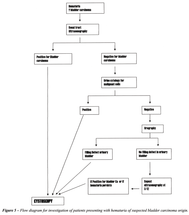
We present a flow diagram (Figure-5) that will be helpful in investigating
patients presenting with hematuria of suspected bladder carcinoma origin.
It is hoped that by employing ultrasonography as primary imaging modality
in patients with hematuria more new cases of bladder carcinoma will be
detected especially in developing countries where ultrasonography is easily
available compared with urography. Patients diagnosed to be suffering
from bladder carcinoma by ultrasonography should be scheduled directly
and promptly for cystoscopy and bladder tumor resection.
REFERENCES
- Black RJ, Bary F, Ferlay J, Parkin DM: Cancer incidence and mortality in European Union: cancer registry data and estimates of national incidence for 1990. Eur J Cancer. 1997; 33: 1075-9.
- Parker SL, Tong T, Bolden S, Wingo PA: Cancer statistics.1997: CA Cancer J Clin. 1997; 47: 5-27.
- Gardner BP, Doyle PT: Symptoms of bladder carcinoma. J R Coll Gen Pract.1987; 37: 367.
- Ravi R, Rao RC, Ahlawat R, Berry M: Carcinoma bladder: Comparative evaluation of urinary cytology, excretory urography and ultrasonography. Indian J Cancer. 1990; 27: 55-61.
- Gossel C, Knispel HH, Miller K, Klan R: Is routine excretory urography necessary at first diagnosis of bladder carcinoma? J Urol. 1997; 157: 480-1.
- Herranz-Amo, Diez-Cordero JM, Verdu-Tartajo F, Bueno-Chomon G, Leal-Hernandez F, Bielsa-Carrillo A: Need for intravenous urography in patients with primary transitional carcinoma of the bladder? Eur Urol. 1999; 36: 221-4.
- DeFelippo NP, Fortunato RP, Mellins HZ, Richie JP: Intravenous urography: important adjunct for diagnosis of bladder tumours. Br J Urol. 1984; 56: 502-5.
- Amar A, Das S: Pre-cystoscopic diagnosis of bladder tumour by modified intravenous urography. Br J Urol. 1984; 56: 381-4.
- Corrigan NT, Crooks J, Shand J: Are dedicated bladder films necessary as part of intravenous urography for hematuria? BJU Int. 2000; 87: 806-10.
- Mariani AJ, Mariani MC, Macchioni C, Stams UK, Hariharan A, Moriera C.: The significance of adult hematuria: 1000 hematuria evaluations including a risk-benefit and cost effectiveness analysis. J Urol. 1989: 141: 350-5.
- Teruel JI, Marcen R, Onaindia JM, Serrano A, Quereda C, Ortuno J.: Renal function impairment caused by intravenous urography: A prospective study. Arch Int Med. 1981; 141: 1271-4.
- Katayama H, Yamaguchi K, Kozuka T, Takashima T, Seez P, Matsuura K: Adverse reactions to ionic and non ionic contrast media, A report from the Japanese Committee on the safety of contrast media. Radiology. 1990; 175: 621-8.
- Davidson AJ, Hartman DS, Choyke PL, Wagner BJ: Radiology of the kidney and genitourinary tract.. Philadelphia, WB Saunders, 3rd ed. 1999; pp. 485-515.
- Sanders RL, Hundley SL: Hematuria. In: Sander RL (ed.), Clinical Sonography - Pracical Guide. Boston, LittleBrown. 1991; pp. 321-5.
- Abu-Yousef MM, Narayan AS, Franken EA, Brown RC: Urinary bladder tumors studied by cystosonography. BJ Urol. 1989; 64: 409-11.
- Malone PR, Weston-Underwood J, Aron PM, Wilkinson KW, Joseph AE, Riddle PR: The use of transabdominal ultrasound in the detection of early bladder tumours. Br J Urol. 1986; 58: 520-2.
- Itzchak Y, Singer D, Fischelovitch Y: Ultrasonographic assessment of bladder tumors. I. Tumor detection. J Urol. 1981; 26: 31-3.
- Leung HY, Grifiths DE, Neal DE: Bladder cancer. Postgrad Med J. 1996; 72: 719-24.
- Spencer J, Lindsell D, Mastorakou I: Ultrasonography compared with intravenous urography in the investigation of adults with hematuria. BMJ. 1990: 301:1074-6.
______________________
Received: August 25, 2003
Accepted after revision: May 12, 2004
_______________________
Correspondence address:
Dr. M Rafique
5, Altaf Town, Tariq Road
Multan. Pakistan
E-mail: rafiqanju@ hotmail.com
EDITORIAL COMMENT
In
this study, ultrasonography was effective in showing obstruction and involvement
of the lower ureter by the bladder tumor. Ultrasonography does not adequately
evaluate the mid or upper ureter or the upper collecting system and calices.
Regarding excretory urography; it is not,
in most uroradiologist’s opinion, an adequate examination for bladder
carcinoma, and most will add the caveat of “cystoscopy is necessary
to adequately evaluate the bladder for tumor” or something to that
effect. The intravenous urography, done well (i.e. with nephrotomography),
does provide excellent evaluation of the ureters and upper collecting
system and that is its role; it thereby precludes the need for retrograde
ureteropyelography either at the time of cystoscopy or later if the cystoscopy
is negative.
But, in many countries, this approach of
ultrasonography as the initial evaluation of patients with hematuria and
suspected bladder cancers makes considerable sense as optimizing provision
of health care, recognizing the limitations of ultrasonography and the
need for a process so that patients with a “negative” ultrasonography
do not escape adequate evaluation and followup.
In many countries where computed tomography
(CT) scanning is readily available, the CT-urogram (multi-phase CT with
noncontrast of abdomen and pelvis for calculi, nephrogram phase of the
kidneys, and delayed imaging of the kidneys and ureters) is becoming the
gold standard in evaluating patients suspected of having “surgical”
hematuria not due to simple stone disease. However, this “high-tech”
approach has disadvantages, i.e. higher radiation dose, high cost and
impact on health care costs and, of course, availability.
Regarding the evaluation of bladder cancer,
it is my impression that cystoscopy and biopsy is the gold standard. Newer
magnetic resonance imaging techniques may ultimately be helpful, but confirmation
of efficacy is still in progress.
The paper presents a nice flow diagram of
patient management.
Dr.
William H. Bush, Jr.
Director, Genitourinary Radiology
University of Washington Medical Center
Seattle, Washington, USA