LAPAROSCOPIC
RADICAL PROSTATECTOMY BY EXTRAPERITONEAL ACCESS WITH DUPLICATION OF THE
OPEN TECHNIQUE
(
Download pdf )
M. TOBIAS-MACHADO, PEDRO FORSETO JR., JIMMY A. MEDINA, MARCELO WATANABE, ROBERTO V. JULIANO, ERIC R. WROCLAWSKI
Discipline of Urology, Medicine School of ABC, Santo André, São Paulo, Brazil
ABSTRACT
Introduction:
The laparoscopic radical prostatectomy is a continually developing technique.
Transperitoneal access has been preferred by the majority of centers that
employ this technique. Endoscopic extraperitoneal access is used by a
few groups, nevertheless it is currently receiving a higher acceptance.
In general, the antegrade technique is used, with dissection from the
bladder neck to the prostate apex.
The objective of the present paper is to
describe the extraperitoneal technique with reproduction of the open surgery’s
surgical steps.
Surgical Technique: With this technique,
the dissection of the prostate apex is performed and, following the section
of the urethra while preserving the sphincteric apparatus, the Foley catheter
is externally tied and internally recovered, which allows cranial traction,
similarly to the way it is performed in conventional surgery. The retroprostatic
space is posteriorly dissected and the seminal vesicles are identified
by anterior and posterior approach, obtaining with this method an optimal
exposure of the posterolateral pedicles and the prostate contour. The
initial impression is that this technique does not present higher bleeding
rate or difficulty level when compared with antegrade surgery. Potential
advantages of this technique would be the greater familiarity with surgical
steps, isolated extraperitoneal drainage of urine and secretions and a
good definition of prostate limits and lateral pedicles, which are critical
factors for preserving the neurovascular bundles and avoiding positive
surgical margins. A higher number of cases and a long-term follow-up will
demonstrate its actual value as a technical option for endoscopic access
to the prostate.
Key
words: prostatic neoplasms; prostatectomy; laparoscopy
Int Braz J Urol. 2004; 30: 221-6
INTRODUCTION
Laparoscopic
radical prostatectomy has become an option for treatment of localized
prostate cancer in some centers. The majority of laparoscopists prefer
the transperitoneal technique that was standardized by Guilleneau &
Vallencien (1).
The endoscopic extraperitoneal technique
performed by some groups promotes antegrade dissection, from the bladder
neck to the prostate apex (2-4). Our objective was to describe the extraperitoneal
technique that was initiated in our institution in 2002 with duplication
of open surgery’s surgical steps, discussing potential advantages
and initial impressions obtained after its use in 25 patients.
SURGICAL TECHNIQUE
1. Patient
is positioned in horizontal dorsal decubitus, with Y-shaped abduction
of lower limbs on the table;
2. Display of the surgical team. The surgeon operates on the left side,
the camera is positioned at the upper end of the table, and the assistant
stand at the patient’s right side. During suture, for improved comfort,
the surgeon and the camera switch places;
3. Umbilical incision measuring 1.5 cm up to the Retzius space;
4. Creation of extraperitoneal space through digital dissection and modified
balloon dilator (handicraft);
5. Hasson trocar (10 mm) through the umbilical incision for the 0-grade
optics;
6. Installation of pneumoretroperitonium with CO2 tension of 15 mmHg;
7. Introduction of another 4 working trocars (2 pararectal external measuring
10 mm, and 2 in iliac fossa measuring 5 mm) under direct view, in an arciform
shape, taking care in order to avoid peritoneal lesion (Figure-1);

8. Exeresis of pre-prostatic fat with monopolar cautery for proper identification
of prostate, bladder and puboprostatic ligaments;
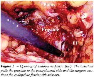
9. Bilateral opening of endopelvic fascia with scissors, following previous
contralateral traction of the prostate (Figure-2);
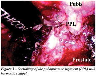
10. Identification and sectioning of puboprostatic ligaments (Figure-3);
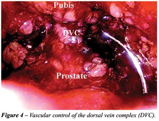
11. Vascular control of dorsal vein complex of the penis with a X-stitch
using 2-0 polyglactine suture with CT-1 needle (Figure-4) and control
of the retrograde blood flow with harmonic or bipolar scalpel, or polymer
clip (Hem-o-lock®) (Figure 5). Applying the clip makes the subsequent
identification of the bladder neck easier for reconstruction, a surgical
step that is often arduous when we choose to preserve the bladder neck;
12. Apical dissection with preservation of the sphincteric apparatus;
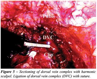
13. Sectioning of the dorsal vein complex of the penis with electrocautery
or harmonic scalpel, until the urethra is viewed (Figure-5);
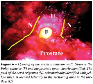
14. Opening of the urethral anterior wall with scissors (Figure-6). Section
is performed after perfectly identifying the limits of the prostate apex
and urethra, thus avoiding positive margins;
15. The catheter balloon is filled with 20 mL of distilled water. The
Foley catheter is externally pulled for subsequent knot application with
0-cotton suture including drainage and balloon routes;
16. Sectioning of the catheter close to the previously applied knot;
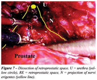
17. Recovery of the remaining stump of the Foley catheter, through endoscopic
view in the extraperitoneal space (Figure-7);
18. Posterior section of the urethra and recto-urethral muscle following
cranial traction of the stent by the assistant;
19. Blunt retroprostatic dissection up to the most proximal point as feasible;
20. Identification and opening of the posterior layer of the Denovilliers
fascia (Figure-8). At this time it is possible to identify the pre-rectal
fat. Analogically to open surgery, we know that the neurovascular bundle
lies laterally and under the fascia, which makes nervous preservation
easier during ligation of the prostatic pedicle, which is performed by
posterior access;
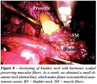
21. Sectioning of the bladder neck, with preservation of muscular fibers
whenever possible. The dissection is started with harmonic or bipolar
scalpel and upon reaching the urethral mucosa, it is sectioned with scissors
(Figure-9);
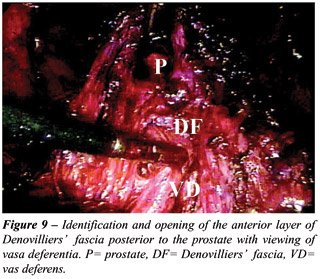
22. Identification and opening of the anterior layer of Denovilliers’
fascia, posterior to the prostate with visualization of vasa deferentia;
23. Identification and sectioning of vasa deferentia with harmonic or
monopolar scalpel;
24. Superior traction of the vasa deferentia by the assistant in order
to release the seminal vesicles. At this time, we preferred to use harmonic
or bipolar scalpel in order to avoid dissipation of thermal energy that
could damage the nervi erigentes;
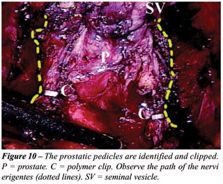
25. The assistant performs the lateral and superior traction of previously
mobilized (released) prostate, enabling the clear identification of the
prostatic pedicles and the prostate capsular limits. The control of the
prostatic pedicles is performed with harmonic or bipolar scalpel. Alternatively
polymer clips (Hem-o-lock®) can be used (Figure-10);
26. Exeresis and entrapment of the specimen that is located in right iliac
fossa;
27. Vesicoureteral anastomosis is initiated with the patient in Trendelemburg
position in order to improve the visualization of the urethra. The surgeon
works with the pararectal 10-mm trocars at the upper end of the table.
We perform a continuous 3-0 polyglecaprone (monocryl®) suture with
SH needle. We use two 13-cm sutures, one colorless and the other one violet,
externally tied by the distal end. Suture begins at 6 o’clock position
in the bladder directed inwards and each of the sutures rises toward 12
o’clock position, where a single internal knot is made (5);
28. Drainage with Penrose though one of the 5-mm ports;
29. Removal of the specimen by enlargement of the umbilical port and closure
of the incisions;
COMMENTS
Laparoscopic
radical prostatectomy is a laborious procedure with a long learning curve.
The most significant series in literature, where it was possible to standardize
and systemize the technique, use transperitoneal access (1).
The endoscopic extraperitoneal technique
was initially described by Raboy et al. where, after creating the space
and ligating the dorsal vein complex, the dissection was performed from
the bladder neck to the prostate apex (antegrade). The author reported
that this technical option resulted from the higher possibility of bleeding
and technical difficulty if the early sectioning of the complex was performed
(2). This observation is contrary to the results obtained in our initial
series of 25 patients, where none required hemotransfusion or conversion
to open surgery.
In our setting, Andreoni et al. were the
first authors to report laparoscopic radical prostatectomy using the antegrade
technique (3). Potential advantages of the extraperitoneal access are
the non-manipulation of abdominal viscera, reducing the risk of direct
or distant lesions, keeping the drainage of secretions isolated from the
peritoneal cavity, greater familiarity with local anatomy, with the Trendelemburg
exaggerated position being unnecessary (frequently required in the transperitoneal
technique). As disadvantages it presents a working space with lower gas
content, requiring greater adaptation for instrument movements and aspiration
of secretions and smoke. If the space is not properly developed in its
lateral area, according to previous descriptions, a higher tension in
the vesicoureteral anastomosis can occur. Peritoneal perforation hampers,
but does not prevent the surgery from being completed. If the progression
in dissection is hard, it is possible to operate by transperitoneal approach
following wide peritoneal opening (2-4).
Our initial impression is that transperitoneal
and extraperitoneal techniques are equivalent concerning surgical time,
blood loss, complications and post-operative recovery. However, in the
extraperitoneal technique, the presence of urinary fistula shows a better
outcome, since there is no urine drainage to the peritoneal cavity, thus
avoiding prolonged paralytic ileus.
As original modifications, in addition to
the retrograde dissection as described in the open technique, we used
a polymer clip in order to avoid venous reflux from the dorsal complex,
which aids in the subsequent identification of the bladder neck during
suture. The external handling and sectioning of the Foley catheter enabled
the internal and superior traction by the assistant, similarly to the
open technique for accessing the posterior aspect of the prostate. Such
dissection makes the identification of lateral prostatic pedicles quite
easier following the dissection of the bladder neck. The accurate identification
of the prostate limits is fundamental for a proper preservation on the
neurovascular bundles and to avoid the occurrence of positive margins.
Recently, Dubernard et al. (2003) described
the first series of 143 patients using retrograde laparoscopic extraperitoneal
technique. The authors conclude that in spite of presenting only preliminary
functional results, the technique is promising and can potentially become
the method of choice for laparoscopic radical prostatectomy (5).
From this initial work, we concluded that
extraperitoneal access is feasible, being possible to practically duplicate
surgical steps of the open surgery. The actual role and advantages of
this surgery when compared with laparoscopic transperitoneal technique
waits for future assessments in prospective studies with a higher number
of cases.
REFERENCES
- Guillonneau B, Vallancien G: Laparoscopic radical prostatectomy: the Montsouris technique. J Urol. 2000; 163: 1643-9.
- Raboy A, Ferzli G, Albert P: Initial experience with extraperitoneal endoscopic radical retropubic prostatectomy. Urology. 1997; 50: 849-53.
- Andreoni C, Gattas N, Srougi M: Initial experience with extraperitoneal endoscopic radical retropubic prostatectomy. Int Braz J Urol. 2001; 27: 563-5.
- Bollens R, Bossche MV, Roumeguere TH, Damoun A, Ekane S, Hoffmann P, et al.: Extraperitoneal laparoscopic radical prostatectomy. Eur Urol. 2001; 40: 65-9.
- Dubernard P, Benchetrit S, Chaffange P, Hamza T, Van Box, Som P.: Retrograde extraperitoneal laparoscopic prostatectomy (R.E.I.P). Simplified technique (based on a series of 143 cases). Prog. Urol. 2003; 13: 163-74.
________________________
Received: December 2, 2003
Accepted after revision: May 31, 2004
_______________________
Correspondence address:
Dr. Marcos Tobias-Machado
Rua Graúna, 104 / 131
Moema, São Paulo, SP, 04514-000, Brazil
Fax: + 55 11 5533-5227
E-mail: tobias-machado@uol.com.br