TOTAL
BLADDER REPLACEMENT WITH DE-EPITHELIALIZED ILEUM. EXPERIMENTAL STUDY IN
DOGS
(
Download pdf )
FÁBIO O. VILAR, LUIZ A. P. DE ARAÚJO, SALVADOR V.C. LIMA
Nucleus of Experimental Surgery, Department of Surgery, Federal University of Pernambuco, Recife, PE, Brazil
ABSTRACT
Objective:
To assess the value of the silicone modeler in preventing graft retraction
in dogs undergoing bladder replacement with de-epithelialized ileum.
Materials and Methods: Twelve female dogs
underwent total cystectomy and bladder replacement by neobladder made
of demucosalized ileal segment, comparing the group with modeler (group
I) and the group without modeler (group II). Cystometry data, graft epithelization
and radiological assessment (cystography and excretory urography) were
analyzed.
Results: Neobladder capacity, at 2 months,
ranged from 50 to 250 mL (mean 191 mL) and from 5 to 60 mL (mean 22 mL)
and at 6 months, from 60 to 270 mL (mean 202.5 mL) and from 5 to 75 mL
(mean- 30.5 mL), respectively in groups I and II, with a statistically
significant difference between groups. After 30 days, postoperatively
the presence of transitional epithelium was observed in all fragments
obtained by biopsy.
Conclusion: The use of the intravesical
silicone modeler prevented the retraction of the neobladder of de-epithelialized
ileum.
Key
words: bladder; ileum; epithelium; prostheses and implants
Int Braz J Urol. 2004; 30: 237-44
INTRODUCTION
Efforts
to augment or replace the bladder are old (1,2), and most commonly digestive
tract segments were used for this purpose (3). Considering, however, the
characteristics of intestinal mucosa, structurally and functionally distinct
from the bladder, problems such as production of secretions, infection,
electrolytic changes and even developing of tumors still await for a definitive
solution (4). Thus, in order to overcome such difficulties, some authors
suggested using de-epithelialized flaps of digestive tract (5-7). Martin
used a Foley stent balloon aiming to distend the de-epithelialized graft
(7). Other authors have tried to reproduce experiments with de-epithelialized
segments of the digestive system (8-10).
Given the mucosecreting and absorbing nature
of the digestive epithelium, problems resulting from mucous secretion
and metabolic changes are, sometimes, difficult to solve (11). Intestinal
neobladder could be made with digestive tract segments lacking their original
mucosa (de-epithelialized) over which a layer of transitional epithelium
would develop, whether from the original bladder or from grafted islets
of transitional epithelium (5-7). Our studies with de-epithelialized colon
have allowed for improving bladder capacity through the use of a silicone
modeler placed inside the neobladder and submitted to a slight distension.
This distension allows the de-epithelialized flap not to retract, and
thus it can undergo the epithelization process from the existing bladder
(12).
The present study applies the de-epithelialized
ileal segment for bladder replacement and analyzes the role of the silicone
modeler in preventing graft retraction and in its epithelization.
MATERIALS AND METHODS
Animals
underwent total cystectomy and bladder replacement by neobladder of demucosalized
ileal segment, comparing the group with modeler (group I) and the group
without modeler (group II). Graft epithelization, cystometry data and
radiological assessment (cystography and excretory urography) were analyzed.
The research project was approved by the
Committee for Research Ethics from the Health Science Center of Federal
University of Pernambuco.
Twelve cross-bred female dogs, apparently
healthy, weighting between 13 and 27 kg (mean 16 kg/median 16.5 kg) were
operated, among which 10 survived for longer than 60 days and were used
for this study.
All animals were operated and maintained
at the Nucleus of Experimental Surgery of Federal University of Pernambuco.
On the first postoperative day they were fed with a liquid meal, which
was advanced to solid meals according to each animal’s acceptance.
All animals were maintained on therapeutic antibiotic treatment with gentamicin
(80 mg/day) for 10 days following each manipulation, and from then on,
prophylactic therapy with nitrofurantoin (100 mg/day) until death.
Surgical
Technique
Animals were weighted and underwent puncturing
of the radial vein in one of the front paws, with 19 or 21 butterfly-type
needle. They underwent intravenous anesthesia with ketamine (1 mg/kg),
fentanyl citrate (1 mg/kg) and pentobarbital sodium (25 mg/kg). After
being positioned in dorsal decubitus, the animals were intubated with
an orotracheal tube and maintained under controlled ventilation, using
the muscle relaxant pancuronium bromide (1 mg/kg). All animals received
between 30 and 50 mL/kg/h of 0.9 % physiological saline solution or Ringer
lactate, during surgery.
The access approach was median laparotomy
measuring approximately 20 cm, until the pubic symphysis. Following the
inspection of every abdominal organ, a 45-cm ileal segment with suitable
vascular pedicle was isolated (Figure-1 A), and intestinal transit was
reconstituted by termino-terminal ileum-ileal anastomosis, with continuous
sutures in 2 planes, using 3-0 chromic catgut suture for the mucosa and
4-0 prolene for seromuscular layer.
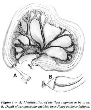
The isolated ileal segment had its seromuscular
layer separated from the mucosa, as following. Inserting a 14 or 16F Foley
catheter within the lumen and insufflating the balloon with 10 mL of distilled
water. Blunt dissection with Kelly forceps, and separation of seromuscular
layer from the mucosa, on the segment ileal supported by the stent balloon,
and longitudinal section on the anti-mesenteric aspect of the seromuscular
layer. In order to start the dissection, close to one of the extremities
of the isolated ileal segment, with the stent balloon inflated, a circular
incision of the seromuscular layer was performed around the entire ileum
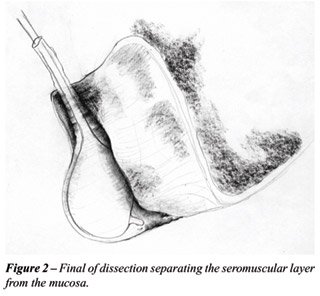
circumference (Figure-1 B) with 15-blade scalpel. Using the Kelly forceps,
the seromuscular layer was separated from the mucosa, with the procedure
being complete when the other extremity was reached (Figure-2). Electrocautery
was used for hemostasia. Simultaneously, the de-epithelialized area was
irrigated with distilled water at a temperature of 5°C, through a
20-mL syringe with insulin needle, in order to promote vasoconstriction.
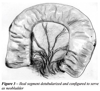
Upon completing the dissection, the mucosa
layer was discarded, the ileal segment of the seromuscular layer was configured
into an U-shape, and the edges were closed by continuous stitches with
4-0 chromic catgut suture, so that it nearly formed a demucosalized bowel
plate (Figure-3).
Bladder was sectioned at the level of the
bladder neck. Ureteral distal ends were dissected and sectioned at the
level of their insertion in the bladder. In the region more proximal to
the urethral orifice, an orifice measuring approximately 1.5 cm in diameter
was confectioned by suturing the edges of the de-epithelialized ileum,
and the anastomosis with the urethra was performed. Suture consisted in
separate stitches in 4-0 chromic catgut.
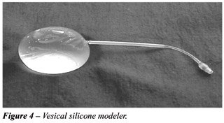
Ureters were anastomosed in the most cephalic
region of the ileal plate, with 5-0 monofilament PDS suture in separate
stitches. An orifice measuring approximately 0.5 cm in diameter was made
in the ileal wall, passing 1.5 cm of ureter that was fixed to the internal
aspect (de-epithelialized aspect), keeping the catheterization with 4F
plastic urethral catheter (Figure-4). The extremity of the catheter was
left inside the urethra in a silicone tube.
The edge of the ileal plate was fixed with
4-0 chromic catgut suture in continuous stitches, so that the neobladder
was configured into a seemingly spherical shape. After sorting within
groups, in the animals from group I, an empty silicone modeler was inserted
(Figures-4 and 5).
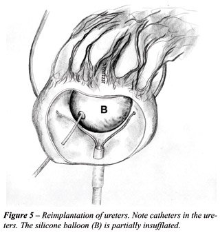
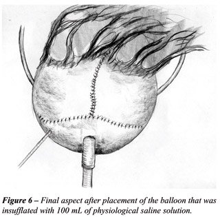
The silicone modeler was inflated and maintained
with 100 mL of physiological saline solution, after completing the suture
and the neobladder confection (Figure-6). The modeler valve was placed
in the subcutaneous tissue of the abdominal wall, close to the incision.
In the animals from group II, the ileal plate was sutured similarly to
group I, though without the modeler.
Closure of the abdominal wall was performed
by planes, with separate stitches. The reversion of neuromuscular block
was achieved with atropine (0.01 mg/kg) and neostigmine (0.03 to 0.07
mg/kg). The orotracheal tube was removed following the return to spontaneous
breathing.
Postoperative
period
For surgical procedures and postoperative
assessment examinations, sedation and analgesia were performed with ketamine
(1 mg/kg) and fentanyl citrate (1 mg/kg) by intravenous approach.
The silicone modeler was removed on the
14th postoperative day through a small abdominal incision measuring approximately
3.0 cm at the level of the modeler valve, which was in the subcutaneous
tissue. After being totally emptied through its valve, the modeler was
pulled and removed. The ureteral catheters were also removed at the same
occasion by pulling them through the urethra.
The morphologic-functional assessment of
the neobladder was performed through radiological study (excretory urography
and cystography), cystometry and cystoscopy with biopsy of the graft wall.
Cystometry and cystoscopy with biopsy were performed on the same occasion,
monthly.
Cystometry
The cystometry was performed before the
biopsy. A 10F nelaton catheter was inserted, the neobladder was emptied,
and residual urine was measured. Through a second nelaton catheter connected
to an external system of the hydration equipment, assembled on a stand
with measuring tape, 0.9% physiological saline solution was infused at
an approximate rate of 25 mL/min. Intravesical tension was measured in
H2O column (cm), from the point zero, which was settled at the level of
the pubic symphysis, with the animal in dorsal decubitus. The container
of physiological saline solution was located at 30-cm height from the
point zero. All infused volumes and corresponding pressures were measured
at every 5 mL of infusion and put on a graphic. The maximum vesical capacity
was considered when fluid extravasation started around the urethral catheter,
while infusing the solution. For comparison between groups, the capacity
at 2 and 6 postoperative months was considered.
Cystoscopy
with Biopsy
Following the cystometry, a cystoscopy with
biopsy was performed, using a 21F sheath cystoscope and flexible biopsy
forceps. Neobladder biopsies were performed on the lateral wall, fundus
and regions close to the ureters. Samples were identified, placed in different
containers, fixed in 10% formalin, and processed as usual for histological
study. When the animal died, the necropsy was performed with histological
analysis of the entire urinary tract.
Cystography and excretory urography were
performed 2 or 3 times during the study. In some animals, the urography
was not performed.
Statistical
Study
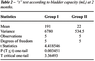
The Student’s t-test was used to compare
neobladder capacity in animals with and without the use of silicone modeler,
with the significance level set at 0.05 or 5%, for assessments at 2 and
6 months.
RESULTS
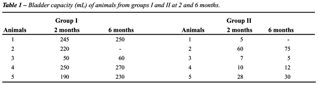
Two animals died within the first month following surgery. The main cause was peritonitis due to urine extravasation. Ten animals, 5 in each group, survived for more than 2 months; and eight (4 in each group) survived for more than 6 months. Mean survival in group I was 268.4 days (median 330) and in group II it was 253.6 days (median 240).
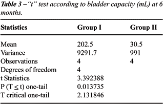
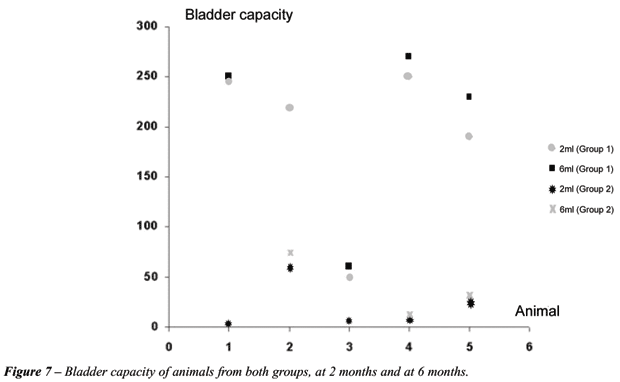
Cystometry
Results for neobladder capacity and statistical
study are presented, respectively, in Tables-1, 2 and 3. The evolutional
bladder capacity for each animal from groups I and II, at 2 and 6 months,
is represented in Figure-7.
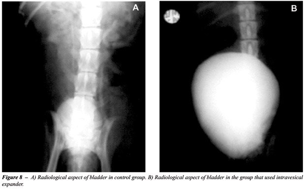
Radiological
Assessment
Cystographies showed suitable and oval-shaped
neobladders in animals from both groups, with neobladders in group I being
visually larger than those in group II (Figures-8 A and B).
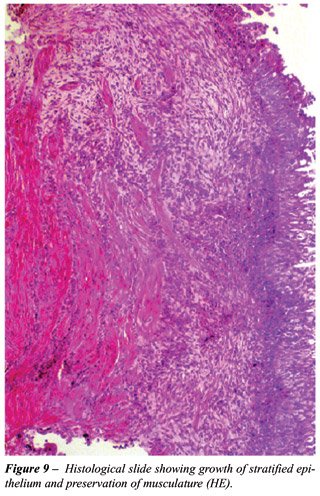
Histological
Study
Transitional epithelium was observed in
all fragments obtained by biopsy, both at 30 days postoperatively, and
in subsequent ones, in all animals from both groups (Figure-9).
DISCUSSION
The
use of de-epithelialized and balloon-protected intestinal segments had
been previously reported in experimental animals. The type of material
employed by the researcher (latex Foley stent balloon) was probably improper
and limited his studies (7).
Studies for bladder augmentation similar
to ours were conducted almost simultaneously by Australian researchers
(8). They used de-epithelialized sheep stomach and reported results similar
to those obtained in our initial studies with sigmoid colon.
Jednak et al. used a model with sigmoid
colon also similar to our initial model, though preserving the submucous
layer of the bowel (13). They studied 16 patients, 14 with neurogenic
bladder and 2 with sequelae from posterior urethral valve and reported
a 2.4 times increase in bladder capacity. Filling pressures decreased
by an average of 50%. Postoperative endoscopic biopsies revealed the presence
of colonic epithelium in 3 cases. Four patients required reintervention,
with 2 requiring a new augmentation. In an effort to extend the technique
of de-epithelialized bowel for use in cases of small bladders or cases
of vesical extrophy, some laboratories tried alternative methods for applying
these grafts. Merguerian et al. used de-epithelialized grafts of sigmoid
colon covered with grafts of cultured transitional epithelium covered
by polygalactine (14). Despite the positive results of “in vitro”
epithelial seeding, there was no growth when it was applied “in
vivo”. More recently, Frey et al. used a similar model in mini-pigs.
De-epithelialized bowel or stomach were grafted with urothelium islets,
removed at the moment of surgery or collected from another animal. Severe
contracture of the intestinal graft was observed in all cases (15).
In our study, catheterization of both ureters
with the purpose of avoiding contact of urine with the graft seems to
have a great importance and facilitates epithelial growth. The idea of
maintaining the vesical modeler for 2 weeks is, to a certain extent, casual,
since there is only one previous report in this sense, however only one
animal was studies (7).
There is a recent study in rats where a
silicone “stent” was used inside an augmented bladder obtaining
results similar to ours (16).
In relation to the mortality observed in
some animals in our study, we attribute it to the fact that dog bladder,
as in the majority of animals, is located intraperitoneally and it favored
urosepsis, which was triggered by urine extravasation. Similar complications
were observed by other researchers in different time periods (5,16).
Despite such mortality, we could study in
detail the surviving animals through monthly bladder biopsies and, considering
that we had animals that survived up to 12 months, the opportunity of
performing multiple biopsies in different occasions on the same animal,
makes the apparently low number of animals in each group to be projected
as a quite more significant number when we imagine that each animal was
assessed several times with repeated cystometry and biopsy. A total of
45 biopsies were performed in the group using the expander and 42 in the
control group.
We histologically demonstrated the growth
of urinary epithelium in the de-epithelialized graft in all studied samples.
The performance of cystographies and excretory
urographies was also important in order to better documenting and comparing
the morphology of confectioned neobladders.
We concluded that the use of the intravesical
silicone modeler prevented retraction of the neobladder of de-epithelialized
ileum.
Prof. Romero Glasner drew the illustrations
contained in this work, and Silimed, Rio de Janeiro,
supplied the vesical modelers.
REFERENCES
- Simon J: Ectopia vesica; operation for temporary success; autopsy. Lancet. 1852; 2: 568-70.
- Mikulicz J: Zur Operation der angeborenen. Belsanesn plate. Zentralb Chir. 1899; 26: 641-3.
- Leong CH, Ong GB: Gastrcystoplasty in dogs. Aust N Z J Surg. 1972; 41: 272-9.
- Nurse DE, Mundy AR: Metabolic complications of cystoplasty. Br J Urol. 1989; 63: 165-8.
- Shoemaker WC: Reversed seromuscular grafts in urinary tract reconstruction. J Urol. 1955; 74: 453-75.
- Filmer RB, Spencer JR: Malignancies in bladder augmentations and intestinal conduits. J Urol. 1990; 143: 671-8.
- Martin LSJ: Uroepithelial lined ileal segment as a bladder replacement, experimental observations and brief review of literature. J Urol. 1959; 82: 633.
- Dewan PA, Lorenz C, Stefanek W, Byard RW: Urothelial lined colocystoplasty in a sheep model. Eur Urol. 1994; 26: 240-6.
- Buson H, Manivel JC, Dayan cM, Long R, Gonzalez R: Seromuscular colocystoplasty lined with urothelium: experimental study. Urology. 1994; 44: 743-8.
- Lutz N, Frey P: Enterocystoplasty using modified pedicled, detubularized, de-epithelialized sigmoid patches in the mini-pig model. J Urol 1995; 154: 893-8.
- Duel BP, Gonzalez R, Barthold JS: Alternatives techniques for augmentation cystoplasty. J Urol. 1998; 159: 998-1005.
- Lima SV, Araujo LA, Montoro M, Maciel A, Vilar FO: The use of demucosalized bowel to augment small contracted bladders. Br J Urol. 1998; 82: 436-9.
- Jednak R, Schimke CM, Barroso JR U, Barthold JS, González R: Further experience with seromuscular colocystoplasty lined with urothelium. J Urol. 2000; 164: 2045-9.
- Merguerian P, Chavez DR, Hakim S: Grafting of cultured uroepithelium and bladder mucosa into de-epithelialized segments of colon in rabbits. J Urol. 1994; 152: 671-4.
- Frey P, Lutz N, Leuba AL: Augmentation cystoplasty using pedicled and de-epithelialized gastric patches in the mini-pig model. Urol. 1997; 156: 608-13.
- Aktu T, Zdem RTÖ, Aartan C, Özer E, Olguner M, Akgür FM: Experimetally prefabricated bladder. J Urol. 2001; 165: 2055-8.
____________________
Received: April 30, 2004
Accepted: May 24, 2004
_______________________
Correspondence address:
Dr. Fábio de Oliveira Vilar
Av. Flor de Santana, 189 / 502
Recife, PE, 52060-290, Brazil
E-mail: urology@salvador.net