DEFECTIVE
URINARY CRYSTALLIZATION INHIBITION AND URINARY STONE FORMATION
(
Download pdf )
MAURICIO CARVALHO, JODY P. LULICH, CARL A. OSBORNE, YASUSHI NAKAGAWA
Kidney Stone Program (YN, MC), Division of Biological Sciences and the Pritzker School of Medicine, University of Chicago, Chicago, Illinois, USA, Department of Small Animal Clinical Sciences (JPL, CAO), College of Veterinary Medicine, University of Minnesota, St. Paul, Minnesota, USA, and Department of Internal Medicine (MC), Federal University of Parana, Curitiba, Parana, Brazil
ABSTRACT
Introduction:
Nephrocalcin (NC) is a glycoprotein produced in the kidney and inhibits
calcium oxalate crystal formation. It has been separated into 4 isoforms
(A, B, C, and D) and found that (A + B) are more abundant than (C + D)
in urine of healthy subjects, but the reverse is seen in human urine of
kidney stone patients. To further examine the role of this protein in
inhibition of urinary crystallization, nephrocalcin isoforms were purified
from 2 genetically pure dog species.
Materials and Methods: We studied healthy
Beagles, known to be non-stone forming dogs, and Mini-Schnauzers, known
to be calcium oxalate stone formers. NC was isolated and purified from
each group. Urinary biochemistry and calcium oxalate crystal growth inhibition
were measured.
Results: Specific crystal growth inhibition
activity was significantly higher in non-stone forming dogs (9.79 ±
2.25 in Beagles vs. 2.75 ± 1.34 of Mini-Schnauzers, p < 0.005).
Dissociation constants toward calcium oxalate monohydrate were 10-fold
different, with Beagles’ isoforms being 10 times stronger inhibitors
compare to those of Mini-Schnauzers’. Isoforms C + D of NC were
the main isoforms isolated in stone-forming dogs.
Conclusion: NC of these two species of dogs
differently affects calcium oxalate crystallization and might have a role
in determining ulterior urinary stone formation.
Key
words: kidney stone; calcium oxalate; crystallization; experiments;
dogs
Int Braz J Urol. 2006; 32: 342-9
INTRODUCTION
Normal
urine is supersaturated with respect to crystalline components, as a consequence
of the essential homeostatic water conservation. This condition suggests
the existence of physiological mechanisms that actively inhibit urinary
crystallization of calcium salts (1). Various inhibitory macromolecules
have been implicated in this process, e.g. osteopontin, crystal matrix
protein, bikunin and nephrocalcin.
Nephrocalcin (NC) was isolated from human
urine and kidney tissues (2), and later found in urine of 9 vertebrates’
species (3). This glycoprotein is produced in proximal tubules in kidney
(4) and its excretion is increased in renal carcinoma patients (5) and
during pregnancy (6). NC has a mol. wt. of 14 kD, and can be separated
into 4 isoforms with different degrees of phosphorylation and amphiphilicities
(7). Healthy subjects excrete more isoforms A and B that are less phosphorylated
and have stronger hydrophobicity properties. In contrast, kidney stone
forming patients excrete more isoforms -C and -D, which have higher degree
of phosphorylation and weaker hydrophobicity. These isoforms of NC coat
the surface of calcium oxalate crystals and control morphology, size and
surface topography of crystals (8).
Evidence suggests that defective inhibitors
can cause nephrolithiasis and NC accounts for a considerable portion of
the inhibitory property of crystallization in urine (9). To examine this
premise, we evaluated two species of pure-breed dogs with different incidences
of kidney stones. We purified NC from urine samples from Beagles dogs,
a non-stone forming species (10) and from Mini-Schnauzer dogs, known for
frequent formation of calcium oxalate stones (11,12). In this report we
compared chemical and physicochemical properties of NCs isolated from
these species.
MATERIALS AND METHODS
Eleven
healthy Beagles (3 neutered males and 8 neutered females, 4.0 ±
0.4 years old, body weight 9.28 ± 0.36 kg), and 7 Mini-Schnauzers
(4 neutered males and 3 neutered females, 2.5 to 10.5 years old, body
weight 6.6 ± 1.9 kg, who had at least one urinary stone) were selected.
They were housed in individual cages under the conditions of controlled
lighting and temperature, at the College of Veterinary Medicine, the University
of Minnesota, according to the principles outlined in the National Institutes
of Health “Guide for the Care and Use of Laboratory Animals”.
At the beginning of collection period, the
urine from the bladders of dogs was emptied by transurethral catheterization.
They were then housed in metabolic cages to facilitate complete collection
of voided urine. Water was accessible throughout the collection period.
Urine was collected in plastic containers surrounded by ice and stored
in capped plastic containers with thymol at 4º C. To ensure complete
removal of urine, dogs were catheterized at the end of 24 h. To minimize
catheter induced bacterial urinary tract infection, cefadroxilÒ
was administered orally (20 mg/kg, q 12 h) during the 24 h period of urine
collection (13). Refrigerated urine samples were warmed at room temperature.
Urine pH was measured by using a Beckman pHmeter. Calcium, citrate, creatinine,
oxalate, phosphate, and uric acid were determined by using a Beckman CX-5
autoanalyzer. Protein was determined in urine by micro-Lowry method using
Folin-Ciocalteu Phenol reagent (14). Bovine serum albumin was used as
a calibration standard with a concentration range between 10 to 50 mg.
NC was isolated and purified by the method
previously described (15). Briefly, urine was diluted 3-fold by distilled
water, pH adjusted to 7.3, and added 1/10 volume of DEAE-cellulose pre-equilibrated
in 0.05 M Tris-HCl, pH 7.3, then stirred gently for 30 min at room temperature.
The DEAE cellulose was separated by filtering through Whatman #1 filter
paper with a Buchner funnel. The DEAE-cellulose cake was then washed with
1 L of 0.05 M Tris-HCl, pH 7.3 containing 0.1 M NaCl (Buffer-A). NC was
eluted by 200 mL of 0.05 M Tris-HCl, pH 7.3 containing 0.5 M NaCl (Buffer-B)
with gentle stirring for 30 min at room temperature. The filtrated NC
fraction was dialyzed against 10 L of distilled water overnight with 1
change. The dialyzed fraction was further subjected to a DEAE-cellulose
column (2 x 15 cm), and 4 NC isoforms (A,B,C, and D) were isolated by
a linear NaCl gradient using 125 mL each of Buffer-A and Buffer-B. The
salt gradient was monitored by a conductivity meter (Radiometer CDM210).
The quantity of the individual isoform inhibition as measured under the
curve was calculated and expressed as a relative ratio of inhibitory activity
by percentage. Each of the four NC isoforms was further purified by a
molecular sieve column of BioRad P-10 column (2 x 85 cm) using 50% formamide
for separating urobilirubin from nephrocalcin, then followed by Sephacryl
S-200 (1 x 90 cm) using Buffer-A. Purified NC concentration was determined
by an alkaline hydrolysis followed by a ninhydrin reaction (7). Phosphoric
acid content was determined by the method described by Ames (16). The
color was developed by using Fiske-Subbarow reagent and 0.01 M KH2PO4
was used for preparing a calibration standard ranging between 50 to 500μ
moles of phosphate concentration.
Surface tension at the air-water interface
was measured by Lauda film balance (Brinkman Instruments Co., Westbury,
NY) using 0.01 M Tris-HCl, pH 7.4, containing 0.1 M NaCl, and applied
100 μg of protein over the surface of the buffer solution. The protein
film was compressed from the surface area of 327 cm2 to 18
cm2 in 30 min and the pressure changes were monitored and recorded
through a computer.
Calcium oxalate crystal growth inhibition
was measured by either 14C-calcium oxalate incorporation method
(15) or spectrophotometric method measuring decrease of oxalic acid (2).
In brief, 14C-calcium oxalate incorporation assay was done
by mixing 500 μL of sodium acetate buffer (50 mM acetic acid, 5 mM
barbituric acid, 0.15 M NaCl, pH 5.7 containing 0.05 μCi/mL of 14C-oxalic
acid), 500 μL of calcium chloride solution (50 mM acetic acid, 2
mM CaCl2.2 H2O, 0.13 M NaCl, 5 mM barbituric acid,
pH 5.7) and 25 μL of a sample solution. The crystallization is initiated
by adding 100 μL of calcium oxalate monohydrate crystal slurry (1.8
mg/mL in sodium acetate buffer, pH 5.7). After 40 minutes of incubation,
the mixture was centrifuged, and radioactivity was measured in the supernatant.
Inhibitory activity is calculated as the following equation: I = (C40
- Cblank)/(C0 - C40).(Cblank)
radioactivity of buffer solution; Co: radioactivity counts at initial
time, C40: radioactivity count in the supernatant after 40
min incubation. Spectrophotometric assay was performed as following: 1
mL of sodium oxalate was added to acetate buffer (8.75 mM acetic acid
and 90 mM NaCl, pH 5.7) and to a calcium chloride solution in a cuvette,
with an aliquot of the sample solution. While this mixture was stirring,
10 μL of calcium oxalate monohydrate slurry in acetate buffer (0.8
mg calcium oxalate monohydrate/mL of acetate buffer) was collected to
the spectrophotometric analysis. As oxalic acid consumed to forming calcium
oxalate, absorbance at 214 nm decreases. The slope of the curve reflects
the strength of crystal growth inhibitory activity of a sample, and also
the dissociation constant of an isolated inhibitor can be calculated by
plotting a Langmuir isotherm type plot. Amino acid composition was determined
by a Beckman amino acid analyzer (Model 119CL, Beckman Instruments, Palo
Alto, CA), after hydrolysis in an evacuated tube containing 6 N HCl for
24 hrs at 11º C. Neutral sugar analysis was carried out by phenol-H2SO4
method (17). Calibration curve was made by using 5 to 20 μg of glucose
aqueous solution. Molecular weight was determined by HPLC with a molecular
sieve column (TSK-2000SW, ToSoHaas, Montogomeryville, PA). Molecular weight
standards used were BSA, soybean trypsin inhibitor, and cytochrome C.
The solvent used was a Buffer-B, and running conditions were isocratic
mode, flow rate 1.0 mL/min, and detection wavelength at 220 nm.
Results are expressed as means ±
SD. Statistical analyses were performed using Minitab 11.0 software. Group
differences were compared by unpaired t-test and the frequency of nephrocalcin
isoforms by χ2-statistical analysis. A value of p <
0.05 was considered significant.
RESULTS
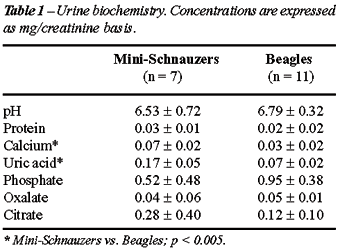
Urine
of individual dogs was analyzed and averaged values of pH, calcium, phosphate,
uric acid, citrate, oxalate, and protein are summarized in Table-1. There
were significant differences in calcium and uric acid excretion between
Mini-Schnauzers and Beagles (p < 0.005).
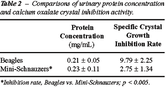
Urinary protein concentration and crystal
growth inhibition activities of both species were compared in Table-2.
The specific inhibitory activity of non-stone forming dogs (Beagles) was
approximately 4 times higher than stone forming dogs (Mini-Schnauzers).
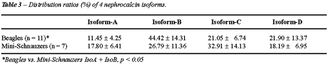
Elution patterns of NC isoforms A,B,C, and
D are summarized in Table-3. Non-stone forming Beagles excreted in their
urine 56% of isoforms (A + B), and 44% of (C + D) isoforms. Particularly,
B isoform peaked in the isoforms isolated. The ratios of isoforms in the
Mini-Schnauzers dogs were 43% of isoforms (A + B) and 57% of isoforms
(C + D). In this group, C was the main isoform isolated.
Carbohydrate content was measured by phenol-sulfonic
acid method and calculated by molar ratio per protein (Table-4). All isoforms
contained 0.01 to 0.02 g per gram of protein. Dogs urinary NC showed high
content of acidic amino acid residues and low content of aromatic and
basic amino acid residues. However, both species showed almost identical
amino acid compositions. On the other hand, phosphate content in Mini-Schnauzers’
NC was significantly higher compared to Beagles (Table-4).
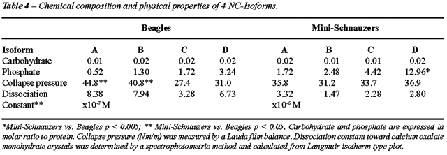
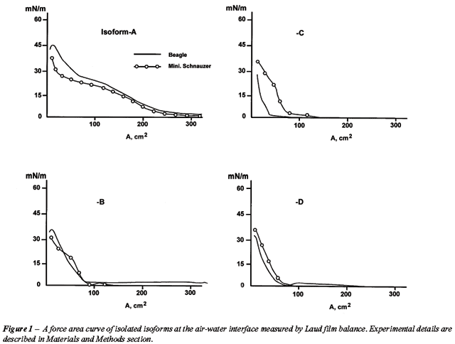
Figure-1 shows typical force area curves
of these isoforms at the air-water interface measured by a Lauda film
balance. Isoform A of the Beagles showed the highest collapsing pressure,
44.8 mN/m, and gradual decreases were seen in B,C and D. Mini-Schnauzers
showed lower collapse pressure in isoforms A and higher collapse pressure
in isoforms C and D when compared to the Beagles’ group. Table-4
summarizes collapsing pressure and dissociation constants of 4 NC isoforms
isolated from both species.
COMMENTS
It
has been accepted that stone formation is a crystallization process, taking
place in supersaturated urine. However, despite nearly universal urine
supersaturation, stones occur in a minority of people. Crystallization
inhibitors, like citrate, proteins and glycosaminoglycans may account
for this discrepancy (18). In this study, using two different species
of dogs with distinct incidences of urinary stones several conclusions
can be reached.
Mini-Schnauzers, a class of dogs with high
formation of kidney stones, excreted more calcium and uric acid in their
urine (p < 0.005) when compared to Beagle’s group, a species
that rarely presents with nephrolithiasis. These electrolytes combine
to promote higher supersaturated urine, certainly predisposing these dogs
to urinary stone formation.
However, we also found a remarkable difference
in the qualitative excretion of urinary NC between these dogs’ breeds.
The NC isolated from the urine of Beagles showed strong inhibitory activity
toward calcium oxalate crystals, but Mini-Schnauzers’ urine has
4 times less inhibitory activity (Table-2). Previously we reported the
same pattern of data in humans with or without urinary stones (7).
Non-stone forming Beagles’ excreted
in their urine more NC isoforms A and B than isoforms C and D when compared
to Mini-Schnauzers (p < 0.05). NC isoforms are calcium-binding proteins,
and binds 4 atoms of calcium ions per one molecule of NC. The Ca2+
binding mode is significantly different between isoform A or B and C or
D: isoforms A-B binds Ca2+ directly through carboxyl groups
of asp and/or glu residues. However, isoforms C-D requires at least two
molecules of water between Ca2+ and carboxyl groups (19). Isoforms
A and B changes its conformation and increase its hydrophobicity upon
binding Ca2+, but isoforms C or D do not. As a consequence
isoforms C and D are more soluble in water and forms a less stable monolayer
at the interface of air-water, and the collapsing pressure is lower. Less
hydrophobicity of isoforms C and D might be related to a higher content
of phosphate residues. In resume, we can say that in the current study
we found the presence of a larger quantity of “good” inhibitors
in Beagles’ urine when compared to the group of Mini-Schnauzers
dogs. Our classification of “good” inhibitors is based on
different hydrophobicity, charge of the molecule and phosphate content.
We can speculate at this moment that the isoforms that acts as strong
(A-B) or weak (C-D) inhibitors of calcium oxalate crystallization are
the same protein with different post-transcriptional modifications.
Finally, we found another indication of
better inhibitory performance in the NC isolated from the urine of Beagles’
group. When compared to the stone forming Mini-Schnauzer’s dogs,
dissociation constants toward CaOx monohydrate crystals for Beagles NC
isoforms are 10-fold higher than those of stone forming dogs’ isoforms.
This means that they are at least 10-fold stronger in binding capacity
to the CaOx crystal surface when compared to those of the Mini-Schnauzer
NC.
We would like to point some limitations
of our work. We did not study others inhibitors like glycosaminoglycans
or Tamm-Horsfall protein, for example. However, we (20,21) have showed
that almost 80% of inhibitory crystallization of calcium oxalate in urine
is due to NC. Also, we made some inter-species analysis without using
controls in each group. Nonetheless, this work can be considered as a
pilot study and supported by these findings inter- and intra-species assessments
will be the theme of a future analysis. Finally, our findings are not
necessarily applicable to human nephrolithiasis. But in some cases animals’
models of nephrolithiasis have close pathogenetic association to kidney
stone formation in humans (22,23).
CONCLUSION
Nephrocalcin isolated from urine samples of Beagles dogs, a non-stone forming species, and from Mini-Schnauzer dogs, known for frequent formation of calcium oxalate stones, differently affected calcium oxalate crystallization and might have a role in determining urinary stone formation.
ACKNOWLEDGMENT
Yasushi Nakagawa thanks Prof. W. Cho, Chemistry Department, the University of Illinois at Chicago, for using a Lauda film balance and for useful suggestions.
CONFLICT OF INTEREST AND FINANCIAL SUPPORT
This research was supported by Morris Animal Foundation, Englewood, CO and Hill’s Nutrition Inc., Topeka, KS (JPL and CAO, University of Minnesota).
REFERENCES
- Mazzali M, Kipari T, Ophascharoensuk V, Wesson JA, Johnson R, Hughes J: Osteopontin—a molecule for all seasons. QJM. 2002; 95: 3-13.
- Nakagawa Y, Ahmed M, Hall SL, Deganello S, Coe FL.: Isolation from human calcium oxalate renal stones of nephrocalcin, a glycoprotein inhibitor of calcium oxalate crystal growth. Evidence that nephrocalcin from patients with calcium oxalate nephrolithiasis is deficient in gamma-carboxyglutamic acid. J Clin Invest. 1987; 79: 1782-7.
- Nakagawa Y, Renz CL, Ahmed M, Coe FL: Isolation of nephrocalcin from kidney tissue of nine vertebrate species. Am J Physiol. 1991; 260: F243-8.
- Sirivongs D, Nakagawa Y, Vishny WK, Favus MJ, Coe FL: Evidence that mouse renal proximal tubule cells produce nephrocalcin. Am J Physiol. 1989; 257: F390-8.
- Nakagawa Y, Sirivongs D, Novy MB, Netzer MF, Michaels E, Vogelzang NJ, et al.: Nephrocalcin: biosynthesis by human renal carcinoma cells in vitro and in vivo. Cancer Res. 1992; 52: 1573-9.
- Davison JM, Nakagawa Y, Coe FL, Lindheimer MD: Increases in urinary inhibitor activity and excretion of an inhibitor of crystalluria in pregnancy: a defense against the hypercalciuria of normal gestation. Hypertens Pregnancy. 1993; 12: 25-35.
- Nakagawa Y, Abram V, Parks JH, Lau HS, Kawooya JK, Coe FL: Urine glycoprotein crystal growth inhibitors. Evidence for a molecular abnormality in calcium oxalate nephrolithiasis. J Clin Invest. 1985; 76: 1455-62.
- Kurutz JW, Carvalho M, Nakagawa Y: Nephrocalcin isoforms coat crystal surfaces and differentially affect calcium oxalate monohydrate crystal morphology, growth, and aggregation. J Cryst Growth. 2003; 255: 392-402.
- Carvalho M, Mulinari RA, Nakagawa Y: Role of Tamm-Horsfall protein and uromodulin in calcium oxalate crystallization. Braz J Med Biol Res. 2002; 35: 1165-72.
- Kaspar LV, Poole CM, Norris WP: Incidence of struvite urinary calculi in two ancestral lines of beagles. Lab Anim Sci. 1978; 28: 545-50.
- Houston DM, Moore AE, Favrin MG, Hoff B: Canine urolithiasis: A look at over 16 000 urolith submissions to the Canadian Veterinary Urolith Centre from February 1998 to April 2003. Can Vet J. 2004; 45: 223-30.
- Lulich JP, Osborne CA, Unger LK, Sanna J, Clinton CW, Davenport MP: Prevalence of calcium oxalate uroliths in miniature schnauzers. Am J Vet Res. 1991; 52: 1579-82.
- Osborne, CA: Techniques of urine collection and preservation. In: Osborne, CA and Finco DR (ed.), Canine and feline Nephrology and Urology. Philadelphia, PA, Williams and Wilkins. 1995; pp. 100-21.
- Peterson GL: Determination of total protein. Methods Enzymol. 1983; 91: 95-119.
- Nakagawa Y, Abram V, Kezdy FJ, Kaiser ET, Coe FL: Purification and characterization of the principal inhibitor of calcium oxalate monohydrate crystal growth in human urine. J Biol Chem. 1983; 258: 12594-600.
- Ames BN, Dubin D: The role of polyamines in the neutralization of bacterophage deoxyribonucleic acid. J Biol Chem. 1960; 235: 769-75.
- Chaplin MF: Monosaccharides. In: Chaplin MF, Kennedy JF (ed.), Carbohydrate analysis. Washington, DC, IRL Press. 1986; pp. 1-36.
- Carvalho M, Vieira MA: Changes in calcium oxalate crystal morphology as a function of supersaturation. Int Braz J Urol. 2004; 30: 205-8; discussion 209.
- Mustafi D, Nakagawa Y: Characterization of Ca(2+)-binding sites in the kidney stone inhibitor glycoprotein nephrocalcin using vanadyl ions: different metal binding properties in strong and weak inhibitor proteins revealed by EPR and ENDOR. Biochemistry. 1996; 35: 14703-9.
- Coe FL, Nakagawa Y, Asplin J, Parks JH: Role of nephrocalcin in inhibition of calcium oxalate crystallization and nephrolithiasis. Miner Electrolyte Metab. 1994; 20: 378-84.
- Worcester EM, Nakagawa Y, Wabner CL, Kumar S, Coe FL: Crystal adsorption and growth slowing by nephrocalcin, albumin, and Tamm-Horsfall protein. Am J Physiol. 1988; 255: F1197-205.
- Carvalho M, Lulich JP, Osborne CA, Nakagawa Y: Role of urinary inhibitors of crystallization in uric acid nephrolithiasis: Dalmatian dog model. Urology. 2003; 62: 566-70.
- Ombra MN, Casula S, Biino G, Maestrale G, Cardia F, Melis P, et al.: Urinary glycosaminoglycans as risk factors for uric acid nephrolithiasis: case control study in a Sardinian genetic isolate. Urology. 2003; 62: 416-20.
____________________
Accepted
after revision:
February 27, 2006
________________________
Correspondence address:
Dr. Mauricio Carvalho
Hospital de Clínicas da UFPR
Departamento de Clínica Médica
Rua General Carneiro, 181 – 10o. andar
Curitiba, Paraná, 80060-900, Brazil
E-mail: carvalho@mais.sul.com.br
EDITORIAL COMMENT
Nucleation,
growth and aggregation are the principal crystallization mechanisms in
stone development (1). Urine contains compounds that modulate such processes
as well as the attachment of crystals to renal epithelial cells. While
nucleation promotion activity is most likely sustained by membrane lipids,
most of the inhibitory aggregation activity resides in macromolecules
such as glycoproteins and glycosaminoglycans (2). Inhibitory proteins
found in urine include nephrocalcin, Tamm-Horsfall glycoprotein (THG),
prothrombin fragment 1, bikunin (uronic acid-rich protein), osteopontin,
inter-a-trypsin inhibitor, among others. Most of the molecules are anionic,
with many acidic amino acid residues, frequently contain post-translational
modifications such as phosphorylation and glycosylation, and appear to
exert their effects by binding to calcium oxalate surface (3). The specific
structural motifs that favor crystal binding and inhibition are not yet
known. A number of proteins are made by renal epithelial cells, whereas
others gain access to the urine by glomerular filtration. In a number
of cases, abnormalities of protein structure or function have been found
in stone formers. It is not yet known what proportion of stone formers
have an abnormality of inhibitor function (3).
Nephrocalcin, first described by Nakagawa
et al. (4), has subsequently been shown in studies by the same group,
to be deficient in gamma-carboxyglutamic acid in patients with calcium
oxalate nephrolithiasis (5,6), hence reducing its ability to inhibit CaOx
crystallization. According to Ryall (7), the fact that this protein has
not been sequenced yet may raise some questions about it being possibly
related to bikunin, a fragment of inter-a-trypsin inhibitor (8).
In this issue, Carvalho et al. compared
biochemical and physicochemical properties of NC in 2 species of dogs
that form (Mini-Schnauzers) or do not form (Beagles) calcium oxalate stones.
They concluded that the specific crystal growth inhibition activity was
significantly higher in the latter, which also possessed the isoforms
usually not implicated in stone formation. Although these findings may
not be applicable to human nephrolithiasis, it sheds further light into
the understanding of how crystallization inhibitors may ultimately affect
stone formation.
REFERENCES
- Kavanagh JP: Enlargement of a lower pole calcium oxalate stone: a theoretical examination of the role of crystal nucleation, growth, and aggregation. J Endourol. 1999; 13: 605-10.
- Khan SR, Kok DJ: Modulators of urinary stone formation. Front Biosci. 2004; 9: 1450-82.
- Worcester EM: Inhibitors of stone formation. Semin Nephrol. 1996; 16: 474-86.
- Nakagawa Y, Kaiser ET, Coe FL: Isolation and characterization of calcium oxalate crystal growth inhibitors from human urine. Biochem Biophys Res Commun. 1978; 84: 1038-44.
- Nakagawa Y, Abram V, Parks JH, Lau HS, Kawooya JK, Coe FL: Urine glycoprotein crystal growth inhibitors. Evidence for a molecular abnormality in calcium oxalate nephrolithiasis. J Clin Invest. 1985; 76: 1455-62.
- Nakagawa Y, Ahmed M, Hall SL, Deganello S, Coe FL: Isolation from human calcium oxalate renal stones of nephrocalcin, a glycoprotein inhibitor of calcium oxalate crystal growth. Evidence that nephrocalcin from patients with calcium oxalate nephrolithiasis is deficient in gamma-carboxyglutamic acid. J Clin Invest. 1987; 79: 1782-7.
- Ryall RL: Macromolecules and urolithiasis: parallels and paradoxes. Nephron Physiol. 2004; 98: 37-42.
- Tang Y, Grover PK, Moritz RL, Simpson RJ, Ryall RL: Is nephrocalcin related to the urinary derivative (bikunin) of inter-alpha-trypsin inhibitor? Br J Urol. 1995; 76: 425-30.
Dr.
Ita Pfeferman Heilberg
Associate Professor, Division of Nephrology
Federal University of Sao Paulo, UNIFESP
Sao Paulo, SP, Brazil
E-mail: ipheilberg@nefro.epm.br