RISKS
AND BENEFITS OF THE INTERCOSTAL APPROACH FOR PERCUTANEOUS NEPHROLITHOTRIPSY
(
Download pdf )
ERICH K. LANG, RAJU THOMAS, RODNEY DAVIS, IVAN COLON, WELLMAN CHEUNG, ERUM SETHI, ERNEST RUDMAN, AMER HANANO, LEANN MYERS, ALEXANDER KAGEN
Departments of Radiology (EKL, ES, ER, AH, AK) and Urology (IC), SUNY Downstate School of Medicine, Brooklyn, New York, Department of Urology (RT, RD), Tulane Health Science Center, New Orleans, Louisiana, Department of Biostatistics (LM), Tulane School of Tropical Medicine, New Orleans, Louisiana, and Department of Radiology (EKL), Johns Hopkins Medical Institutions, Baltimore, Maryland, USA
ABSTRACT
Objective:
The objective of our retrospective study was to provide evidence on
the efficacy of the intercostal versus subcostal access route for percutaneous
nephrolithotripsy.
Materials and Methods: 642 patients underwent nephrolithotomy or nephrolithotripsy
from 1996 to 2005. A total of 127 had an intercostal access tract (11th or 12th);
515 had a subcostal access tract.
Results: Major complications included one pneumothorax (1.0%), one arterio-calyceal
fistula (1.0%) and three arteriovenous fistulae (2.7%) for intercostal upper
pole access; two pneumothoraces (1.7%), one arteriovenous fistula (1.0%), one
pseudoaneurysm (1.0%), one ruptured uretero-pelvic junction (1.0%), 4 perforated
ureters (3.4%) for subcostal upper pole access; one hemothorax (1.6%), one colo-calyceal
fistula (1.6%), one AV fistula (1.6%), and two perforated ureters (3.2%) with
subcostal interpolar access. Diffuse bleeding from the tract with a subcostal
interpolar approach occurred 3.2% of the time compared with 2.4% with a lower
pole approach. Staghorn calculi demonstrated similar rates of complications.
Conclusion: Considering the advantages that the intercostal access route offers
the surgeon, it is reasonable to recommend its use after proper pre-procedural
assessment of the anatomy, and particularly the respiratory lung motion.
Key words: kidney; calculi; lithotripsy; nephrostomy, percutaneous; thorax; complications
Int Braz J Urol. 2009; 35: 271-83
INTRODUCTION
Percutaneous
nephrolithotomy and nephrolithotripsy have emerged as procedure of choice
in the management of staghorn calculi and in patients presenting with
a large stone burden (1,2). Extracorporeal shockwave lithotripsy (SWL)
is transposed to problems of potential retained and residual fragments,
with subsequent “steinstrasse” formation (3). Conversely,
percutaneous techniques have attained a stone free status in up to 98.3%
of targeted renal stones (4).
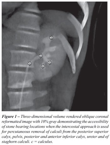
Access via the superior posterior calyx offers optimal exposure to staghorn calculi
as well as multiple calculi in the superior and inferior calyceal groups, renal
pelvis, and upper ureter, and is therefore generally preferred by urologists
(Figures-1 and 2). However, prior publications on the subject have suggested
a higher rate of complications with 11th and 12th intercostal approach (5-8).
The purpose of our study was to compare the rates of complications via the 11th
and 12th intercostal upper pole approaches with those via subcostal access routes.
In addition, we will examine the indications for upper pole versus subcostal
access routes for different stone locations as well as the relevant anatomy that
may increase the rate of complications for a given access route (9-12).
MATERIALS AND METHODS
The study-population consisted of 642 patients, 367 male, 275 female,
ages 15-91 years, 46 years mean age, who underwent percutaneous nephrolithotripsy
and nephrolithotomy from 1996 to 2005 at the Medical Center of Louisiana,
New Orleans V.A. Hospital, Tulane Health Science Center and SUNY, Downstate
Medical School, Brooklyn, N.Y. selection criteria for use of percutaneous
nephrolithotripsy and nephrolithotomy were staghorn calculi involving
the superior calyceal group, a stone mass greater then 2500 mm2, calculi
in superior calyceal group as well as pelvis and upper ureter, stones
in high lying kidneys and sometimes horseshoe kidneys as well as failures
of SWL. A total of 127 had an intercostal (11th or 12th rib, 73 left
and 54 right kidney); and 515 had a subcostal access approach. In 133
patients, a second and third access tract became necessary to reach all
stone bearing areas.
In 255 patients, the calculi were classified as staghorn, in 160 as multiple,
and in 227 as single calculi. Non-contrast computed tomography (CT) examinations
were reviewed to establish the location of calculi and determine an optimal
approach. An intercostal approach was favored for staghorn calculi, multiple
calculi located in the superior posterior, anterior and posterior inferior
calyceal group, pelvis and uretero-pelvic junction (UPJ); a subcostal approach
for solitary calculi in the anterior or posterior inferior, anterior and posterior
interpolar calices and pelvis or combinations thereof. For calculi inaccessible
from the primary access tract, a “Y” tract or a new percutaneous
access tract was developed. Ninety percent of the intercostal procedures were
performed in the last 5 years. Ninety percent of all intercostal and 50% of
all subcostal access procedures were performed by a senior interventional radiologist
(with more than 30 years of experience); the remainder by 2 senior interventional
radiologists (with 5 and 10 years experience respectively) well beyond the
learning curve and always in conjunction with a senior urologist. Informed
consent was obtained in all procedures. The respective institutional review
committees had approved these procedures.
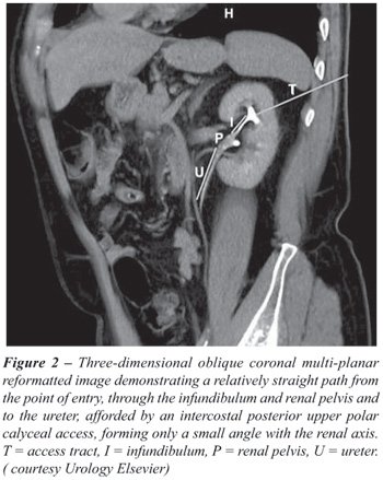
In order to establish a straight tract from the skin to the posterior superior
calyx, infundibulum, and renal pelvis, triangulation was used with the aid
of CT, preferentially 3-dimensional CT reconstructions (12,13), Figure-2. The
angle of entry approximates 30 degrees, with straight-line continuation through
the infundibulum into the pelvis in an anterior-inferior-medial direction (the
renal pelvis being approximately 1.5 cm anterior to the posterior superior
calyx). This trajectory takes advantage of the avascular zone of Brodel. The
patients are then placed on the fluoroscopic table, with the back and flank
prepped and draped using standard surgical technique. Meticulous assessment
of diaphragmatic excursion in inspiration and expiration was obtained to establish
a safe and appropriate point of entry, avoiding the pleura and lung. Puncture
is carried out in mid-expiration to minimize risk of puncture of the lung even
though the needle tract may traverse the lowermost pleura (11,12,14). Infusion
of a 200 mL bolus of N-Saline into the pleural space was another modification
used to prevent puncture of the lung. Because of the known increased risk of
pneumothorax or even calico-pleural fistulae access via the 11th interspace
was avoided whenever possible (4,8,13-16).
The initial access is carried out in the Interventional Radiology Unit under
fluoroscopic control. Local anesthesia, complemented by conscious sedation,
was routinely used. Thereafter a 22-gauge needle is advanced blindly into the
kidney until urine is aspirated, indicating puncture of the collecting system.
Approximately 8 mL of 50% dilute nonionic contrast medium are then injected
to outline the collecting system. Under biplanar fluoroscopic (or rarely CT,
n = 14, or ultrasound, n = 5) guidance, the center of the fornix of the targeted
calyx is accessed with an 18-gauge diamond tip needle (which is the most peripheral
point of the calyx). A 0.038 inch guide wire is then advanced into the renal
pelvis, the tract dilated with 6 and 8F Teflon dilators, and finally a renal
curve Cobra 2 Catheter (Boston Scientific, Boston, MA, USA) is advanced into
the pelvis and ureteropelvic junction is engaged. A glidewire (Boston Scientific,
Natick, MA, USA) is then advanced under fluoroscopic control into the bladder.
Finally, it is replaced through an exchange sheath with super-stiff 0.038 Amplatz
wires (1st working wire in pelvis, and a second safety wire in bladder). At
this point, the patient is transferred to the operating room and the procedure
continued under general anesthesia. The tract is dilated with a high pressure
Pathway balloon (Boston Scientific, Natick, MA, USA) and a 28 - 32F Amplatz
sheath (Bard, Covington, GA, USA) is advanced over the inflated balloon into
the desired calyx. The Amplatz sheath serves to tamponade the tract, keeping
the pressure in the accessed system at a low of 16 mm H2O and allows access
for the rigid nephroscope. Percutaneous stone removal under general anesthesia
is now carried out. At the completion of the procedure, all patients had a
22-G nephrostomy tube and double “J” ureteral stent inserted.
ANATOMIC CONSIDERATIONS
For safe access via the 11th or 12th interspace, location of the posterior
costo-phrenic sulcus particularly, on the left side should be established
in both inspiration and expiration by fluoroscopy (12). In most patients,
a viable trajectory to the posterior superior pole calyx can be achieved
that does not violate the pleural space or lung (Figure-2). Retro-renal
position of the left colon is one condition occurring in approximately
10% of prone patients (12). This could preclude access via a 11th or
12th intercostal approach in those select patients. In some patients,
a large spleen can provide a challenge.
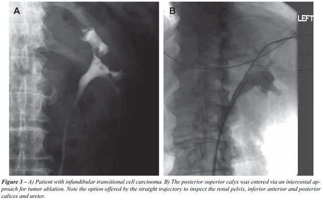
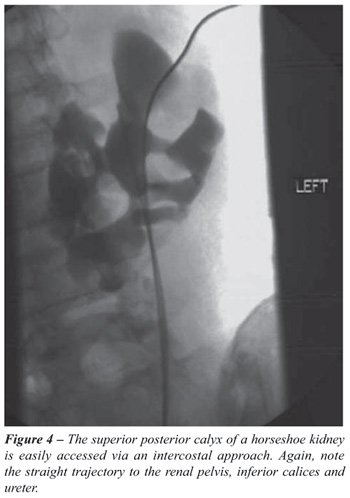
Access to the posterior upper pole calyx affords an almost straight path to
the renal pelvis, upper ureter and both anterior and posterior inferior calyceal
groups (Figures-1 to 4). Even the posterior interpolar calyx may be accessible
via this path without significant angulation. The potential to advance the
Amplatz sheath or nephroscope in a straight path from the posterior upper pole
calyx into the renal pelvis, upper ureter, and anterior and posterior inferior
calyceal groups is of great advantage to the urologist, and reduces the propensity
for injury to the peri-infundibular venous plexus if an angulation of the tract
is necessary to reach the stone-bearing region (15,16). While the infundibulum
of the superior calyceal group tends to be longer, the vascularity of the peri-papillary
and peri-infundibular plexus is less prominent than in the mid calyceal (interpolar
calyceal) group (11). Access via the posterior superior calyceal group makes
calculi in the renal pelvis, upper ureter, and anterior and posterior inferior
calyceal groups accessible, and thus makes this an almost universal access
route. Only the superior anterior calyceal group and anterior and sometimes
posterior interpolar calyceal groups cannot be reached easily via this entry
and hence may mandate separate punctures and access routes if calculi are harbored
in these regions (5,15). Moreover, access from the superior posterior calyceal
group creating a straight path to the pelvis, upper ureter and inferior calyceal
group reduces injury to renal parenchyma by the Amplatz sheath or nephroscope
during respiratory excursion (12,14).
RESULTS
To provide access for nephrolithotripsy of calculi in the posterior
superior calyx, the intercostal access route was chosen in 111 patients,
and the subcostal route in 119 patients (Table-1). A total of 134 of
these patients were treated for a staghorn calculus, another 96 for at
least one calculus lodged in the superior calyx (Table-2). To attain
a near stone free status, the mid calyceal group had to be accessed secondarily
in 44 patients with staghorn calculi (for dendritic stones or residual
debris) and in 11 others (for otherwise inaccessible stones or debris);
as well as the inferior calyx in 15 patients with staghorn calculi and
16 patients with residual calculi or debris (Table-2). Patients with
residual or otherwise inaccessible calculi mandated access to the mid
calyx in 75 patients (12 via intercostal and 63 via subcostal approach)
and in 31 to the inferior calyx (2 via intercostal and 29 via subcostal
approach) (Table-2). An abnormal high location of the kidney mandated
access via the intercostal route in 2 patients, with calculi in the mid
calyx and 2 in the lower pole calyx (Table-2). Overall, the midcalyx
was directly accessed via intercostal route in 12 patients and via a
subcostal approach in 63 patients; the lower pole calyx via an intercostal
route in 4 patients and via a subcostal approach in 333 patients (Table-1).
The major complications we experienced were septic shock and effects to the
vascular system, collecting system, and lungs. Access to the upper pole by
the intercostal route resulted in 1 pneumothorax, 1 arterio-calyceal fistula
and 3 AV fistulae in 111 patients (Table-3). Via a subcostal access route,
we recorded 2 pneumothoraces, 1 AV fistula, 1 pseudoaneurysm, 1 ruptured UPJ,
and 4 perforated ureters in 119 patients (Table-3). The ratio of complication
to no complication was significant (p = 0.0395). In the same group of patients,
we experienced 7 minor complications in the intercostal access group and 23
in the subcostal access group (Table-4). A high incidence of atelectasis (n
= 13) in the subcostal access group as well as a relatively high need for blood
transfusion (n = 2) should be noted (Table-4). For intercostal access to the
mid-calyceal group, we recorded no major complications. However, for subcostal
access, the rate was exceedingly high (5 in 63 patients, 7.8%). We recorded
1 hemothorax, 1 AV fistula, 1 colo-calyceal fistula and 2 perforated ureters
(Table-3). Among minor complications, the need for blood transfusions in the
intercostal access group was high (2 in 12 patients, 16.7%). Minor complications
in the subcostal entry group were high (12 in 63 patients, 19%); again blood
transfusions were among the most frequent of the minor complications, occurring
in 4 patients (6.3%, Table-4). We experienced no major complications with entry
into the lower poles via an intercostal route, however via a subcostal route
there were 5 major complications in 333 patients. Septic shock in 2 patients
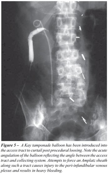
(0.6 %) is noteworthy, since it is easily avoidable (Table-3). Of all minor
complications, diffuse tract bleeding occurring in 8 patients (2.4%), which
deserves special attention (Figure-5). In the subgroup of staghorn calculi,
4 major vascular complications occurred in 102 intercostal accesses to the
upper pole (4%, Table-5). An even higher incidence of major complications occurred
when subcostal access was provided (4 in 32 patients, 12.5%). The incidence
of complications with mid-polar subcostal route access is again very high (5
in 47), Table-5.
COMMENTS
The literature reports a stone-free status attainable by percutaneous
nephrolithotripsy (PCNL) and nephrolithotomy in 64.5-98.3% of patients
(4,14,17,18). Conversely, extracorporeal nephrolithotripsy (SWL), even
if complemented by follow-up medical management, achieves stone-free
results in only about 37% of patients (19-21). Even in locations such
as the lower pole, where SWL had been favored, PCNL rendered 90-95% of
patients stone-free versus SWL (14-63%) (19). Ureteroscopic nephrolithotripsy
by electrohydraulic or Holmium Yag laser likewise cannot match PCNL results
(22). For large stone loads (2500 mm2 or larger), staghorn calculi, calculi
in diverticula and even smaller stones in lower pole calices; PCNL is
now the preferred method. The choice of access tract is based on the
ability to provide good visibility of the stone bearing area and a point
of entry with minimal risk of injury to adjacent organs. Additionally,
the access tract should provide a trajectory projecting without torque
or angulation into the infundibulum and renal pelvis, hence facilitating
atraumatic intraoperative advancement of the Amplatz sheath to the UPJ
or inferior calyceal group (4,6,8,14,23; Figure-1). Intercostal access
via the posterior superior calyx offers the best trajectory via infundibulum
to pelvis, UPJ and inferior calyx (4,10), Figure-2. Lack of angulation
and torque when advancing the Amplatz sheath significantly reduces the
risk of inducing bleeding. However, the preferred intercostal access
route has been incriminated with a higher rate of complications than
the subcostal approach (4,6,16,17). The supra 11th rib approach has a
particularly high rate of complications. Pneumo- and hemothorax, and
calyceal-pleural fistulae have been reported in up to 23.1% (4) The possibility
of both a transthoracic and transpleural trajectory of this type of access
tract, despite attempts to attain a high position of the lung by puncturing
during the expiration phase, predisposes to these complications (4,6,14,24).
The incidence of hydropneumothorax occurring with intercostal access
has been reported at a rate of 4% to 15.3 %, with subcostal access, 0%
to 1.4% (4,7,14,16,24). Similarly, large pleural effusions were reported
in 8% to 12.5% with intercostal approach, but virtually absent with subcostal
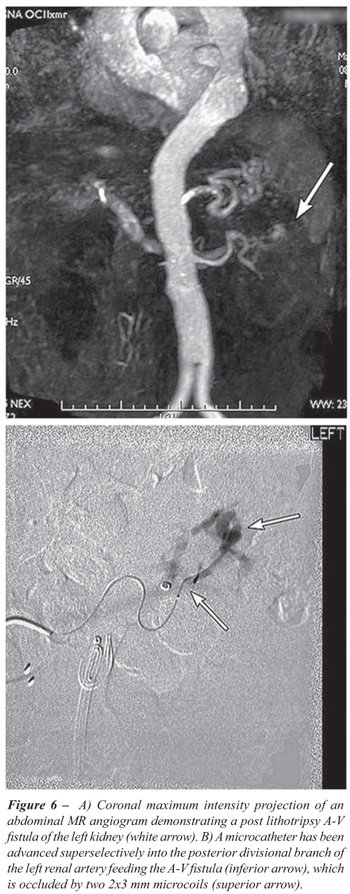
access (6,14,15,24). Moreover, on the basis of anatomic considerations,
the intercostal access route might have a higher chance for injury to
anterior segmental vessels or even anterior and posterior divisional
arteries (4,10,11,14) Figure-6.
In our series we encountered 4 major (3.6%) and 7 minor complications (6.3%)
in 111 patients with intercostal access to the upper pole (p = 0.0395; Table-4).
Interestingly, there was only one pneumothorax (0.9%). There were three vascular
complications reflecting the inherent risk posed by anatomic proximity of vascular
and excretory system in the upper pole (10,11). Both AV fistulae involved posterior
division branches; the arterio-calyceal fistula was between a posterior middivisional
branch and the posterior calyceal infundibulum. The etiology is most likely
secondary to the needle penetrating the infundibulum and passing through the
anteriorly located artery, giving rise to the fistula (10,11). Only 7 minor
injuries were recorded (Table-4). One Steinstrasse caused by multiple fragments
in the lower ureter caused obstruction (Table-4). A renal branch vein thrombosis
also resulted, without late sequellae. Three patients developed atelectasis
on the ipsilateral side, likely related to irritation of the diaphragm and
curtailed respiratory motion. Our incidence of intrathoracic complications
(3.1%) attendant to the access tracts compares favorably to that of the 7.1%
- 12 % range reported in the literature (4,6). Our complication rate for supra
11th rib access is 25%, similar to that of 23.1% of the literature (6,16,24).
Our technique of puncturing lateral to the erector muscle in mid expiration
takes advantage of the higher location of the pleural deflection under these
circumstances, a feature that has been pointed out by Hooper et al. (12).
In 119 subcostal access procedures to the upper pole, we experienced 9 major
complications (Table-3). Pneumothoraces and rupture of UPJ and ureters likely
occurred while manipulating and repositioning the Amplatz sheath during the
stone extraction phase. Vascular injuries involving anterior divisional branches
were also encountered, most likely secondary to excessive needle penetration.
The large incidence of minor complications (22) deserves scrutiny (Table-4).
Atelectasis in 13 patients suggests that the access route might have caused
significant irritation of the diaphragm. Diffuse bleeding from the tract (n
= 2) and need for blood transfusions (n = 2) suggest either injury to the peri-infundibular
plexus, either during the procedure or due to inadequate postoperative tamponade
by the Malecot or Council Catheter. Injury to the peri-infundibular venous
plexus is likely to occur when advancing the Amplatz sheath while negotiating
the angle formed between the subcostal access tract and the infundibulum and
pelvis (8,11,15,16), (Figure-5). Access to the mid-calyx (interpolar calyx)
via a subcostal approach shows a similar high incidence rate of minor complications
(19%), Table-4. The high incidence of diffuse tract bleed requiring transfusion
is again attributable to injury of the peri-infundibular venous plexus. With
access to the lower pole via subcostal route, diffuse tract bleeding, obstruction
and fever are the most common minor complications (14), Table-5. Again, difficulty
adjusting the Amplatz sheath interoperatively may result in injury, failure
to completely evacuate stone debris (and hence Steinstrasse) and infection
and fever. In the subgroup of staghorn calculi, the major complication rate
for subcostal access is almost 10%, but for intercostal access the rate is
only 3% (Table-5). This reflects the advantage of the intercostal approach
when dealing with dendritic extension of stones.
We analyzed our data using logistic regression and predicted complications
for the respective calyx and intercostal or subcostal entry. The complication
rates for upper calyx were 17.3%, for interpolar calyx 44.4%, and for lower
calyx 15.4%. The odds of complication were 3.5 times higher for interpolar
than upper polar entry (p = 0.0003); 5.1 times higher for interpolar than lower
polar entry (p = 0.0001). The odds of complications were 1.9 times higher with
subcostal entry compared to intercostal entry (p = 0.0389).
CONCLUSIONS
The findings of our study support preferential use of intercostal access routes (12th, 11th, 10 th rib space) via the posterior calyx for percutaneous nephrolithotripsy in patients with a large stone load, staghorn calculi, multiple calculi lodged in the posterior superior calyx, pelvis, UPJ, upper ureter, and posterior and anterior inferior calyceal groups. This route offers optimal visibility, easy interoperative advancement and adjustment of the Amplatz sheath and rigid nephroscope, a low rate of procedural complications, reduced operative time and excellent results in removal of targeted stones. For calculi in other locations, separate access tracts or “Y” tracts are advocated.
CONFLICT OF INTEREST
None declared.
REFERENCES
- Duvdevani M, Razvi H, Sofer M, Beiko DT, Nott L, Chew BH, et al.: Third prize: contemporary percutaneous nephrolithotripsy: 1585 procedures in 1338 consecutive patients. J Endourol. 2007; 21: 824-9.
- Wolf JS Jr, Clayman RV: Percutaneous nephrostolithotomy. What is its role in 1997? Urol Clin North Am. 1997; 24: 43-58.
- Osman MM, Alfano Y, Kamp S, Haecker A, Alken P, Michel MS, et al.: 5-year-follow-up of patients with clinically insignificant residual fragments after extracorporeal shockwave lithotripsy. Eur Urol. 2005; 47: 860-4.
- Munver R, Delvecchio FC, Newman GE, Preminger GM: Critical analysis of supracostal access for percutaneous renal surgery. J Urol. 2001; 166: 1242-6.
- Hentschel H, Janitzky V, Weirich T: Percutaneous nephrolithotomy - always effective and free of complications? Aktuelle Urol. 2007; 38: 232-6.
- Radecka E, Brehmer M, Holmgren K, Magnusson A: Complications associated with percutaneous nephrolithotripsy: supra- versus subcostal access. A retrospective study. Acta Radiol. 2003; 44: 447-51.
- Stening SG, Bourne S: Supracostal percutaneous nephrolithotomy for upper pole caliceal calculi. J Endourol. 1998; 12: 359-62.
- Shaban A, Kodera A, El Ghoneimy MN, Orban TZ, Mursi K, Hegazy A: Safety and efficacy of supracostal access in percutaneous renal surgery. J Endourol. 2008; 22: 29-34.
- Preminger GM, Schultz S, Clayman RV, Curry T, Redman HC, Peters PC: Cephalad renal movement during percutaneous nephrostolithotomy. J Urol. 1987; 137: 623-5.
- Sampaio FJ, Mandarim-De-Lacerda CA, De Aragão AH: The collector system of the kidney. Applied anatomy based on the analysis of 3-dimensional casts. J Urol (Paris). 1987; 93: 183-5.
- Sampaio FJ, Aragao AH: Anatomical relationship between the intrarenal arteries and the kidney collecting system. J Urol. 1990; 143: 679-81.
- Hopper KD, Sherman JL, Luethke JM, Ghaed N: The retrorenal colon in the supine and prone patient. Radiology. 1987; 162: 443-6.
- Lojanapiwat B, Prasopsuk S: Upper-pole access for percutaneous nephrolithotomy: comparison of supracostal and infracostal approaches. J Endourol. 2006; 20: 491-4.
- Yadav R, Aron M, Gupta NP, Hemal AK, Seth A, Kolla SB: Safety of supracostal punctures for percutaneous renal surgery. Int J Urol. 2006; 13: 1267-70.
- Sukumar S, Nair B, Ginil KP, Sanjeevan KV, Sanjay BH: Supracostal access for percutaneous nephrolithotomy: less morbid, more effective. Int Urol Nephrol. 2008; 40: 263-7.
- Lashley DB, Fuchs EF: Urologist-acquired renal access for percutaneous renal surgery. Urology. 1998; 51: 927-31.
- Segura JW: Endourology. J Urol. 1984; 132: 1079-84.
- Netto NR Jr, Ikonomidis J, Ikari O, Claro JA: Comparative study of percutaneous access for staghorn calculi. Urology. 2005; 65: 659-62; discussion 662-3.
- Lam HS, Lingeman JE, Mosbaugh PG, Steele RE, Knapp PM, Scott JW, et al.: Evolution of the technique of combination therapy for staghorn calculi: a decreasing role for extracorporeal shock wave lithotripsy. J Urol. 1992; 148: 1058-62.
- Fine JK, Pak CY, Preminger GM: Effect of medical management and residual fragments on recurrent stone formation following shock wave lithotripsy. J Urol. 1995; 153: 27-32; discussion 32-3.
- Kang DE, Maloney MM, Haleblian GE, Springhart WP, Honeycutt EF, Eisenstein EL, et al.: Effect of medical management on recurrent stone formation following percutaneous nephrolithotomy. J Urol. 2007; 177: 1785-8; discussion 1788-9.
- Mariani AJ: Combined electrohydraulic and holmium:YAG laser ureteroscopic nephrolithotripsy of large (greater than 4 cm) renal calculi. J Urol. 2007; 177: 168-73; discussion 173.
- Aron M, Goel R, Kesarwani PK, Seth A, Gupta NP: Upper pole access for complex lower pole renal calculi. BJU Int. 2004; 94: 849-52; discussion 852.
- Narasimham DL, Jacobsson B, Vijayan P, Bhuyan BC, Nyman U, Holmquist B: Percutaneous nephrolithotomy through an intercostal approach. Acta Radiol. 1991; 32: 162-5.
____________________
Accepted after revision:
January 6, 2009
_______________________
Correspondence address:
Dr. Erich K. Lang
Department of Radiology
SUNY Downstate College of Medicine
450 Clarkson Avenue
Box 1198, Brooklyn, NY, 11231, USA
Fax: + 1 718-270-3848
E-mail: erich.lang@downstate.edu
EDITORIAL COMMENT
The mainstay of percutaneous nephrolithotomy is to create a straight
path from the skin to the renal pelvis in order to avoid renal angulations
and consequently damage to the peri-infundibular structures (either vessels
or parenchyma).
Historically at the beginning of percutaneous renal surgery the patient
was in prone position, to avoid colonic puncture, and the access was
routinely
subcostal, to avoid injury to the lungs and pleura, but through the lower calyx
(1,2). However, it has also been recognized that, in selected cases, it could
not provide an optimal access. In this retrospective analysis on a quite large
series of percutaneous nephrolithotomy (PCNL), Dr. Lang and co-authors address
the so-called “intercostal (or supracostal)” approach for PCNL.
This issue is still highly debated in the field of endourology, even if its
related literature remains scarce. Unfortunately, the value of this report
is negatively affected by some major drawbacks.
First, all the inherent biases of a retrospective study are present here and
this should be taken into account when looking at the conclusions.
One of the significant evolutions of the PCNL has been the widespread-though
not universal-renal puncture by a urologist, making it a single-stage procedure
(3). Here, PCNL is presented as a two-step procedure, the first in local anesthesia,
performed by the radiologist, the second in general anesthesia, performed by
the urologist.
The authors emphasize the benefits of the upper pole access and we can agree
with them that it allows a good exposure of most of the calyces and of the
proximal ureter. As the main requirement during PCNL is always the same, a
straight path causing no angulations, if the superior calyx is too high, intrathoracic,
they should not perform a subcostal puncture: a complication will almost certainly
occur.
However, many of the complications reported are related to the lithotripsy
rather than to the puncture. The high rate of ureteral perforation and UPJ
avulsion are not puncture-related but a matter of a wrong operative technique.
It has been previously suggested that a great difference might be between the
supra twelfth rib approach, which is transthoracic but extrapleural, and the
supra eleventh rib access, which is both transthoracic and transpleural. This
issue is not addressed in the paper (4).
To note that the authors do not use the insertion of a ureteral catheter as
a first step of the procedure (as most of us performing PCNL routinely do).
The needle is inserted and the contrast medium is injected without a prior
retrograde dilation of the calyceal system. With this technique, if you do
not correctly target the calyx the risk of fornix rupturing during the path
dilation and subsequent bleeding is increased.
Finally, we would like to remind the authors that supine position for PCNL
has been advocated in the past decade (5). One of its main advantages is that
it might combine the benefits of percutaneous and ureteroscopic intrarenal
surgery in selected cases (6). Thus, large and/or complex urolithiasis can
be treated with a high one-step stone-free rate, unquestionable anesthesiological
advantages, and no additional procedure-related complications (7). In this
regard, some limitations of the standard prone PCNL might be overcome avoiding
a potentially harmful supracostal approach, as the one proposed.
REFERENCES
- Alken P, Hutschenreiter G, Günther R, Marberger M: Percutaneous stone manipulation. J Urol. 1981; 125: 463-6.
- Gupta R, Kumar A, Kapoor R, Srivastava A, Mandhani A: Prospective evaluation of safety and efficacy of the supracostal approach for percutaneous nephrolithotomy. BJU Int. 2002; 90: 809-13.
- Smith AD: Percutaneous punctures--is this the endourologist’s turf? J Urol. 1994; 152: 1982-3.
- Munver R, Delvecchio FC, Newman GE, Preminger GM: Critical analysis of supracostal access for percutaneous renal surgery. J Urol. 2001; 166: 1242-6.
- Valdivia Uría JG, Valle Gerhold J, López López JA, Villarroya Rodriguez S, Ambroj Navarro C, Ramirez Fabián M, et al.: Technique and complications of percutaneous nephroscopy: experience with 557 patients in the supine position. J Urol. 1998; 160: 1975-8.Autorino R, Giannarini G: Prone or supine: is this the question? Eur Urol. 2008; 54: 1216-8.
- Scoffone CM, Cracco CM, Cossu M, Grande S, Poggio M, Scarpa RM: Endoscopic combined intrarenal surgery in galdakao-modified supine valdivia position: a new standard for percutaneous nephrolithotomy? Eur Urol. 2008; 54: 1393-403.
Dr.
Riccardo Autorino &
Dr. Marco De Sio
Division of Urology
Second University of Naples
Naples, Italy
E-mail: ricautor@tin.it
EDITORIAL COMMENT
The authors compare a series of patients with percutaneous
nephrolithotripsy (PCNL) tracts above the 12th and 11th ribs to a group
with access established
in a subcostal location. Complications including those related to the
thorax are no greater when the access is an intercostal upper pole approach.
We as well have seen a shift to an upper pole approach over the last
several years and this is now at least as common as a lower pole approach
at our centre (1). For these reasons the authors emphasize including
less torquing of the working sheath, working “downhill” on
most large stones and ready access to more of the collecting system.
They do not mention hydrothorax as a specific thoracic complication however
this may be a more common adverse consequence of a high approach as pneumothorax.
Attention to maintaining the working sheath in the collecting system
throughout the procedure is an important technical point. We also are
more likely to place a stent at the conclusion of the PCNL procedure
with a supracostal access to assure antegrade urine drainage postoperatively
and prevent fluid accumulation in the chest. In such instances, it is
important to remove the urethral catheter after the percutaneous tube
is removed to prevent retrograde extravasation of urine.
Access in this series was obtained in the radiology department. I believe that
similar results with a low rate of thoracic complications can be achieved when
the urologist obtains the percutaneous access in the operative room using C-arm
fluoroscopic guidance (2).
Finally, flexible nephroscopy combined as needed with intracorporeal lithotripsy
is an important adjunct to minimize the need for additional tracts when performing
PCNL.
REFERENCE
- Duvdevani M, Razvi H, Sofer M, Beiko DT, Nott L, Chew BH, et al.: Third prize: contemporary percutaneous nephrolithotripsy: 1585 procedures in 1338 consecutive patients. J Endourol. 2007; 21: 824-9.
- Watterson JD, Soon S, Jana K: Access related complications during percutaneous nephrolithotomy: urology versus radiology at a single academic institution. J Urol. 2006; 176: 142-5.
Dr. John Denstedt
Richard Ivey Professor and Chair
Department of Surgery
The University of Western Ontario
London, ON, Canada
E-mail: john.denstedt@sjhc.london.on.ca