RECOVERY
OF SPERMATOGENESIS AFTER MICROSURGICAL SUBINGUINAL VARICOCELE REPAIR IN
AZOOSPERMIC MEN BASED ON TESTICULAR HISTOLOGY
(
Download pdf )
SANDRO C. ESTEVES, SIDNEY GLINA
Androfert (SCE), Center for Male Infertility, Campinas, Sao Paulo, and Department of Urology (SG), Hospital Ipiranga, Sao Paulo, SP, Brazil
ABSTRACT
Objective:
Analyze whether testicular histologic patterns from a group of azoospermic
men with varicocele is predictive of treatment outcome after subinguinal
microsurgical varicocele repair.
Materials and Methods: Seventeen azoospermic
men underwent bilateral open single testis biopsy and microsurgical subinguinal
repair of clinical varicoceles.
Results: Histopathology of testicular biopsies
revealed hypospermatogenesis (HYPO) in 6 men, maturation arrest (MA) in
5, and Sertoli cell-only (SCO) in 6. Overall, presence of spermatozoa
in the ejaculates was achieved in 47% (8/17) of men after varicocele repair,
but only 35% (6/17) of them had motile sperm in their ejaculates. Only
men with testicular histology revealing HYPO (5/6) or maturation arrest
(3/5) had improvement after surgery. Median (25% - 75% percentile) postoperative
motile sperm count for both groups were 0.9 X 10⁶/mL (0.1-1.8 X 10⁶/mL)
and 0.7 X 10⁶/mL (0.1-1.1), respectively (p = 0.87). The mean time for
appearance of spermatozoa in the ejaculates was 5 months (3 to 9 months).
One (HYPO) of 8 (12.5%) men who improved after surgery contributed to
an unassisted pregnancy. Postoperative testicular biopsies obtained from
patients who had no improvement after surgery revealed that testicular
histology diagnosis remained unchanged. Successful testicular sperm retrieval
for intracytoplasmic sperm injection (ICSI) was achieved in 4 of 9 (44.4%)
individuals who did not improve after surgery, including 1 man with testicular
histology exhibiting SCO.
Conclusions: Microsurgical varicocele repair
in nonobstructive azoospermic men with clinical varicoceles can result
in sperm appearance in the ejaculate when hypospermatogenesis or maturation
arrest is found on testicular histology diagnosis.
Key
words: varicocele; microsurgery; azoospermia; testis; histology
Int Braz J Urol. 2005; 31: 541-8
INTRODUCTION
Azoospermia
and severe oligozoospermia in association with varicocele is reported
to range from 4.3% to 13.3% (1). Testicular histopathology in severe oligozoospermic
and azoospermic patients are often bilateral and range from various degrees
of hypospermatogenesis to Sertoli cell-only pattern (2,3). Although few
controlled studies have evaluated the outcome of varicocele repair in
infertile men, most of them support a favorable effect of surgical correction
on general sperm quality and fertility (4-6). The beneficial effect of
varicocele repair in azoospermic patients, on the other hand, remains
controversial. While some studies have documented recovery of spermatogenesis
and unassisted pregnancies after surgery (1,7-11), others associated the
presence of varicoceles as incidental findings (12) or with a limited
role in azoospermia (13).
For azoospermic men with varicoceles, even
modest induction of spermatogenesis leading to the presence of motile
sperm in the ejaculate after varicocele repair could allow these men to
establish a pregnancy on their partners, either unassisted or assisted,
thus expanding the couple’s reproductive options. Identification
of who will benefit from surgery may have profound clinical impact, since
induction of spermatogenesis is not achieved in all individuals after
varicocelectomy. The purpose of this study was to evaluate treatment outcome
after subinguinal microsurgical varicocele repair in relation to testicular
histopathology in a group of nonobstructed azoospermic men with clinical
varicoceles.
MATERIALS AND METHODS
Patients
We reviewed the charts of 256 infertile
men who underwent surgical repair of clinical varicoceles from August
1996 to September 2003. Seventeen of 256 (6.6%) men, with a median of
32 years-old (19 - 45 years), who presented with clinical varicoceles
and nonobstructive azoospermia, were included in this retrospective study.
All men had a history of primary infertility of at least 1 year duration
(median 23.6 months, range 13-96 months). Other causes of azoospermia
were ruled out. Varicoceles were identified on physical examination and
graded as large (grade 3, visible when standing), moderate-sized (grade
2, visible with Valsalva’s maneuver when standing) and small (grade
1, palpable with Valsalva’s maneuver when standing). Only men with
clinical unilateral or bilateral varicoceles were included. Testicular
volume was assessed using the Prader orchidometer. A testicular volume
< 20 mL was considered diminished. At least 2 preoperative semen analyses
were obtained and evaluated according to the WHO criteria (14). All semen
analyses confirmed the absence of sperm in the centrifuged pellet. All
men had ejaculate volumes > 1.5 cc, alkaline seminal fluid pH, and
reproductive ductal structures palpably normal. Hormonal profile included
serum testing for follicle-stimulating hormone (FSH), luteinizing hormone
(LH), testosterone, and prolactin. Thirty G-banded metaphases were analyzed
by high-resolution Giemsa karyotype in 15 men. All of them were cytogenetically
normal. Polymerase chain Yq microdeletion screening for AZFa, AZFb and
AZFc was done in 12 individuals. Deletions of Yq were not observed in
any of them.
The median preoperative hormone levels were:
FSH = 14.0 mUI/mL (25% - 75% percentile, range 6.5 - 34.6 mUI/mL), LH
= 5.8 mUI/mL (25% - 75% percentile, range 3.5 - 14.5 mUI/mL), prolactin
= 7.4 ng/mL (25% - 75% percentile, range 4.2 - 16.0 ng/mL), and testosterone
= 544.3 ng/dL (25% - 75% percentile, range 285.0 - 864.0 ng/dL). Ten men
(59%) had elevated serum FSH levels (normal range: 1.0 -10.0 mUI/mL).
Bilateral and unilateral left-sided procedures have been done in 11 (65%)
and 6 men respectively. Varicoceles were large in 9 (53%) men and moderate-sized
in 8. Diminished testicular volume has been found bilaterally in 6 (35%)
men and unilaterally in 7.
Microsurgical
Varicocele Repair
All subjects underwent testicular artery
and lymphatic-sparing subinguinal varicocele repair. Briefly, a 2.5-cm
skin incision was made over the external inguinal ring. The subcutaneous
tissue was separated until the exposure of spermatic cord. The cord was
elevated with a Babcock clamp and the posterior cremasteric veins were
ligated and transected. A Penrose drain was placed behind the cord without
tension. The cremasteric fascia was then opened to expose the cord structures
and the dissection proceeded using either operating microscope with 6-16X
magnification (15 patients) or loupes with 2.5X magnification (2 patients).
Dilated cremasteric veins within the fascia were ligated and transected.
Lymphatics and arteries were identified and preserved. Whenever necessary,
the cord structures were sprayed with papaverine hydrochloride to increase
the arterial beat. All dilated veins of the spermatic cord were identified,
tagged with vessel loops, then ligated and transected. Vasal veins were
ligated only if they exceed 2 mm in diameter. Sclerosis of additional
veins was not used. The incision was closed with absorbable sutures. Procedures
were performed on an outpatient basis using either regional or local anesthesia
in combination with short-acting sedation. The surgical technique used
in this study was nearly identical to that described previously (15).
After surgery, semen samples were obtained at 2 - 4 month intervals and
evaluated according to the WHO criteria (14). The mean postoperative follow-up
duration was 18.9 ± 5.3 months. An average was computed for each
seminal parameter, and then used for statistical purposes.
Testis
Biopsy
Open bilateral diagnostic testis biopsies
were performed in all subjects at the same time of varicocele repair.
For this, a 1-cm transverse incision was made at the anterior scrotal
skin. The scrotal tunics were incised until identification of the tunica
albuginea. The testis was manipulated to expose a relatively avascular
area, where a 5-mm incision was made over the tunica. By producing counter
pressure on the posterior surface of the testis, testicular tissue was
evaginated into the incision, and a single piece measuring approximately
3 x 3 x 3 mm was excised with a wet and sharp Iris scissors using a no-touch
method. The specimens were transferred to the Bouin’s solution and
afterwards stained with hematoxylin and eosin, and histologically sectioned.
Testicular biopsies were classified as follows: (a) Sertoli cell-only
(SCO), (b) maturation arrest and (c) hypospermatogenesis (HYPO). At least
50 seminiferous tubules were evaluated on each testis. Sertoli cell-only
category indicated that germinative cells were absent. Maturation arrest
(MA) category was defined as absence of mature spermatozoa, despite normal
early stages of spermatogenesis. Hypospermatogenesis indicated that all
stages of the spermatogenic cycle were present, including mature sperm,
but there was a proportional reduction in the number of all germ cells
at each level.
Microdissection testicular sperm extraction
(micro-TESE) (16) for procurement of spermatozoa within the testis has
been performed bilaterally at least 6 months postoperatively (median:
9 months; range: 6-15 months) in all men who showed no improvement after
surgery. Concomitantly, a single piece measuring approximately 3 x 3 x
3 mm was excised without using microsurgery from a surrounding area of
the microdissected samples for testicular histology diagnosis. Testicular
sperm extraction with microdissection has been chosen in such cases because
of its concept, i.e., a microscope-guided testis biopsy that has been
shown to significantly improve sperm yield with minimal tissue excision
(16).
Statistical
Analysis
Preoperative hormone levels and testicular
size were compared among the groups and between the patients who did and
did not improve after varicocele repair. After varicocelectomy, sperm
parameters were compared between the groups with testicular histology
diagnosis of hypospermatogenesis and maturation arrest. Due to the large
variation observed in clinical and sperm parameters, our data are presented
as median and percentiles 25% and 75%, which better describes our patient
population. Non-parametric tests were used for statistical analysis because
our data were drawn from not normally distributed population (17). Kruskal-Wallis
test was used to compare FSH levels and total testicular volume among
groups. Mann-Whitney rank-sum test was used to compare sperm count, total
number of motile sperm, sperm viability and normal forms between hypospermatogenesis
and maturation arrest groups. The Mann-Whitney test was also used to compare
FSH levels between patients who improved or not after surgery. Although
nonparametric tests are not as powerful as parametric methods for statistical
evaluation, they are more reliable when analyzing data from not normally
distributed populations (17).
Pairwise comparisons using the Fisher exact
test were performed to analyze statistical differences between spermatogenesis
recovery rates. The Fisher exact test was used instead of the Chi-square
test due to the small number of patients in each group (17).
Sperm retrieval success rates were not compared
due to the extremely low number of subjects in each group. A value of
< 0.05 was considered statistically significant. Statistical calculations
were performed using computer software (Statisticaâ, Stasoft, Tulsa,
OK).
RESULTS
Hypospermatogenesis
(HYPO) was identified on diagnostic testis biopsy in 6 men, maturation
arrest (MA) in 5 and Sertoli cell-only (SCO) in 6. Overall, presence of
spermatozoa in the ejaculates was achieved in 47% (8/17) of men after
varicocele repair, but only 35% (6/17) of them had motile sperm in their
ejaculates. Only men with testicular histology revealing HYPO (5/6) or
maturation arrest (3/5) had improvement after surgery. Median (25% - 75%
percentile) motile sperm count for both groups were 0.9 X 10⁶/mL (0.1
- 1.8 X 10⁶/mL) and 0.7 X 10⁶/mL (0.1 - 1.1), respectively (p = 0.87),
Table-1. The mean time for appearance of spermatozoa in the ejaculates
was 5 months (range 3 - 6 months). One (HYPO) of 8 men who improved after
surgery contributed to an unassisted pregnancy which occurred 6 months
after surgery. Median (25% - 75% percentile) motile sperm count for this
man during the follow-up period was 1.5 X 10⁶/mL (1.1 - 1.8 X 10⁶/mL).
None of the patients who had sperm in the ejaculates after varicocele
repair returned to be azoospermic during the follow-up period.
Preoperative serum FSH levels were 10.9
(3.2 - 21.2) mUI/mL and 19.5 (7.5 - 31.8) mUI/mL in men who did and did
not show recovery of spermatogenesis after varicocele repair (p = 0.22).
FSH levels in men with HYPO, MA and SCO were not significantly different
(Table-1). Appearance of sperm in the ejaculates was observed in 6 (46%)
of 13 men with testes of reduced volume and in 2 (50%) of 4 men with normal-sized
testes (p = 0.99). Combined testicular volume (right plus left sides)
in men with HYPO, MA and SCO were not significantly different (Table-1).
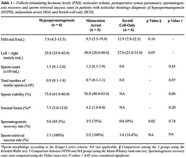
Appearance of spermatozoa in the ejaculate
was not achieved in any men with testicular histology diagnosis of SCO.
These individuals (n = 6) as well as the ones with testicular histology
diagnosis of HYPO (n = 1) and MA (n = 2) underwent postoperative bilateral
open single testis biopsy concomitant with microsurgical-guided sperm
retrieval (Micro-TESE) for intracytoplasmic sperm injection (ICSI) (n
= 6) or for diagnostic purposes only (n = 3). Postoperative testicular
histology diagnosis was unchanged in comparison to preoperative ones.
Successful testicular sperm retrieval using Micro-TESE was achieved in
4 of 9 (44.4%) individuals who did not improve after surgery, including
one who had testicular histology diagnosis of SCO (Table-1).
COMMENTS
Recovery
of spermatogenesis is possible after surgical repair of clinical varicoceles
in men with nonobstructive azoospermia. Few studies have shown that nonobstructive
azoospermic patients with clinical varicoceles can benefit from varicocelectomy
(7-12). These studies reported improvement of semen parameters in up to
50%, including rare cases of spontaneous pregnancies. Matthews et al.
(7), studying 22 men, found that 54% presented sperm in the ejaculate
postoperatively. Although diagnostic testicular biopsy was not available
for many of them, those men most likely to benefit had either hypospermatogenesis
or maturation arrest. Kim et al., studying 28 patients, demonstrated that
testicular histology was the most important predictive factor on outcome
(8). In their study, patients with Sertoli cell-only pattern and maturation
arrest at spermatocyte stage have not shown improvement; however, 50%
of the individuals with maturation arrest at spermatid stage and 55% of
them with hypospermatogenesis achieved postoperative improvement with
appearance of sperm in their ejaculates (8). Pasqualotto et al., on the
other hand, reported that improvement in semen quality after varicocelectomy
may be possible even in azoospermic patients who present germ cell aplasia
in a single large testis biopsy (10). In comparison, our series demonstrated
postoperative return of sperm in the ejaculate in 47% of men after varicocele
repair. We found that testicular histology diagnosis from a single large
testis biopsy was the most important predictive factor on outcome. Only
men with testicular histology revealing hypospermatogenesis or maturation
arrest had improvement after surgery. All patients with Sertoli cell-only
pattern still remained azoospermic after varicocelectomy. In our series,
testicular volume and preoperative serum FSH levels were not predictive
of treatment outcome, and these results were confirmed by others (9,11).
Interestingly, in our series, despite the
induction of spermatogenesis in men with hypospermatogenesis and maturation
arrest, we found that semen parameters still remained severely abnormal
after varicocele repair. Severe oligozoospermia and teratozoospermia have
been observed in all individuals after repeated routine semen analyses.
In addition, 25% (2/8) of men who improved after surgery presented with
only immotile sperm in their ejaculates. Therefore, it is likely that
advanced assisted reproductive techniques will be required for most couples
to initiate a pregnancy, as shown in a recent study by Schlegel &
Kaufmann who reported that only 9.6% men after varicocele repair had adequate
motile sperm in the ejaculate for ICSI (13). The latter does not diminish
the clinical impact of our findings because even modest improvements in
semen quality after varicocele repair may expand the couple’s reproductive
options. Although our series is small, one couple achieved an unassisted
pregnancy, which would have been otherwise impossible if the varicocelectomy
had not been performed. Matthews et al. reported that 9% of azoospermic
men who improved after varicocele repair contributed to unassisted pregnancies
(7). Czaplicki et al. (1), Kim et al. (8) and Pasqualotto et al. (2) also
reported unassisted pregnancies after varicocelectomy in azoospermic patients.
Although spermatozoa have been consistently
found in repeated semen analyses during the follow-up period, we have
observed that appearance of sperm within the ejaculates may not be immediate.
The clinician should be advised that it may take up to 6 months after
varicocelectomy to consider that varicocele repair has not been able to
recover spermatogenesis. Pasqualotto et al., on the other hand, reported
that most of their patients relapsed into azoospermia 6 months after recovery
of spermatogenesis; therefore, information of the possibility of sperm
cryopreservation is also given for such individuals (10). In our series,
none of our patients relapsed into azoospermia during the mean follow-up
period of 18 months. However, patient population between studies may be
distinct. While 4 out of 5 germ cell aplasia patients of the authors’
study recovered spermatogenesis after surgery, none of ours with similar
histology did. Most of our patients who recovered after surgery had hypospermatogenesis
on testicular histology, and it is possible that these patients may have
a better long-term prognosis in terms of sperm production maintenance
than those with SCO who eventually improve after surgery.
The only possible option for nonobstructive
azoospermic men to have their own biological children is invasive testicular
sperm retrieval, such as testicular sperm extraction (TESE) associated
with ICSI. Retrieval techniques fail to obtain sperm for ICSI in 25-50%
of men with spermatogenic failure (18-19), and clinical parameters including
testicular size and FSH levels do not accurately predict whether or not
sperm will be recovered during testicular exploration (18). Schlegel et
al. suggested that the ability to obtain sperm is dependent on the presence
of at least one area of spermatogenic activity on a diagnostic testicular
biopsy (18). Even when the procedure is successful, the number of sperm
harvested is extremely low, thus limiting the feasibility of cryopreservation
of exceeded spermatozoa from a TESE-ICSI cycle. In addition, some individuals
have to undergo repeated biopsies that may injury testicular vascular
supply, thereby causing loss of parenchyma (20).
As discussed previously, even though most
nonobstructive azoospermic men who benefit from varicocele repair will
still require in vitro fertilization in association with intracytoplasmic
sperm injection (ICSI) to achieve pregnancy, the procedure can be performed
using ejaculated sperm, which is technically easier and provides better
results than using sperm harvested from testicular sperm extraction (TESE)
(21,22). Furthermore, it avoids the risk of ICSI cycle cancellation by
an unsuccessful TESE or the use of donor backup (21).
In our study, postoperative testicular biopsy
concomitant with microsurgical-guided sperm retrieval (Micro-TESE) have
been performed in all individuals who remained azoospermic after varicocele
repair. Although testicular histology diagnosis remained unchanged in
comparison to preoperative ones, these findings must be taken into consideration
with caution because single biopsies have the limitation to represent
the predominant testicular pattern only. However, we cannot exclude that
some degree of improvement in spermatogenesis may occur within the testis
which are difficult to identify under standard pathology examination.
In this regard, North et al. have recently demonstrated in a very elegant
study using microthermic evaluation and histomorphometry that meiotic
abnormalities can be reversible in azoospermic men with bilateral varicocele
treated by microsurgical correction (23).
In our series, successful testicular sperm
retrieval using Micro-TESE was achieved in all hypospermatogenesis and
maturation arrest patients, and in 1 out of 6 SCO patients (44.4%) who
did not improve after surgery. We believe that a possible explanation
for these findings may be the fact that microdissected samples, which
are guided-biopsies based on tubule diameter, were able to extract focal
areas of complete spermatogenesis rather than the random parenchyma extraction
obtained from standard biopsies. During microdissection, testicular parenchyma
simultaneously extracted for diagnosis (single biopsies) and for sperm
procurement may reflect distinct areas of spermatogenesis, based on the
current knowledge on spermatogenesis heterogeneity. Although comparison
within the same area would be preferable, in most of our cases microdissected
samples were extracted for sperm procurement during an ICSI cycle, and
histological analyses of part of such material could limit the patient
chance of having sperm found for ICSI, thus limiting the pregnancy success
rate.
Therefore, testicular sperm retrieval for
intracytoplasmic sperm injection can be successfully attempted in nonobstructive
azoospermic men with clinical varicoceles who fail to improve after varicocelectomy.
Ability to find spermatozoa within the testis of such individuals is related
to the existence of focal areas of spermatogenesis, which may not be identified
in a single testis biopsy (9,19). Schlegel et al. have demonstrated that
testicular sperm retrieval using microsurgery-guided biopsies (Micro-TESE)
optimizes the chance of finding the focal areas of normal spermatogenesis.
Micro-TESE has also shown to provide better sperm yields with minimum
tissue excision (16).
Of utmost importance is the fact that 15-20%
of nonobstructive azoospermic patients have deletions of the Y chromosome
(Yq) or karyotypic anomalies (24). In addition, 17% of men with varicoceles
and severe oligozoospermia or azoospermia have deletions of Yq (25). It
is possible that the presence of varicocele in men with germ cell aplasia
is coincidental. Spermatogenic failure in such individuals may be related
to an underlying genetic defect rather than varicocele-induced testicular
damage. However, it is also possible that spermatogenic impairment related
to genetic defects may be more serious if a varicocele is present. Therefore,
genetic testing prior to considering varicocelectomy seems appropriate
for a proper diagnosis and counseling. Repair of clinical varicoceles
in men with testicular failure and genetic abnormalities, such as Yq microdeletions
or Klinefelter karyotype, is currently controversial, and more data are
needed to allow firm conclusions.
In our study, none of the patients who had
genetic screening presented Y chromosome or karyotype abnormalities. In
addition, no association between successful outcome and clinical parameters
such as FSH levels, testicular volume, unilateral or bilateral varicocele
repair were apparent. Novel methods are under investigation for their
ability to predict the presence of testicular spermatozoa in azoospermic
men with varicoceles, pre- and post-varicocelectomy, such as testicular
tissue telomerase assay (25).
CONCLUSIONS
Our observations suggest that microsurgical varicocele repair in nonobstructive azoospermic men with clinical varicoceles can result in sperm appearance in the ejaculate when hypospermatogenesis or maturation arrest is present on testicular histology diagnosis. We believe that testicular histology may be helpful to select men who are candidates for varicocele repair, rather than resorting to testicular sperm extraction in preparation for assisted reproductive technology. Counseling is important for such individuals because poor sperm quality is expected when recovery of spermatogenesis is achieved after varicocele repair, and it is likely that assisted reproductive techniques will be required for such couples to initiate a pregnancy.
CONFLICT OF INTEREST
None declared.
REFERENCES
- Czaplicki M, Bablok L, Janczewski Z: Varicocelectomy in patients with azoospermia. Arch Androl. 1979; 3: 51-5.
- Pasqualotto FF, Lucon AM, de Goes PM, Hallak J, Sobreiro B, Pasqualotto EB, et al.: Testicular growth, sperm concentration, percent motility, and pregnancy outcome after varicocelectomy based on testicular histology. Fertil Steril. 2005; 83: 362-6.
- Agger P, Johnsen SG: Quantitative evaluation of testicular biopsies in varicocele. Fertil Steril. 1978; 29: 52-7.
- Madgar I, Weissenberg R, Lunenfeld B, Karasik A, Goldwasser B: Controlled trial of high spermatic vein ligation for varicocele in infertile men. Fertil Steril. 1995; 63: 120-4.
- Schlesinger MH, Wilets IF, Nagler HM: Treatment outcome after varicocelectomy. A critical analysis. Urol Clin North Am. 1994; 21: 517-29.
- Marks JL, McMahon R, Lipshultz LI: Predictive parameters of successful varicocele repair. J Urol. 1986; 136: 609-12.
- Matthews GJ, Matthews ED, Goldstein M: Induction of spermatogenesis and achievement of pregnancy after microsurgical varicocelectomy in men with azoospermia and severe oligoasthenospermia. Fertil Steril. 1998; 70: 71-5.
- Kim ED, Leibman BB, Grinblat DM, Lipshultz LI: Varicocele repair improves semen parameters in azoospermic men with spermatogenic failure. J Urol. 1999; 162: 737-40.
- Kadioglu A, Tefekli A, Cayan S, Kandirali E, Erdemir F, Tellaloglu S: Microsurgical inguinal varicocele repair in azoospermic men. Urology. 2001; 57: 328-33.
- Pasqualotto FF, Lucon AM, Hallak J, Goes PM, Saldanha LB, Arap S: Induction of spermatogenesis in azoospermic men after varicocele repair. Hum Reprod. 2003; 18: 108-12.
- Cakan M, Altug U: Induction of spermatogenesis by inguinal varicocele repair in azoospermic men. Arch Androl. 2004; 50: 145-50.
- Mehan DJ: Results of ligation of internal spermatic vein in the treatment of infertility in azoospermic patients. Fertil Steril. 1976; 27: 110-4.
- Schlegel PN, Kaufmann J: Role of varicocelectomy in men with nonobstructive azoospermia. Fertil Steril. 2004; 81: 1585-8.
- World Health Organization (WHO) Laboratory Manual for the Examination of Human Semen and Sperm-Cervical Mucus Interaction, 3rd (ed.), Cambridge: The Press Syndicate of the University of Cambridge. 1992.
- Marmar JL, DeBenedictis TJ, Praiss D: The management of varicoceles by microdissection of the spermatic cord at the external inguinal ring. Fertil Steril. 1985; 43: 583-8.
- Schlegel PN: Testicular sperm extraction: microdissection improves sperm yield with minimal tissue excision. Hum Reprod. 1999; 14: 131-5.
- Glantz SA: Primer of Biostatistics. New York, McGraw Hill. 1997; pp. 323-99.
- Tournaye H, Liu J, Nagy PZ, Camus M, Goossens A, Silber S, et al.: Correlation between testicular histology and outcome after intracytoplasmic sperm injection using testicular spermatozoa. Hum Reprod. 1996; 11: 127-32.
- Schlegel PN, Palermo GD, Goldstein M, Menendez S, Zaninovic N, Veeck LL, et al.: Testicular sperm extraction with intracytoplasmic sperm injection for nonobstructive azoospermia. Urology. 1997; 49: 435-40.
- Schlegel PN, Su LM: Physiological consequences of testicular sperm extraction. Hum Reprod. 1997; 12: 1688-92.
- Aboulghar MA, Mansour RT, Serour GI, Fahmy I, Kamal A, Tawab NA, et al.: Fertilization and pregnancy rates after intracytoplasmic sperm injection using ejaculate semen and surgically retrieved sperm. Fertil Steril. 1997; 68: 108-11.
- Verza S Jr, Esteves SC: Sperm defect severity rather than sperm source is associated with lower fertilization rates after intracytoplasmic sperm injection. Fertil Steril 2004; 82 (Suppl. 2): 172.
- North MO, Lellei I, Rives N, Erdei E, Dittmar A, Barbet JP, et al.: Reversible meiotic abnormalities in azoospermic men with bilateral varicocele after microsurgical correction. Cell Mol Biol (Noisy-le-grand). 2004; 50: 281-9.
- Foresta C, Ferlin A, Garolla A, Moro E, Pistorello M, Barbaux S, et al.: High frequency of well-defined Y-chromosome deletions in idiopathic Sertoli cell-only syndrome. Hum Reprod. 1998; 13: 302-7.
- Moro E, Marin P, Rossi A, Garolla A, Ferlin A: Y chromosome microdeletions in infertile men with varicocele. Mol Cell Endocrinol. 2000; 161: 67-71.
____________________
Received: May 20, 2005
Accepted after revision: October 10, 2005
________________________
Correspondence address:
Dr. Sandro Esteves
Av. Dr. Heitor Penteado, 1464
13075-460, Campinas, São Paulo, Brazil
Fax: + 55 19 3294-6992
E-mail:s.esteves@androfert.com.br