PUDENDAL
SOMATOSENSORY EVOKED POTENTIALS IN NORMAL WOMEN
(
Download pdf )
GERALDO A. CAVALCANTI, HOMERO BRUSCHINI, GILBERTO M. MANZANO, KARLO F. NUNES, LYDIA M. GIULIANO, JOAO A. NOBREGA, MIGUEL SROUGI
Divisions of Urology and Neurology, Federal University of Sao Paulo, UNIFESP and University of Sao Paulo, USP, Sao Paulo, Brazil
ABSTRACT
Objective:
Somatosensory evoked potential (SSEP) is an electrophysiological test
used to evaluate sensory innervations in peripheral and central neuropathies.
Pudendal SSEP has been studied in dysfunctions related to the lower urinary
tract and pelvic floor. Although some authors have already described technical
details pertaining to the method, the standardization and the influence
of physiological variables in normative values have not yet been established,
especially for women. The aim of the study was to describe normal values
of the pudendal SSEP and to compare technical details with those described
by other authors.
Materials and Methods: The clitoral sensory
threshold and pudendal SSEP latency was accomplished in 38 normal volunteers.
The results obtained from stimulation performed on each side of the clitoris
were compared to ages, body mass index (BMI) and number of pregnancies.
Results: The values of clitoral sensory
threshold and P1 latency with clitoral left stimulation were respectively,
3.64 ± 1.01 mA and 37.68 ± 2.60 ms. Results obtained with
clitoral right stimulation were 3.84 ± 1.53 mA and 37.42 ±
3.12 ms, respectively. There were no correlations between clitoral sensory
threshold and P1 latency with age, BMI or height of the volunteers. A
significant difference was found in P1 latency between nulliparous women
and volunteers who had been previously submitted to cesarean section.
Conclusions: The SSEP latency represents
an accessible and reproducible method to investigate the afferent pathways
from the genitourinary tract. These results could be used as normative
values in studies involving genitourinary neuropathies in order to better
clarify voiding and sexual dysfunctions in females.
Key
words: neurophysiology; pelvic floor; evoked potentials; electrodiagnosis
Int Braz J Urol. 2007; 33: 815-21
INTRODUCTION
The
pudendal nerve is responsible for motor innervation of the urethral and
anal sphincters as well as other muscles of the pelvic floor. Its sensory
branch innervates the clitoris, distal urethra and vulvar labia (1). Electrical
stimulation of sensory receptors generates action potentials, which travel
through the peripheral nerve and spinal cord to the sensorimotor cortex
(2). This influx of impulses evokes a cortical response, which can be
recorded by surface electrodes placed above the scalp overlaying the somatosensory
cortex.
The clinical use of evoked potentials has
reported on its ability to demonstrate abnormalities in sensory function
when the clinical history and physical or neurological examination are
insufficient for diagnosis, contributing to the definition of the anatomical
distribution of the pathology and to the monitoring of alterations during
the evolution of neurological diseases (3).
Measurement of somatosensory evoked potentials
(SSEP) is the only technique currently available to investigate objectively
the afferent pathways from the genitourinary tract to the brain. Its use
represents an important tool for the evaluation of disorders affecting
sensory innervations like peripheral neuropathies, spinal cord disorders
and some supraspinal diseases. There are currently several indications
for the use of SSEP to evaluate peripheral nerve disease: conduction measurements
along normal or diseased nerves not easily accessible to standard electromyographic
methods; to document axonal continuity when a sensory nerve action potential
cannot be recorded; evaluation of radiculopathies, especially when sensory
signs or symptoms predominate as well as plexopathies (4). Clinical studies
using pudendal SSEP have been reported (5-7), but characteristics and
normative values in normal women have been incorrectly described in short
samples and not considering factors such as age, body mass and obstetric
history.
The objective of this study was to establish
reference latencies of clitoral sensory threshold and pudendal SSEP in
normal women, observing physiologic factors, which could potentially influence
the electrophysiological parameters.
MATERIALS AND METHODS
A
prospective study was performed on 38 female volunteers without urogenital
dysfunctions and prolapses, urinary incontinence or previous pelvic or
vaginal surgery (excepting cesarean section), after approval by the local
Ethics Committee. Those with diabetes, renal insufficiency, alcoholism,
previous or current neurological pathologies, interstitial cystitis, urinary
infections in the last six months, voiding symptoms, pregnancy or using
cardiac pacemaker were excluded from the study. Tests were performed in
lithotomy position with a Neuropack sigma (Nihon-Kohden) evoked response
unit. Volunteers’ characteristics are described in Table-1.
Women rested comfortably on a bed with pillow
to minimize electromyography interference from neck muscles. Stimulation
was performed with the cathode placed adjacent to the clitoris on the
left and on the right, respectively, at 3 and 9 o’clock positions.
The anode was placed between the labia minora and labia majora on the
same side. Clitoral sensory threshold was considered as the intensity
necessary for the patient to first realize the stimulus. Volunteers received
square wave pulses 0.2 milliseconds (ms) in duration, frequencies of 4.7
hertz (Hz) increasing the intensity until 2 to 3 times the sensory threshold.
The recording was done with surface electrodes
placed in the midline of the scalp, 2 cm behind the vertex region. A reference
electrode was placed in the midline of the forehead at the Fz region according
to the 10-20 International System (8). A ground electrode was placed between
these two electrodes. In some cases, the recordings were also obtained
at P3 and P4 regions. Before the electrodes placement, the skin was gently
scraped and prepared. Resistance was kept at less than 5.0 kOhms. A filter
setting from 5-3000 hertz was used for all SSEP recordings. The first
100 ms after the stimulus were analyzed, considering for study at least
250 to 500 responses. The P1 latency or first positive deflections in
the waveform (also referred as P40) was measured using electronic cursors
on the screen of the machine. Only SSEP latency have been taken into account
because amplitude values depend on a variety of technical and biological
factors and are therefore less reliable than latencies.
The results are demonstrated as average
± standard deviation (SD). The t test was used to compare sensory
thresholds and the pudendal SSEP latency with stimulation of both sides.
The Pearson correlation coefficient was used to investigate the correlation
between sensory threshold and pudendal SSEP of each side to age, body
mass index [BMI = weight / (height)2] and height. The variance
analysis was used to compare the sensory threshold and the pudendal SSEP
latency of both sides between nulliparous and vaginal or cesarean groups
with age and parity matched. In all statistical tests results were considered
significant at 5% (a = 0.05) level.
RESULTS
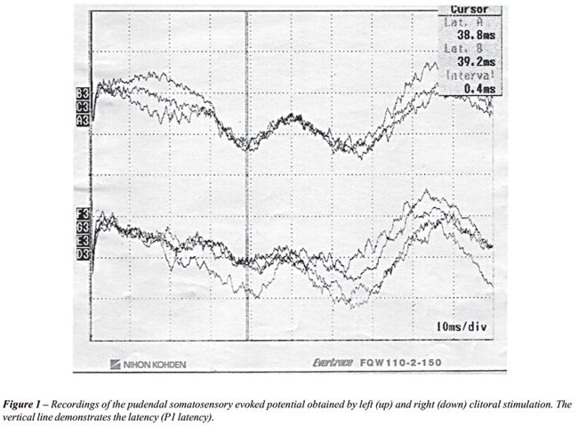
Stimulus
intensity between 2 and 3 times the perception threshold was well tolerated
by the subjects. The cortical responses appeared as identifiable W-shaped
waveforms (Figure-1). The mean clitoral sensory threshold obtained was
3.64 ± 1.01 mA (n = 34) on the left side and 3.84 ± 1.53
mA (n = 33) on the right. There were no differences for the sensory thresholds
obtained on both sides (p = 0.43). The mean P1 latency obtained after
left and right clitoral stimulation were 37.68 ± 2.60 ms (n = 36)
and 37.42 ± 3.12 ms (n = 35) respectively. There were no significant
differences in latency between the sides (p = 0.86). There were no correlations
between sensory thresholds and SSEP latencies according to age, BMI and
height of volunteers (Table-2). The sensory threshold and P1 latency in
relation to obstetric history are demonstrated in Table-3. A significant
difference of the P1 latency between nulliparous women and volunteers
submitted to cesarean section was detected. There was no difference in
the sensory threshold among the groups. There was also no difference in
SSEP latency between nulliparous and volunteers who had vaginal deliveries
or between vaginal delivery group and cesarean section group (Table-3).
COMMENTS
Evoked
potentials are used clinically to provide assessment of functional abnormality
in nerve conductions and to monitor its progression mainly in patients
whose neurological disorders are diagnosed or suspected by suggestive
clinical history or physical examination. The pudendal SSEP have been
used in pelvic dysfunctions and showed responses with prolonged latencies
or not recordable in subjects with multiple sclerosis and bladder or sexual
dysfunctions (5,7,9,10). However, this method has been rarely studied
on healthy women. Haldeman et al. (11) and Guerit et al. (12) published
their observations made on only 5 volunteers and other studies were done
in a maximum of 14 women (1,13,14). Only recent studies have described
reference latencies in a large sample, which included 77 healthy women
(15). Normative values are necessary to discuss technical aspects of the
methodology before its use in research or clinical practice, in a similar
way described before for pudendo-anal reflex latency (16). In the present
study, this method was applied to a significant number of volunteers,
furthermore considering the influences of age, height, BMI and obstetric
history.
Haldeman et al. (10) demonstrated that the
amplitude of the pudendal SSEP was maximal over the sensory cortex in
the midline (Cz-2cm) for both men and women; they also showed
that the latencies and waveform were similar to those obtained following
tibial nerve stimulation at the ankle.
Vodusek et al. (13) emphasized that the
awkwardness of stimulation may be a major obstacle in applying this diagnostic
procedure to females. The clitoral stimulation accomplished with a conventional
bipolar stimulator was well tolerated by the volunteers, being easily
performed. The placement of the anode on the labia majora / minora instead
of the pubis, as described by other authors (14) has the advantage of
not obtaining the SSEP by addition of stimulation of other peripheral
nerves of the region such as the ilioinguinal.
The stimulus frequency of 4.7 Hz was used
in order to reduce the noise caused by 60 Hz frequency, which was used
in previous study (11). The W waveforms as well as the central recording
site are similar as previously described (1,9,11,13). Despite the short
distance between the stimulus sites, only some volunteers presented mild
discrepancy between both sides in P1 latency, not reaching average difference.
This could represent only physiological differences. The laterality of
pudendal nerve stimulation cannot be ascertained according to the closeness
of the cathode sites of stimulation and to the existence of only one dorsal
nerve of the clitoris. This could reflect identical P1 latencies for the
both sides of stimulation in some cases. Besides unilateral stimulation,
medial or bilateral clitoral stimulations have been performed for eliciting
pudendal SSEP in women (15).
The recordings obtained between P3 and P4
were not always as clear as those observed from the midline of the scalp.
However, in cases when the response in Cz’-Fz demonstrated low signal
to noise ratio, the responses obtained in the parietal area were used
to define the P1 latency.
There was no difference in the sensory threshold
and P1 latency obtained from each side and there was no correlation between
these parameters and age, BMI or height of the volunteers. The influence
of height on the SSEP is described especially when studying peripheral
nerves of the lower limbs (e.g. the posterior tibial nerve). Some authors
observed a positive correlation of pudendal SSEP latency with height in
men (5). Although we have not found this correlation, it is reasonable
to suppose its existence, nevertheless minimized if compared to the posterior
tibial nerve. Since the height’s difference of our volunteers was
relatively small, this effect may not have been significant to be detected.
According to this assumption, we can explain the fact that the mean latency
obtained in this study is lower than in other studies accomplished in
European and American women (11,13,14). Comparative studies with different
pelvic floor pathologies in shorter women must be interpreted carefully.
Similar differences in varied ethnic groups are also demonstrated in P40
component obtained after stimulation of the posterior tibial nerve (17).
A longer P1 latency was observed in the
cesarean section group when compared to the nulliparous women group. This
result contradicts the expectation that neurological lesions of the pelvic
floor occur after vaginal delivery and cesarean section has a protective
factor to the pelvic floor structure (18). The reasons for this discovery
are speculative at this moment. Evidence of lesion in women’s pudendal
motor innervation submitted to salvage cesarean section has already been
described (19). An explanation for this would be the time spent waiting
for vaginal delivery to occur before opting for cesarean section. Groutz
et al. found that elective cesarean section was associated with a significantly
lower prevalence of postpartum urinary dysfunction than those who had
spontaneous vaginal delivery or cesarean section performed for obstructed
labor (20). However, other studies are necessary to clarify if this result
is a clinically relevant finding. The study of SSEP in women should be
carefully analyzed in those with a history of cesarean section. There
was no difference between women who have been submitted to vaginal delivery
and nulliparous. However, there was a tendency for longer latencies in
vaginal delivery group that could reach significance if larger sample
had been studied.
A critical reading of the literature reports
suggests that the sensitivity of the test is low in assessment of axonal
lesions. Some authors admit that the presence of an abnormal pudendal
SSEP in an individual patient is, as a rule, accompanied by other neurological
deficits and that the necessity to measure the latency may be questioned
(21). However, according to other reports, the ability to demonstrate
and document a dysfunction of the nervous system could be fundamental
in validating clinical symptoms and signs (2,6).
In conclusion, the SSEP represents a reproducible
and accessible method of evaluating the afferent pathways of the pudendal
nerve in women. The SSEP latencies obtained in these healthy women are
within the ranges currently reported in literature. We found that there
is a statistically significant difference in the latencies when comparing
nulliparous women to those with Cesarean section, but its clinical significance
is unknown. These results could be used as normative values in studies
involving genitourinary neuropathies in order to better understand voiding
and sexual dysfunctions in females.
ACKNOWLEDGEMENT
Support by grant # 99/11546-5 from the Sao Paulo Foundation for Research Support (FAPESP).
CONFLICT OF INTEREST
None declared.
REFERENCES
- Opsomer RJ, Guerit JM, Wese FX, Van Cangh PJ: Pudendal cortical somatosensory evoked potentials. J Urol. 1986; 135: 1216-8.
- Vodusek DB: Evoked potential testing. Urol Clin North Am. 1996; 23: 427-46.
- Chiappa KH. Principles of evoked potentials. In: Keith H. Chiappa (ed.), Evoked potentials in clinical medicine (3rd ed). Philadelphia, Lippincott-Raven Publishers. 1997; pp. 1-30.
- Eisen A: The use of somatosensory evoked potentials for the evaluation of the peripheral nervous system. Neurol Clin. 1988; 6: 825-38.
- Tackmann W, Porst H, van Ahlen H: Bulbocavernosus reflex latencies and somatosensory evoked potentials after pudendal nerve stimulation in the diagnosis of impotence. J Neurol. 1988; 235: 219-25.
- Klausner AP, Batra AK: Pudendal nerve somatosensory evoked potentials in patients with voiding and/or erectile dysfunction: correlating test results with clinical findings. J Urol. 1996; 156: 1425-7.
- Kirkeby HJ, Poulsen EU, Petersen T, Dorup J: Erectile dysfunction in multiple sclerosis. Neurology. 1988; 38: 1366-71.
- Jasper HH: Report of the committee on methods of clinical examination in electroencephalography. Electroenceph Clin Neurophysiol. 1958; 10: 370-5.
- Haldeman S, Bradley WE, Bhatia N: Evoked responses from the pudendal nerve. J Urol. 1982; 128: 974-80.
- Ertekin C, Akyurekli O, Gurses AN, Turgut H. The value of somatosensory-evoked potentials and bulbocavernosus reflex in patients with impotence. Acta Neurol Scand. 1985; 71: 48-53.
- Haldeman S, Bradley WE, Bhatia NN, Johnson BK. Cortical evoked potentials on stimulation of pudendal nerve in women. Urology. 1983; 21: 590-3.
- Guerit JM, Opsomer RJ: Bit-mapped imaging of somatosensory evoked potentials after stimulation of the posterior tibial nerves and dorsal nerve of the penis/clitoris. Electroencephalogr Clin Neurophysiol. 1991; 80: 228-37.
- Vodusek DB: Pudendal SEP and bulbocavernosus reflex in women. Electroencephalogr Clin Neurophysiol. 1990; 77: 134-6.
- Loening-Baucke V, Read NW, Yamada T, Barker AT: Evaluation of the motor and sensory components of the pudendal nerve. Electroencephalogr Clin Neurophysiol. 1994; 93: 35-41.
- Yang CC, Kromm BG: New technique in female pudendal somatosensory evoked potential testing. Somatosens Mot Res. 2004; 21: 9-14.
- Cavalcanti Gde A, Manzano GM, Bruschini H, Giuliano LM, Srougi M, Nobrega JA: Pudendo-anal reflex in normal women. Arq Neuropsiquiatr. 2004; 62: 839-43.
- Chabot RJ, John ER: Normative Evoked Potential Data. In: Lopes da Silva FH, Storm van Leeuwen W, Rémond A (eds.), Handbook of Electroencephalography and Clinical Neurophysiology - revised series - vol. 2. Amsterdam, Elsevier Science. 1986; pp. 263-309.
- Sultan AH, Kamm MA, Hudson CN: Pudendal nerve damage during labour: prospective study before and after childbirth. Br J Obstet Gynaecol. 1994; 101:22-8.
- Snooks SJ, Swash M, Mathers SE, Henry MM: Effect of vaginal delivery on the pelvic floor: a 5-year follow-up. Br J Surg. 1990; 77: 1358-60.
- Groutz A, Rimon E, Peled S, Gold R, Pauzner D, Lessing JB, Gordon D: Cesarean section: does it really prevent the development of postpartum stress urinary incontinence? A prospective study of 363 women one year after their first delivery. Neurourol Urodyn. 2004; 23: 2-6.
- Delodovici ML, Fowler CJ: Clinical value of the pudendal somatosensory evoked potential. Electroencephalogr Clin Neurophysiol. 1995; 96: 509-15.
____________________
Accepted
after revision:
July 6, 2007
_______________________
Correspondence address:
Dr. Homero Bruschini
Rua Barata Ribeiro, 414 / 35
São Paulo, SP, 01308-000, Brazil
Fax: + 55 11 3218-8283
E-mail: bruschini@uol.com.br