IMAGING
OF THE ACUTE SCROTUM: IS THERE A PLACE FOR CONTRAST-ENHANCED ULTRASONOGRAPHY?
(
Download pdf )
doi: 10.1590/S1677-55382009000600008
H. MOSCHOURIS, K. STAMATIOU, E. LAMPROPOULOU, D. KALIKIS, D. MATSAIDONIS
Departments of Radiology (HM, EL, DK DM) and Urology (KE), General Hospital of Pireas Tzaneio, Pireas, Greece
ABSTRACT
Purpose:
To present and evaluate the findings of contrast-enhanced ultrasonography
(CEUS) in typical cases of acute painful scrotum.
Materials and Methods: Nineteen patients
aged from 19 to 61 years old were included in the study. All patients
underwent grey-scale and color Doppler ultrasonography (US) of the scrotum,
followed by imaging after i.v. administration of 2.4 mL of a second generation
ultrasound contrast agent (microbubbles of sulphur hexafluoride). ? dedicated,
contrast-sensitive technique was used (Contrast Tissue Imaging - CnTI).
The diagnosis was confirmed surgically in 6 cases while in the remaining
8 cases it was based on the combination of clinical, imaging and laboratory
findings.
Results: The final diagnosis was testicular
torsion (n = 4), epididymitis (n = 2, one of the cases complicated by
abscess), testicular abscess (n = 1), scrotal abscess (n = 1), testicular
trauma of varying severity (n = 6). Five out of 19 cases were true negatives:
neither clinical examination nor laboratory tests revealed any pathology.
CEUS showed complete lack of enhancement in all cases of torsion, permitting
a rapid and definitive diagnosis. In the cases of infection complicated
by abscesses, CEUS delineated the lesions much better than the combination
of B-mode/Color Doppler US. The severely traumatized testicles showed
minimal, inhomogeneous or patchy enhancement, while cases of minor trauma
showed no significant enhancement defects. Hematomas were presented as
non-enhancing lesions.
Conclusion: Generally, there was no advantage
over Doppler US as has been previously shown. However, CEUS can be used
supplementary to traditional Doppler US in the investigation of blunt
testicular trauma especially when there is uncertainty in diagnosis after
appropriate clinical and radiographic evaluations occurs. Further studies
are required to clearly define the indications of this method.
Key
words: testis; epididymis; torsion; trauma; color Doppler; ultrasound
Int Braz J Urol. 2009; 35: 692-705
INTRODUCTION
Due
to the great efficacy of the existing facilities of modern ultrasonography
(US) such as high frequency transducers, color and power Doppler, ultrasonography
is considered the imaging modality of choice for evaluating acute and
non acute scrotal disease (1,2). Many of these disease processes, including
testicular torsion, epididymo-orchitis, and intratesticular tumor, produce
the common symptom of pain at presentation, and differentiation of these
conditions and disorders is important in determining the appropriate treatment.
US with a high-frequency transducer helps to better characterize intra-scrotal
lesions, and in many instances the findings suggest diagnoses that are
more specific. High-frequency US in its present state can be useful to
identify certain benign intratesticular lesions, resulting in testes-sparing
surgery (3). Nevertheless, a percentage of inconclusive sonograms -often
causing medico-legal problems- are still reported in the literature (4).
Imaging modalities such as scintigraphy
and magnetic resonance imaging (MRI) of the scrotum, which can be applied
when ultrasonography proves inadequate, could provide a more accurate
diagnosis in the evaluation of testicular perfusion than color Doppler
US (5,6). In particular MRI offers useful, and in some cases decisive,
information, as it is capable of revealing unexpected findings and elucidating
complex aspects. Additionally, MRI helps improve patient management, with
an overall reduction in costs (5). Unfortunately, being specialized, relatively
expensive and not always available it is not routinely used for the evaluation
of acute painful scrotum but remains an ideal choice for second-line investigation.
From the view point that US is more convenient
and easier to be performed in the emergency clinical settings than MRI,
a contrast-enhanced ultrasound study would be the ideal tool in the assessment
of testicular perfusion in patients with acute scrotal symptoms.
Under those circumstances, the investigation
of the role of contrast-enhanced ultrasonography (CEUS) as a practical
complementary tool in the study of the acute painful scrotum is worth
exploring. To our knowledge, to date no similar CEUS study focusing exclusively
on representative urgent, painful situations of acute painful scrotum
has been reported in the literature.. Only two prior studies using this
method have been previously performed in very small numbers of patients,
and no advantage over Doppler US has been shown (7,8). In contrast, a
recent experimental study by Liang et al. demonstrated that CEUS was superior
to color Doppler ultrasound in evaluating the perfusion of testicular
ischemia (9).
The present study was carried out to illustrate
the findings of CEUS in a variety of cases of acute scrotal pathology
in order to evaluate its possible role in the investigation of painful
scrotum. Our objective was also to analyze the advantages and limitations
of this method and suggest potential indications.
MATERIALS AND METHODS
This
controlled prospective study was carried-out at the General Hospital of
Pireas, “Tzaneio” (Pireas, Greece) from April 2008 to November
2008. The study group included 20 consecutive cases with acute painful
scrotum who presented at the outpatient department of our hospital. The
exclusion criteria were a previous surgery of the testis, which may confuse
the radiological image, and history of allergic reaction to any contrast
agent. Only one out of the 20 patients had a previous surgery of the testis.
The remaining nineteen patients who fulfilled the inclusion criteria were
initially investigated with conventional scrotum ultrasonography and color
and power Doppler.. CEUS was performed immediately afterwards. Both the
basic examination and CEUS were performed with an Esaote Big GPX (Esaote,
Genoa, Italy) ultrasound device in the radiology department by two experienced
ultrasonographers.
A 2.4 mL of a second generation ultrasound
contrast agent (microbubbles of sulphur hexafluoride, SonoVue, Bracco,
Milan Italy) were injected as a bolus in the forearm vein followed by
5 mL of normal saline. Both the affected and the normal side of the scrotum
were imaged with a contrast-specific technique (Contrast Tuned Imaging-
CnTI) combined with low mechanical index (MI = 0.07-0.08) in order to
obtain a satisfactory shading of the testicle for at least 2.5-3 minutes.
A high acoustic power “flash” causing instant disruption of
the microtubules was applied when deemed necessary by the sonographer.
The examination was performed with a linear 5-7.5 MHz transducer. Patients
with profound scrotal swelling were also studied with a convex 2.5-5 MHz
transducer. Those with pathological ultrasound and/or clinical findings
were further evaluated and treated. For study purposes, a uniform registration
form was used for recording findings of Doppler US and CEUS from each
patient. Representative images from the baseline examination and the entire
CEUS examination were stored as digital archives in the computer database.
In all cases, the reported findings of CEUS were correlated with those
of the grey-scale and colored Doppler. Comparison with the normal side
made the identification of pathologic enhancement easier. Final diagnosis
was based upon clinical and laboratory examination and/or surgical exploration.
Upon the final evaluation of the CEUS technique, images were examined
by separate examiners blinded to the other’s results. Finally, the
recorded images were reviewed to reach a consensus.
The study was approved by the locally appointed
Ethics Committee, and all informed patients gave their written signed
consent. The research protocol was performed in accordance with the International
Conference on Harmonisation Guideline for Good Clinical Practice (1996),
which represents the international ethical and scientific quality standard
for designing, conducting, recording, and reporting trials that involve
participation of human subjects.
RESULTS
Five
out of 19 cases were true negatives: neither clinical examination nor
laboratory testing revealed any pathology. Similarly, both the basic examination
and CEUS were normal Therefore, they were excluded from the study. Contrast-enhanced
examination was successfully completed and its quality was considered
adequate for inclusion in the study in the remaining 14 patients with
abnormal ultrasound findings. No patient suffered any adverse reactions
despite the fact almost all patients were not on an empty stomach. In
all the 14 cases both the baseline ultrasound examination and CEUS diagnosis
was in accordance with that of clinical/laboratory examination and surgery/pathology
findings.
The definite diagnosis was confirmed upon
immediate surgical exploration in 6 cases: testicular rupture (n = 2),
testicular torsion (n = 4). In the remaining 8 cases, diagnosis was based
on the combination of clinical, imaging and laboratory findings: epididymitis
(n = 2), testicular trauma (n = 4) testicular abscess (n = 1), scrotal
abscess (n = 1).
More precisely, among 4 patients diagnosed
with testicular torsion one (19 yo) was finally found with incomplete
torsion, two with complete torsion (24 and 27 yo) and the last with abscess
due to testicular necrosis (22 yo). In all cases, the involved testicle
was edematous, enlarged and painful on palpation. The cremasteric reflex
was absent and no relief of pain upon elevation of scrotum was observed.
Both cases with complete torsion were characterized by scrotal erythema
and unilateral position of the testicle, while abscess due to testicular
necrosis was accompanied by edema involving the entire scrotum and fever.
One additional testicular abscess case,
as a result of epididymitis no response to the medical treatment, was
also diagnosed in a 65 year-old diabetic male. Edema involving the entire
scrotum and fever were present.
Five out the six cases of blunt testicular
trauma were unilateral and isolated while the remaining one was bilateral.
All were associated with motor vehicle accident. Two (28 and 44 yo) out
of the six patients exhibited symptoms of extreme scrotal pain associated
with nausea and vomiting and were finally discovered with major testicular
trauma upon surgical exploration. The remaining patients who experienced
mild scrotal pain were diagnosed with minimal testicular trauma. Physical
examination revealed a swollen, severely tender testicle with a visible
hematoma in all cases. Scrotal ecchymosis was present in two cases.
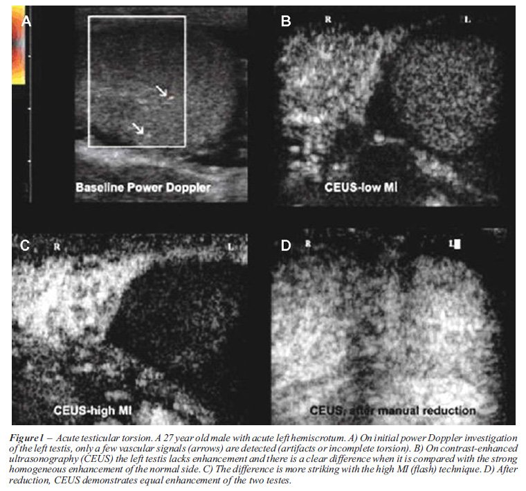
In all testicular torsion cases, both the
baseline US examination and CEUS equally diagnosed the underlying pathology.
While Power-Doppler showed lack of vascularization of the affected testes,
contrast-enhanced US confirmed the absence of macro- and microvascularization
(Figure-1), but failed to add any relevant information in the study of
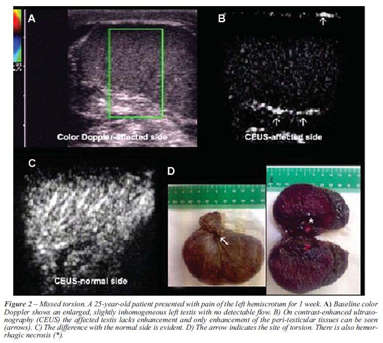
complete testicular torsion. In chronic (missed) torsion peri-testicular
tissues displayed increased vascularity on CEUS (Figure-2), while, in
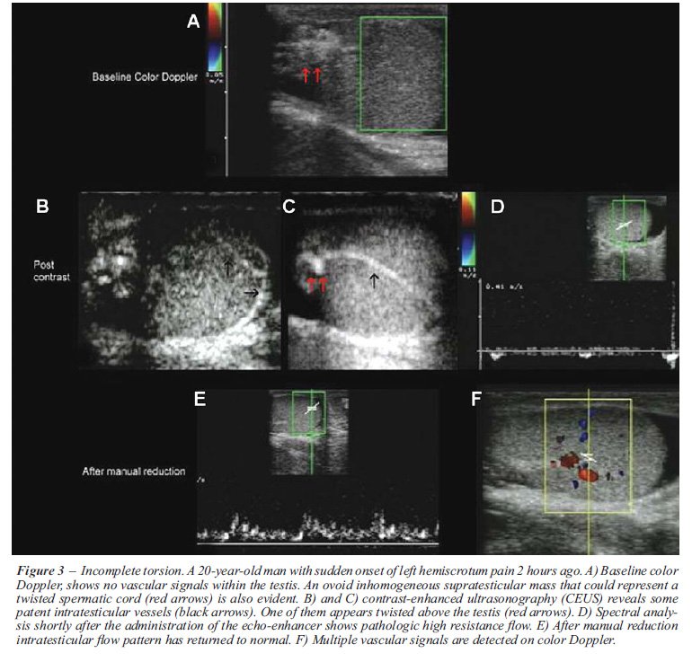
the study of incomplete torsion, CEUS showed a clear difference in the
degree of enhancement between the normal and affected side (Figure-3).
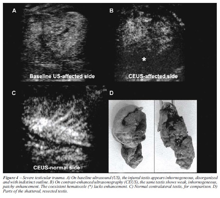
CEUS was proved more accurate in the investigation
of major testicular trauma, providing essential information on the viability
of the affected organ. On conventional US, intra-parenchymal lacerations
were visualized as linear or deliquescent non echogenic lesions. Only
one scrotal sonogram showed a fractured testis with a disrupted tunica
albuginea and testicular contents surrounded by tunica vaginalis, while
in the remaining surgically treated testes (n = 2) the conventional US
failed to clearly determine the presence and extend of tunica albuginea’s
eruption. When examined with contrast-enhanced technique surgically treated
ruptured testes showed lack of enhancement in almost all their extent
(Figure-4). On the contrary, conservatively treated testis showed a decreased,
non-homogeneous, partial, contrast enhancement. Minimal testicular traumatic
lesions were depicted as small hypoechoic vaguely dispersed areas, within
a more echogenic normal testicular parenchyma which was not accompanied
by a serious disorder of the testicular vascularity (Figure-5). Minimal
traumatic injuries of the testis were almost uniformly presented as intratesticular
hematoma in conventional US.

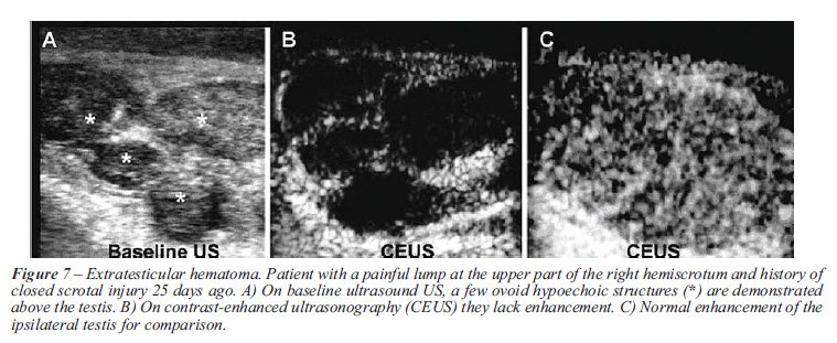
The conventional ultrasound diagnosis was
in accordance with that of CEUS in the remaining positive cases; however,
CEUS offered higher definition in the visualization of testis and scrotal
abscess (Figures-6 and 7). In all testicular abscess cases a peripheral,
target like enhancement and evident central liquefaction was shown. In
one out of the three cases with testicular abscess (a 69 yo diabetic patient),
contrast enhanced US (52 sec from injection) also identified a multiple,
echoic internal septae, with evident septal enhancement, which was missed
in the baseline examination. Finally, CEUS ruled out the diagnosis of
a tumor like small subcapsular hematoma, which mimicked a solid nodular
lesion on baseline, where US demonstrated the absence of enhancement.

COMMENTS
In
the late 90’s an experimental study by Coley et al., demonstrated
contrast-enhanced US, even with the use of first-generation sonographic
contrast media, to be more accurate than color and power Doppler in the
visualization of testis vascularity (8). Nevertheless, most researchers
have mainly focused on the application of contrast-enhanced US to the
study of focal renal or hepatic parenchymal lesions and no imaging studies
of the scrotum using sonographic contrast media have been published over
the last ten years. Recently, Catalano et al., using similar sonographic
contrast media and technique with that of our study, evaluated the use
of contrast-enhanced US in emergency radiology (7). Despite the large
number of patients, only 8 out of the 126 cases investigated by the authors
with the real-time contrast-specific US, concerned acute painful scrotum
situations [testicular torsion (n = 6), epididymitis (n = 1), testicular
trauma (n = 4)]. Although, no advantage over Doppler US has been shown
it seems that real-time contrast-specific US is an effective technique
in emergency imaging. Its role should not be considered as a replacement
of Doppler US but as a useful integration of conventional US. In fact,
compared to baseline sonography there is a loss of image quality with
images appearing grainy, but this is widely compensated by the possibility
of evaluating the area of interest in real time, which is impossible with
intermittent-mode high mechanical-index techniques (10,11).
Our experience showed that CEUS fails to
further increase the diagnostic accuracy of conventional grey scale and
Doppler ultrasound in the investigation of testicular torsion; both Doppler
US and CEUS findings were equally in accordance with that of surgical
exploration in patients with suspicious testicular torsion. It should
be mentioned, however, that CEUS is not susceptible to artifacts that
hinder a Color Doppler (and especially a Power Doppler) examination (11).
For example, in incomplete, less than 360-degrees torsion cases the affected
side often maintains some vascularity and comparison of color Doppler
signals with the normal side can be difficult. In this case, CEUS can
facilitate diagnosis by showing a clear difference in the degree of enhancement
between the normal and affected side. Similarly, in chronic (missed) torsion,
no testis enhancement on CEUS facilitates to interpret weak color Doppler
signals when increased vascularity of the peri-testicular tissues occurs
(4,12,13). In addition, since CEUS requires no parameter optimization
for the detection of slow flow, or flow in small vessels, it may prove
easier and faster than color Doppler in the diagnosis of acute testicular
torsion (14).
Detection of flow in testes of small children
is another challenge for ultrasonography (13). Currently, the key clinical
dilemma still remains to avoid surgery, especially in children, when clinical
findings are suspicious despite negative color Doppler ultrasound (CDU)
results (14,15). On the other hand, color Doppler may fail to demonstrate
normal flow in testicles with a volume of less than l cc, leading thus
to a false positive diagnosis of testicular torsion (13,16). Therefore,
CEUS as it is not susceptible to artifacts can, at least theoretically,
facilitate the diagnosis of torsion in pediatric patients; however, the
safety of SonoVue® in children has not been currently established.
Grayscale US findings of epididymo-orchits
are enlarged hypoechoic epididymis and testis. These findings are non-specific
and often are indistinguishable from testicular torsion (17). In contrast,
combination of grey-scale US and color Doppler is helpful in evaluating
patients with infection thus preventing unnecessary surgical exploration.
Since US with color Doppler is effective in demonstrating the morphologic
and hemodynamic changes in epididymitis and orchitis, CEUS does not seem
to add to the diagnostic accuracy of the emergency ultrasound study. However,
CEUS could play a role in the investigation of the complications of both
epididymitis and orchitis. In fact, advanced epididymo-orchitis may cause
testicular infarction as a result of extrinsic compression of testicular
vascular supply by enlarged epididymis and spermatic cord and pyocele
therefore; intratesticular vascularity is decreased (2). In addition,
severe epididymo-orchitis may also cause testicular abscess and scrotal
abscess, which is demonstrated as a complex echoic mass. These findings
suggest the need for surgical exploration. In contrast, CEUS can confirm
or exclude the presence of abscess in cases of serious, persistent inflammation
(epididymitis or epididymo-orchitis) (12,17). In fact, CEUS findings are
diagnostic: epididymal, testicular and scrotal abscesses show a typical
strong rim enhancement and non-enhancing contents while enhancing septa
may be demonstrated within the lesions (18). Additional information offered
by CEUS may alter the management or outcome of patients with scrotal abscess
permitting urologists to avoid surgery, especially in ageing patients
with other significant life-limiting medical conditions. In our study,
a left orchiectomy was performed in the patient with scrotal abscess and
testicular torsion, while scrotal abscess in patient with epididymitis
was managed by incision and drainage.
According to the perspective of the authors
CEUS could be a useful complement to clinical and baseline sonographic
examination, when it is not clear whether a traumatized scrotum requires
surgical exploration (when, for example, a hematocele without an obvious
testicular rupture is demonstrated). When compared with conventional ultrasound
CEUS was proved more accurate in the investigation of major testicular
trauma, providing essential information on the viability of the affected
organ. In fact, the intensity and homogeneity of testicular perfusion
on CEUS correlates with the severity of the trauma. Minor trauma (in the
form of small contusions) usually causes no significant enhancement deficits.
On the other hand, severely fractured testicles show weak, inhomogeneous,
or patchy enhancement. Complete lack of enhancement may be seen in trauma-associated
torsion or in cases of completely disorganized testicles. Similarly, hematomas
(intra- or extra-testicular) and hematoceles show no enhancement. Therefore,
CEUS improves their delineation and the differential diagnosis from other
focal lesions. To our knowledge if rupture of the testis is detected sonographically,
immediate surgical exploration is indicated. In contrast, sonographically
detected hematoceles without visible signs of rupture should be treated
conservatively. Since inappropriately protracted expectant management
promotes testicular infection, atrophy, and necrosis, adequate preoperative
diagnosis is essential for the viability of wounded testis (19). In our
study, two out of 6 cases with testicular trauma were finally found with
minimal lesion in CEUS and thus avoided unnecessary surgery.
Based on the above described findings, the
possible indications for the CEUS investigation of acute scrotum can be
summarized as follows: suspected testicular torsion, when color Doppler
findings are equivocal (due to artifacts, or incomplete torsion), inflammation
of the contents of the scrotum, when a complication is suspected, and
scrotal trauma, in order to confirm a testicular damage that requires
surgical intervention.
Two important limitations of CEUS should
be underlined: as the low spatial resolution of the method, compared to
baseline US and the additional cost of the contrast-agent (5). Nevertheless,
it should be mentioned that CEUS is no more than an effective complement
to grey-scale and color Doppler ultrasonography, therefore it is mandatory
to perform a detailed initial examination of the scrotum with conventional
US, in order not to miss subtle findings that may not be detected on the
following CEUS examination. As regards the cost of the contrast-agent
it can be reduced by injecting a part of the standard dose. In our study,
only 2.4 mL (i.e. half of the standard dose) of the echo-enhancer were
used, and provided satisfactory tissue enhancement for at least 3 minutes.
It is probable that even smaller doses can guarantee an accurate diagnostic
examination. According to Catalano et al., an important concern regarding
the use of CEUS is the additional time needed for the contrast-agent preparation,
administration and performance of contrast-enhanced US scan (5). Our limited
experience however has shown that CEUS of the scrotum can easily be accomplished
in the emergency department, immediately after the baseline sonographic
examination, and with a minimum delay (approx. 5 min.) in the diagnostic
work-up of the patient. Such a delay is insignificant in the investigation
of acute scrotum, as it does not seem to alter the patient outcome.
CONCLUSION
Although current experience on the use of sonographic contrast media in the imaging study of acute painful scrotum is limited, it seems that CEUS can be used supplementary to traditional Doppler US in the investigation of blunt testicular trauma especially when uncertainty in diagnosis after appropriate clinical and radiographic evaluations occurs. In several selected acute scrotum cases, real-time contrast-specific ultrasound may also increase the efficacy of ultrasonographic diagnosis. The use of second-generation sonographic contrast media along with the newly-introduced low mechanical index software allows dynamic exploration of organ perfusion, with identification of both the macrocirculation and the lesion microcirculation. Therefore, CEUS can constitute a valuable integration of conventional grey-scale and color Doppler ultrasonography, particularly when these yield unclear, poorly visible findings. Further studies are required to clearly define the indications of this method.
CONFLICT OF INTEREST
None declared.
REFERENCES
- Deurdulian C, Mittelstaedt CA, Chong WK, Fielding JR: US of acute scrotal trauma: optimal technique, imaging findings, and management. Radiographics. 2007; 27: 357-69.
- Pavlica P, Barozzi L: Imaging of the acute scrotum. Eur Radiol. 2001; 11: 220-8.
- Dogra VS, Gottlieb RH, Oka M, Rubens DJ: Sonography of the scrotum. Radiology. 2003; 227: 18-36.
- Steinhardt GF, Boyarsky S, Mackey R: Testicular torsion: pitfalls of color Doppler sonography. J Urol. 1993; 150: 461-2.
- Watanabe Y, Dohke M, Ohkubo K, Ishimori T, Amoh Y, Okumura A, et al.: Scrotal disorders: evaluation of testicular enhancement patterns at dynamic contrast-enhanced subtraction MR imaging. Radiology. 2000; 217: 219-27.
- Paltiel HJ, Connolly LP, Atala A, Paltiel AD, Zurakowski D, Treves ST: Acute scrotal symptoms in boys with an indeterminate clinical presentation: comparison of color Doppler sonography and scintigraphy. Radiology. 1998; 207: 223-31.
- Catalano O, Lobianco R, Sandomenico F, Mattace Raso M, Siani A: Real-time, contrast-enhanced sonographic imaging in emergency radiology. Radiol Med. 2004; 108: 454-69.
- Coley BD, Frush DP, Babcock DS, O’Hara SM, Lewis AG, Gelfand MJ, et al.: Acute testicular torsion: comparison of unenhanced and contrast-enhanced power Doppler US, color Doppler US, and radionuclide imaging. Radiology. 1996; 199: 441-6.
- Liang RX, Xue ES, Lin LW, Yu L, Chen S, Yu LY, et al.: Correlation between sonographic appearance of experimental testicular ischemia and histological changes of the testis after reperfusion. Zhonghua Nan Ke Xue. 2009; 15: 115-21.
- Lencioni R, Cioni D, Bartolozzi C: Tissue harmonic and contrast-specific imaging: back to gray scale in ultrasound. Eur Radiol. 2002; 12: 151-65.
- Bauer A, Solbiati L, Weissman N: Ultrasound imaging with SonoVue: low mechanical index real-time imaging. Acad Radiol. 2002; 9 (Suppl 2): S282-4.
- Berman JM, Beidle TR, Kunberger LE, Letourneau JG: Sonographic evaluation of acute intrascrotal pathology. AJR Am J Roentgenol. 1996; 166: 857-61.
- Aso C, Enríquez G, Fité M, Torán N, Piró C, Piqueras J, et al.: Gray-scale and color Doppler sonography of scrotal disorders in children: an update. Radiographics. 2005; 25: 1197-214.
- Pepe P, Panella P, Pennisi M, Aragona F: Does color Doppler sonography improve the clinical assessment of patients with acute scrotum? Eur J Radiol. 2006; 60: 120-4.
- Luker GD, Siegel MJ: Color Doppler sonography of the scrotum in children. AJR Am J Roentgenol. 1994; 163: 649-55.
- Bader TR, Kammerhuber F, Herneth AM: Testicular blood flow in boys as assessed at color Doppler and power Doppler sonography. Radiology. 1997; 202: 559-64. Erratum in: Radiology 1997; 203: 580.
- Feld R, Middleton WD: Recent advances in sonography of the testis and scrotum. Radiol Clin North Am. 1992; 30: 1033-51.
- Muttarak M, Lojanapiwat B: The painful scrotum: an ultrasonographical approach to diagnosis. Singapore Med J. 2005; 46: 352-7.
- Buckley JC, McAninch JW: Use of ultrasonography for the diagnosis of testicular injuries in blunt scrotal trauma. J Urol. 2006; 175: 175-8.
____________________
Accepted after revision:
July 27, 2009
_______________________
Correspondence address:
Dr. Stamatiou Konstantinos
Department of Urology
General Hospital of Pireas “Tzaneio”
2 Salepoula str.
18536 Piraeus, Greece
Fax: + 302 1042-96987
E-mail: stamatiouk@gmail.com
EDITORIAL COMMENT
The paper by Moschouris et al. describes
their experience using contrast-enhanced ultrasound (CEUS) to augment
Doppler US (US) in the evaluation of 19 cases of acute scrotum. In cases
of suspected torsion, CEUS provided no additional benefit beyond DUS.
The authors state that CEUS provides improved images of vascular flow
and may have a role in cases of incomplete torsion or when the DUS is
equivocal, but they do acknowledge that based on the current evidence,
CEUS does not have a clinical role in the evaluation of suspected torsion.
In cases of testicular or scrotal abscess, the authors again state that
CEUS may provide better delineation of abscess from inflamed parenchyma,
but with only 3 cases of abscess (none of which were diagnosed solely
by CEUS) they do not have evidence to support that claim. Finally, in
cases of testicular trauma that authors state that CEUS may be more specific
in ruling out testicular rupture than conventional US, stating that in
2 of their cases unnecessary surgery was avoided by re-assuring CEUS findings
when US was inconclusive. However, recent evidence suggests that magnetic
resonance imaging (MRI) is very accurate in cases of testicular trauma
when US is inconclusive (1), and is warranted when considering expectant
management of a patient with possible testicular rupture. Furthermore,
while avoiding surgical exploration of the scrotum is a laudable goal,
the more critical issue in testicular trauma is identifying testicular
rupture, and for this purpose conventional US is very sensitive (2). If
future larger studies demonstrate that CEUS is as accurate as MRI in cases
of testicular trauma, then it may have a role as a more cost-effective
alternative, but at this time, CEUS does not have a clinical role in evaluation
of the acute scrotum.
REFERENCES
- Kim SH, Park S, Choi SH, Jeong WK, Choi JH: The efficacy of magnetic resonance imaging for the diagnosis of testicular rupture: a prospective preliminary study. J Trauma. 2009; 66: 239-42.
- Guichard G, El Ammari J, Del Coro C, Cellarier D, Loock PY, Chabannes E, et al.: Accuracy of ultrasonography in diagnosis of testicular rupture after blunt scrotal trauma. Urology. 2008; 71: 52-6.
Dr.
Jeffrey Tiemstra
Associate Professor
Department of Family Medicine
UIC College of Medicine
Chicago, Illinois, USA
E-mail: jtiemstr@uic.edu
EDITORIAL COMMENT
I
would like congratulate Moschouris H et al. for this beautiful study.
This study does have its limitations but also opens up new opportunities
for research in scrotal pathology.
There is a need to improve the sensitivity and specificity of traditional
ultrasound. These experimental imaging modalities include contrast-enhanced
ultrasound, dynamic contrast magnetic resonance (MR), and near-infrared
(NIR) imaging. Particular interest lies in the evaluation of pediatric
testicular torsion.
The sensitivity and specificity of the current
ultrasound machines is excellent in detecting absence of blood flow in
testicular torsion patients and may not require ultrasound contrast enhancement
in its evaluation. Color flow Doppler alone has a sensitivity of 86%,
specificity of 100%, and accuracy of 97% in diagnosing testicular torsion
(1). Blood flow within the testis can be quantified using spectral Doppler
(2). Color Doppler sonography often has difficulty demonstrating perfusion
within the normal pediatric testis. While still in its infancy, contrast-enhanced
ultrasound has the potential to provide improved sensitivity in detecting
flow in the pediatric testis (3). In an experimental study using 35 rabbits,
Paltiel et al. (4) demonstrated that contrast-enhanced pulse-inversion
sonography could reliably detect altered levels of testicular perfusion
when compared to radiolabeled microsphere perfusion measurements.
Vasculitis involving the testes is uncommon,
but may be seen in patients with polyarteritis nodosa (3,4) and systemic
lupus erythematosus (5). Color Doppler evaluation may reveal the absence
of testicular blood flow, mimicking torsion (5). It is in these patients
with vasculitis where blood flow is so small that its beyond the resolution
of the ultrasound machine to detect it that ultrasound contrast enhancement
may play a significant role in their diagnosis (6,7).
Additionally, contrast enhanced sonography
has a definite role in scrotal examinations, especially for characterizing
small tumors less than two centimeters in size. Ultrasound contrast agents
can play a significant role in testicular trauma to identify testicular
contusions or hematomas and exclude testicular masses by demonstrating
the presence of blood flow in them.
Near infrared fluorescence (NIRF) has also
been performed with the intravenous administration of indocyanine green.
In one preliminary study, NIRF was able to reliably detect vascular flow
obstruction within the torsed testis of an adult male Sprague-Dawley rat
model [Personal communication with VS Dogra et al. Abstract presented
at World Congress of Endourology, Cleveland 2006]. This method can improve
the sensitivity in detecting early testicular torsion and may increase
testis salvage rate. As future investigations are completed, NIR spectroscopy
and fluorescence will likely become a fast and cost-effective method in
the initial evaluation of testicular torsion in the emergency setting.
REFERENCES
- Burks DD, Markey BJ, Burkhard TK, Balsara ZN, Haluszka MM, Canning DA: Suspected testicular torsion and ischemia: evaluation with color Doppler sonography. Radiology. 1990; 175: 815-21.
- Scoutt LM, Zawin ML, Taylor KJ: Doppler US. Part II. Clinical applications. Radiology. 1990; 174: 309-19.
- Coley BD, Frush DP, Babcock DS, O’Hara SM, Lewis AG, Gelfand MJ, et al.: Acute testicular torsion: comparison of unenhanced and contrast-enhanced power Doppler US, color Doppler US, and radionuclide imaging. Radiology. 1996; 199: 441-6.
- Paltiel HJ, Kalish LA, Susaeta RA, Frauscher F, O’Kane PL, Freitas-Filho LG: Pulse-inversion US imaging of testicular ischemia: quantitative and qualitative analyses in a rabbit model. Radiology. 2006; 239: 718-29.
- Suty JM, Hubert J, Duquenne M, Weryha G, Mangin P: Bilateral testicular ischemia in vasculitis. Differential diagnosis with torsion and the value of color Doppler ultrasonography. Prog Urol. 1995; 5: 586-9.
- Leibovici D, Strauss S, Sharon A: Acute, painful, swollen testis in polyarteritis nodosa: a diagnostic problem. Harefuah. 1999; 136: 938-9.
- Dogra VS, Bhatt S, Rubens DJ: Sonographic evaluation of testicular torsion. Ultrasound Clin. 2008: 1; 55-66.
Dr.
Vikram S. Dogra
Department of Imaging Sciences
University of Rochester School of Medicine
Rochester, New York, USA
E-mail: vikram_dogra@urmc.rochester.edu
EDITORIAL COMMENT
The aim
of the study is very interesting, but several points must be implemented
and improved.
The number of patients is too low; with the particular reference to the
patients submitted to surgical exploration because of suspicious testicular
torsion (four cases). The key clinical dilemma remains to avoid surgery,
especially in children, when clinical findings are suspicious despite
negative color Doppler ultrasound (CDU) results. Although CEUS evaluation
after medium administration seems to be useful and accurate to detect
intraparenchimal vascularization, a more consistent number of patients
is needed. CEUS evaluation, as testicular scintigraphy, introduces a quantitative
parameter and could improve CDU that in most cases is an operator depending
procedure. In conclusion, an excellent idea supported (until now) by an
anecdotal series that needs further studies.
Dr.
Pietro Pepe
Department of Urology
Cannizzaro Hospital
Catania, Italy
E-mail: piepepe@hotmail.com
EDITORIAL COMMENT
This paper
demonstrates the usefulness of contrast-enhanced ultrasonography in the
diagnosis of acute scrotum by determining testicular perfusion based on
contrast enhancement of testicles through the administration of microbubble
contrast media.
Today, color and power Doppler ultrasonography (US) is the initial imaging
modality used in the evaluation of symptomatic scrotum. It is readily
available and noninvasive, and the testis itself is highly amenable to
sonographic examination. However, scrotal US has some flaws and problems.
Doppler US results are highly dependent on operators' skill, and detectability
of blood flow depends on patient’s age, testicular volume, and sensitivity
of the US equipment used. Therefore, in a variety of clinical settings,
inconclusive results of color and power Doppler US need another imaging
method or modality for accurate and confirmatory assessment of scrotal
disorders.
Use of contrast media in US seems to be a great idea because testicular
perfusion can be much more conspicuous with contrast-enhanced US than
only with Doppler US, as reported in this article. In magnetic resonance
imaging (MRI), Watanabe et al. (reference #5) first showed the usefulness
of contrast-enhanced imaging in the evaluation of acute scrotum. With
use of contrast agents, US is expected to provide as accurate qualitative
information about testicular blood flow as MRI.
The difference between this new contrast-enhanced US and MRI could be
semi-quantitative analysis of testicular perfusion. MRI imaging performs
dynamic contrast-enhanced examination, which allows for semi-quantitative
assessment of blood flow. Though US could not provide semi-quantitative
information, US can be more convenient and easier to be performed in the
emergency clinical settings than MRI.
As the authors described, this new method can be robust and useful to
differentiate testicular torsion from other pathologies such as acute
epididymitis which can be treated with antibiotics conservatively. In
blunt trauma, this method could provide accurate information about the
severity of testicular damage, rupture of tunica albuginea, and the presence
or absence of hematoma. This method would also have a potential to characterize
testicular neoplasm.
In the near future, contrast-enhanced US could hopefully be performed
as the initial and robust imaging modality by the urologists and radiologists
on the front line of medical care.
Dr.
Yuji Watanabe
Radiologist-in-Chief
Department of Radiology
Kurashiki Central Hospital
Miwa, Kurashiki, Japan
E-mail: yw5904@kchnet.or.jp