PREVALENCE
OF HIGH-GRADE PROSTATIC INTRAEPITHELIAL NEOPLASIA AND ITS RELATIONSHIP
TO SERUM PROSTATE SPECIFIC ANTIGEN
(
Download pdf )
HYUNG L. KIM, XIMING J. YANG
Departments
of Surgery, Urology and Pathology, The University of Chicago,
Chicago, Illinois, USA
ABSTRACT
Objective:
We assessed the incidence of high-grade prostatic intraepithelial neoplasia
(HGPIN) in prostates removed for bladder cancer, and evaluated its correlation
with preoperative serum prostate specific antigen (PSA).
Materials and Methods: We reviewed the histology
of the prostate from men who underwent cystoprostatectomy for bladder
cancer without known prostate pathology prior to surgery. Preoperative
serum PSA levels in patients with HGPIN were analyzed.
Results: 61 cystoprostatectomy specimens
from May 1992 to April 1999 were reviewed. High-grade prostatic intraepithelial
neoplasia (PIN) was found in 46/61 (75%) of the prostate specimens, including
21/21 (100%) patients with prostatic cancer, and 25/40 (63%) patients
without prostatic cancer. The mean serum PSA in men with PIN without evidence
of prostatic adenocarcinoma was 1.9 ng/mL (SD=2.026).
Conclusion: The incidence of isolated high-grade
PIN was 63%. The presence of high-grade PIN does not result in a significant
elevation of serum PSA.
Key words:
prostate; pathology; prostate-specific antigen; prostatic intraepithelial
neoplasia; prevalence
Int Braz J Urol. 2002; 28: 413-7
INTRODUCTION
Prostatic
intraepithelial neoplasia (PIN) is defined by neoplastic growth of epithelial
cells within preexisting benign prostatic acini or ducts. High-grade PIN
is considered a premalignant lesion (1,2) and the development of high-grade
PIN may precede the onset of prostatic adenocarcinoma by more than ten
years (3). PIN is frequently seen in association with prostatic adenocarcinoma
(1). Thus, the finding of isolated high-grade PIN on a needle core biopsy
is generally accepted as an indication to repeat biopsy (4-6).
The correlation between prostatic adenocarcinoma
and serum PSA has been extensively studied. However, only a few studies
have focused on the relationship between PIN and PSA. Some studies suggest
that PIN causes serum PSA elevation (7), while other studies dispute this
relationship (8,9). To most directly assess the relationship between PIN
and PSA, PSA levels can be correlated with the finding of isolated PIN,
where the presence of adenocarcinoma has been excluded by an examination
of the entire prostate specimen. Previous studies have been based on either
prostate biopsy tissue, which is subject to sampling error, or radical
prostatectomy specimens, which contain adenocarcinoma. However, the present
study is based on an examination of the complete prostate obtained during
cystoprostatectomy in men with urothelial carcinoma. We assessed the effect
of high-grade PIN on serum PSA by evaluating the preoperative PSA in men
with high-grade PIN without any evidence of prostatic adenocarcinoma (isolated
high-grade PIN).
MATERIALS AND METHODS
Using
a database at the University of Chicago, Department of Pathology, 61 men
who underwent cystoprostatectomy for urothelial carcinoma of the bladder
between May 1992 and April 1999 were identified. None of the patients
had a known history of prostate pathology prior to surgery. At the time
of surgery, all specimens were fixed in formalin within one hour of surgery.
The prostates were serially sectioned at 4mm intervals. The histology
of the prostate was reviewed and specifically assessed for the presence
of PIN and adenocarcinoma of the prostate by a single pathologist (XJY)
with expertise in prostate pathology.
Criteria for defining high-grade PIN included
(1) epithelial cell proliferation within the ducts and acini, forming
pseudostratified layers, (2) enlargement, elongation, irregularity and
hyperchromasia of the nuclei, and (3) multiple prominent nucleoli. High-grade
PIN was classified as “focal” if there were three or fewer separate
foci/acini of high-grade PIN, and as “extensive” if there were
more than three foci/acini of high-grade PIN on different sections. Adenocarcinomas
were graded using the Gleason system. Preoperative serum PSA, obtained
within 1.5 years prior to surgery, was available for 34 patients. Chi-square
analysis and logistic regression analysis were performed using computer
software, Minitab Release 12.
RESULTS
The mean age of the patients was 69 years. High-grade PIN was found in 46/61 (75%) prostates examined. The high-grade PIN was noted in 21/21 (100%) patients who had prostatic adenocarcinoma and in 25/40 (63%) patients who had no evidence of prostatic adenocarcinoma. PIN was considered focal in 26/61 (43%), and extensive in 20/61 (33%) patients. Of the patients with focal PIN, 8/26 (31%) had prostatic adenocarcinoma, and of the patients with extensive PIN, 13/20 (65%) had prostatic adenocarcinoma (p=0.021). The majority (95%) of patients with prostatic adenocarcinomas had tumors categorized as Gleason score six or less. The mean PSA in 21 men with isolated high-grade PIN (without prostatic adenocarcinoma) was 1.9 ng/mL (Range 0.7-8 ng/mL; SD=2.03). The serum PSA levels did not correlate with the categorization of PIN as focal or extensive (p=0.485).
DISCUSSION
In
our study, high-grade PIN was seen in 75% of all prostates examined, and
in 63% of prostates without associated prostatic adenocarcinoma. The true
prevalence of PIN and adenocarcinoma may be even higher, since any review
of pathologic specimens is subject to sampling error. None of the patients
without high-grade PIN had evidence of prostatic adenocarcinoma. To determine
the prevalence of PIN in the general population, other investigators have
examined prostate specimens obtained from needle biopsies, transurethral
resections (TUR), autopsies, or radical prostatectomy specimens removed
for treatment of prostate cancer (Table-1). The incidence of isolated
high-grade PIN on needle biopsy was reported to be from 0.7% to 20% (10).
Needle core biopsies sample less than 1% of the total prostate volume
and, therefore, sampling error is inherent to the procedure. The frequency
of isolated high-grade PIN found in tissues obtained from transurethral
resection of the prostate ranges from 2.8% to 3.2% (11,12). However, most
of the prostate tissue removed during a TUR is from the transition zone,
while PIN is most frequently found in the peripheral zone.
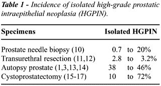
Studies based on examination of the entire
prostate identified a higher frequency of isolated PIN. In a review of
autopsy specimens from men over the age of 50 years, McNeal and Bostwick
found PIN in 82% of men with prostatic adenocarcinoma, and 43% of men
with benign prostates (1). In other studies based on prostates obtained
at autopsy, the incidence of high-grade PIN ranged from 38%-46% (3,13,14).
Other studies using cystoprostatectomy specimens have reported incidences
of isolated high-grade PIN ranging from 10 to 72% (Table-2) (15-17). The
lower incidence of high-grade PIN reported in studies based on cystoprostatectomy
specimens when compared to studies based on autopsy findings, may be related
to differences in patient populations, sampling methods, or diagnostic
criteria applied by the pathologists. In our study, isolated high-grade
PIN was seen in 63% of the cystoprostatectomy specimens, and this is consistent
with a recent report by Troncoso et al. (17). Troncoso et al. reported
that the incidence of high-grade PIN in patients with prostate cancer
was 100% (61/61), and the incidence of isolated high-grade PIN was 72%
(28/39).
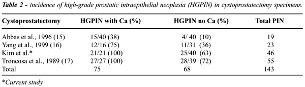
Therefore, isolated high-grade PIN is not
an uncommon finding. With this present study included, there are at least
four studies assessing the incidence of PIN in cystoprostatectomy specimens.
The combined incidence in these studies of high-grade PIN found in association
with prostate cancer is 52% (75/143), while the incidence of isolated
high-grade PIN is 48% (68/143) (Table-2). Although the finding of high-grade
PIN on prostate needle biopsy has been considered an indication to repeat
biopsy, the cystoprostatectomy results underscore the fact that nearly
half of all patients with high-grade PIN may not have associated adenocarcinoma.
PSA is produced by both benign and malignant
epithelial cells of the prostate. It is well documented that the presence
of prostatic adenocarcinoma can cause elevation of serum PSA. The mechanism
by which PSA is released into the serum is not completely understood.
It may be related to the disruption of the basement membrane caused by
tumor invasion or other destructive processes, such as inflammation or
infarct. The basement membrane within PIN is intact. Therefore, it is
reasonable to believe that PSA produced by neoplastic cells in PIN is
not released into the serum at clinically significant levels. Our study
provides direct evidence that PIN does not result in a significant elevation
of serum PSA. Isolated PIN was not associated with an elevated serum PSA
and the serum PSA did not correlate with the extent of PIN found in the
prostate.
Previous studies assessing the relationship
between high-grade PIN and serum PSA have reached conflicting conclusions.
In these studies, the effect of PIN on PSA was analyzed either using prostate
needle biopsy specimens, where the true incidence of PIN is often underestimated,
or from prostatectomy specimens, where the effect of PIN and adenocarcinoma
on serum PSA may be difficult to distinguish. By reviewing needle biopsy
specimens, Brawer & Lange suggested that the presence of PIN might
result in an elevation of serum PSA (7). However, this study could not
exclude the presence of associated prostate cancer, which could have accounted
for the elevation of PSA. Ronnett et al. reviewed 65 radical prostatectomy
specimens with small volume of cancer and found that the volume of PIN
did not correlate with preoperative serum PSA levels (9). Similarly, by
studying 195 radical prostatectomy specimens removed for prostate cancer,
Alexander et al. found no correlation between the presence of PIN in the
specimen and preoperative serum PSA (8).
CONCLUSIONS
The prevalence of isolated high-grade PIN was 63% in patients undergoing cystoprostatectomy for bladder cancer. Extensive high-grade PIN (65%) was more likely to be associated with prostatic adenocarcinoma than focal high-grade PIN (31%). Isolated high-grade PIN is not an uncommon finding. However, as high-grade PIN does not appear to result in a significant elevation of serum PSA, prostatic adenocarcinoma must be ruled out as the source for an elevated PSA in a patient with a high serum PSA and an isolated high-grade PIN on needle biopsy.
REFERENCES
- McNeal JE, Bostwick DG: Intraductal dysplasia: a premalignant lesion of the prostate. Hum Pathol. 1986; 17: 64-71.
- Bostwick DG, Brawer MK: Prostatic intra-epithelial neoplasia and early invasion in prostate cancer. Cancer 1987; 59: 788-94.
- Sakr WA, Haas GP, Cassin BF, Pontes JE, Crissman JD: The frequency of carcinoma and intraepithelial neoplasia of the prostate in young male patients (see comments). J Urol. 1993; 150: 379-85.
- Bostwick DG, Qian J, Frankel K: The incidence of high grade prostatic intraepithelial neoplasia in needle biopsies. J Urol. 1995; 154: 1791-4.
- Wills ML, Hamper UM, Partin AW, Epstein JI: Incidence of high-grade prostatic intraepithelial neoplasia in sextant needle biopsy specimens. Urology 1997; 49: 367-73.
- Langer JE, Rovner ES, Coleman BG, Yin D, Arger PH, Malkowicz SB, et al.: Strategy for repeat biopsy of patients with prostatic intraepithelial neoplasia detected by prostate needle biopsy. J Urol. 1996; 155: 228-31.
- Brawer MK, Lange PH: Prostate-specific antigen and premalignant change: implications for early detection. CA Cancer J Clin. 1989; 39: 361-75.
- Alexander EE, Qian J, Wollan PC, Myers RP, Bostwick DG: Prostatic intraepithelial neoplasia does not appear to raise serum prostate-specific antigen concentration. Urology 1996; 47: 693-8.
- Ronnett BM, Carmichael MJ, Carter HB, Epstein JI: Does high grade prostatic intraepithelial neoplasia result in elevated serum prostate specific antigen levels? J Urol. 1993; 150: 386-9.
- Alsikafi NF, Brendler CB, Gerber GS, Yang XJ: High grade prostatic intraepithelial neoplasia with adjacent atypia is associated with a higher incidence of cancer on subsequent needle biopsy than high grade PIN alone. Urology 2001; 57: 296-300.
- Gaudin PB, Sesterhenn IA, Wojno KJ, Mostofi FK, Epstein JI: Incidence and clinical significance of high-grade prostatic intraepithelial neoplasia in TURP specimens. Urology 1997; 49: 558-63.
- Pacelli A, Bostwick DG: Clinical significance of high-grade prostatic intraepithelial neoplasia in transurethral resection specimens. Urology. 1997; 50: 355-9.
- Oyasu R, Bahnson RR, Nowels K, Garnett JE: Cytological atypia in the prostate gland: frequency, distribution and possible relevance to carcinoma. J Urol. 1986; 135: 959-962,
- Kovi J, Mostofi FK, Heshmat MY, Enterline JP: Large acinar atypical hyperplasia and carcinoma of the prostate. Cancer 1988; 61: 555-61.
- Abbas F, Hochberg D, Civantos F, Soloway M: Incidental prostatic adenocarcinoma in patients undergoing radical cystoprostatectomy for bladder cancer. Eur Urol. 1996; 30: 322-6.
- Yang CR, Ou YC, Ho HC, Kao YL, Cheng CL, Chen JT, et al.: Unsuspected prostate carcinoma and prostatic intraepithelial neoplasm in Taiwanese patients undergoing cystoprostatectomy. Mol Urol. 1999; 3: 33-9.
- Troncoso P, Babaian RJ, Ro JY, Grignon DJ, Eschenbach AC, Ayala AG: Prostatic intraepithelial neoplasia and invasive prostatic adenocarcinoma in cystoprostatectomy specimens. Urology 1989; 34: 52-6.
____________________
Received: May 25, 2002
Accepted after revision: August 28, 2002
_______________________
Correspondence address:
Dr. Ximing J. Yang
Department of Pathology, MC 6101
The University of Chicago
5841 S. Maryland Ave.
Chicago, Illinois, 60637, USA
Fax: + 1 773 702-1001
E-mail: xyang@mcis.bsd.uchicago.edu
The
problem addressed in this article is significant, and the effort involved
in completing this project is appreciated. High-grade prostatic intraepithelial
neoplasia (HGPIN) is considered to be the most likely precursor of peripheral
zone adenocarcinoma, and its presence on needle biopsy is associated with
an approximately 30% likelihood of finding adenocarcinoma on subsequent
biopsies. In the current study, the authors stated that adenocarcinoma
must be ruled out in patients with a high serum PSA and an isolated high-grade
PIN on needle biopsy, as HGPIN by itself does not seem to elevate the
levels of serum PSA because it does not disrupt the basal membrane.
Numerous studies have assessed the relationship
of HGPIN to prostate cancer, but usually they have been carried out in
needle core biopsies, that are prone to sampling variation. This review
was made in cistoprostatectomy specimens from patients with bladder carcinoma,
avoiding the sampling error that can exist in needles biopsies or transurethral
resections.
Routine use of serum PSA has increased the
detection rate of prostatic adenocarcinoma. Therefore, the diagnosis of
isolated HGPIN without carcinoma in a patient with increased levels of
serum PSA should force the urologist to carry out a systematic biopsy
of the prostate to rule out adenocarcinoma.
Dr. Carlos Álvarez-Álvarez
Pathology Department
Policlinic of Vigo
Vigo, Spain