METABOLIC
ASSESSMENT IN PATIENTS WITH URINARY LITHIASIS
(
Download pdf )
CARMEN R. AMARO, JOSE GOLDBERG, JOAO L. AMARO, CARLOS R. PADOVANI
General Hospital,
Botucatu Medical School, and Institute of Biosciences of Botucatu, State
University of
Sao Paulo, UNESP, Botucatu, Sao Paulo, Brazil
ABSTRACT
Introduction:
Metabolic investigation in patients with urinary lithiasis is very important
for preventing recurrence of disease. The objective of this work was to
diagnose and to determine the prevalence of metabolic disorders, to assess
the quality of the water consumed and volume of diuresis as potential
risk factors for this pathology.
Patients and Methods: We studied 182 patients
older than 12 years. We included patients with history and/or imaging
tests confirming at least 2 stones, with creatinine clearance ³ 60
mL/min and negative urine culture. The protocol consisted in the collection
of 2, 24-hour urine samples, for dosing Ca, P, uric acid, Na, K, Mg, Ox
and Ci, glycemia and serum levels of Ca, P, Uric acid, Na, K, Cl, Mg,
U and Cr, urinary pH and urinary acidification test.
Results: 158 patients fulfilled the inclusion
criteria. Among these, 151 (95.5%) presented metabolic changes, with 94
(62.2%) presenting isolated metabolic change and 57 (37.8%) had mixed
changes. The main disorders detected were hypercalciuria (74%), hypocitraturia
(37.3%), hyperoxaluria (24.1%), hypomagnesuria (21%), hyperuricosuria
(20.2%), primary hyperparathyroidism (1.8%) secondary hyperparathyroidism
(0.6%) and renal tubular acidosis (0.6).
Conclusion: Metabolic change was diagnosed
in 95.5% of patients. These results warrant the metabolic study and follow-up
in patients with recurrent lithiasis in order to decrease the recurrence
rate through specific treatments, modification in alimentary and behavioral
habits.
Key
words: urolithiasis; risk factors; salts; metabolic disease;
follow-up studies
Int Braz J Urol. 2005; 31: 29-33
INTRODUCTION
Urolithiasis
is the third most common cause of urinary tract disease, predominating
in the male gender in a proportion of approximately 2:1 (1), and is characterized
by a recurrence rate around 50% (2), reaching 70% within 10 years (3).
It affects from 1 to 20% of population, and it is estimated that in Brazil
there are 5 to 15% of lithiasis patients.
Several factors are related to a predisposition
to urinary lithiasis, such as race, age, gender, occupation, sedentarism,
hygienic-dietetic issues, geographic and climatic aspects, hereditariness
and metabolic changes (4,5).
The assessment of hygienic dietetic aspects
and the diagnosis of potential metabolic changes are factors on which
we can interfere, modifying the progression of this pathology that is
characterized by high recurrence.
The objective of the present study was to
diagnose and to determine the prevalence of metabolic changes present
in patients with urinary lithiasis, as well as to assess the quality of
water consumed and the diuresis volume as potential risk factors for this
pathology.
MATERIALS AND METHODS
A
prospective study was conducted with 182 patients aged over 12 years old,
in the period from February 2000 to December 2001.
Inclusion criteria were previous history
of lithiasis with spontaneous, endoscopic or surgical elimination of 2
or more stones, or at least 2 stones currently present in imaging tests,
creatinine clearance of ³ 60 mL/min, absence of proteinuria and negative
urine culture. The protocol for metabolic investigation consisted in collection
of two 24-hour urine samples for dosing calcium (Ca), phosphorus (P),
uric acid, sodium (Na), potassium (K), creatinine (Cr), magnesium (Mg),
citrate (Ci), oxalate (Ox) and cystine. Serum levels of fasting glycemia,
calcium, phosphorus, uric acid, sodium, potassium, urea (U), creatinine,
magnesium and chlorine (Cl) were measured as well. Fasting urinary pH
was measured with 12-h water restriction, in pHmeter. The urinary acidification
test was performed whenever renal tubular acidosis was suspected. Recurrence
rate of lithiasis was estimated by dividing the number of formed stones
by the length of disease.
Water hardness was assessed from data monthly
provided by the Sao Paulo State Basic Sanitation Company through concentrations
of magnesium and calcium carbonate, throughout the study period.
Serum dosages were obtained from blood collected
in dry tube with separator gel, waiting for 30 minutes and centrifuged
by 10 minutes at 300 rpm. Calcium was determined in serum by colorimetric
test, O-cresolphtalein method using the automation system Technicon RA-XT
(normal values 8.5-10.5 mg%). Urinary calcium was determined by the colorimetric
test, performed in dry-chemical automation system (Arsenazo), Vitros 750
J&J (normal values < 300 mg/24h for male and < 250 mg/24h for
females, or 4 m/kg/24h. Phosphorus was dosed by the UV ammonium molybdate
test, performed with the automation system Technicon RA-XT (normal values
2-4.5 mg% for males and 1.3-6.1 mg% for females in serum, and 340-1000
mg/24h in urine). Serum and urinary uric acid was dosed by the colorimetric
enzymatic test, performed on the automation system Technicon RA-XT (normal
blood values 2.5-7.2 for males and 1.5-6.0 for females and normal urinary
values 300-850 mg/24h). Serum creatinine was determined by the enzymatic
2-point rate assay, dry-chemical automation system, Vitros 750 J&J
(normal values 0.6-1.4 mg%). Chlorine was determined by the potentiometer
test, performed with dry-chemical automation system, Vitros 750 J&J
(normal values 95-106 mEq/L). Serum and urinary sodium was dosed by the
potentiometer test, performed with dry-chemical automation system, Vitros
750 J&J (normal values 147-153 mEq/L in blood and 40-220 mEq/24h in
urine). Potassium was measured by the potentiometer test, performed with
dry-chemical automation system, Vitros 750 J&J (normal values 3.6-5,0
mEq/L in blood and 30-110 mEq/24h in urine). Serum urea was dosed by the
colorimetric test, performed with dry-chemical automation system, Vitros
750 J&J (normal values 15-40 mg%). Magnesium was measured by colorimetric
test, using xylidine blue, performed with the automation system Technicon
RA-XT (normal values 1.6-2.5 mg% in blood and 48-152 mg/24h in urine).
Proteinuria was dosed by Pyrogallol red method, with automation system
Technicon RA-XT (normal values up to 140 mg/24h). Urinary citrate was
measured by the enzymatic method, automation system Cobas (normal values
> 320 mg/24h for females and > 290 mg/24h for males). Urinary oxalate
was dosed by end-point colorimetric enzymatic method, manual technique
with Sigma kit (normal values 4-44 mg/24h). Urinary cystine was measured
by qualitative method through cyanide-nitroprusside reaction, manual technique.
Urinary pH was measured in pHmeter, Micronal B371.
Results were presented as mean, median,
percentage and in the Student’s “t” parametric analysis.
P < 0.05 was considered as statistically significant.
RESULTS
Of
the 182 patients under study, 158 fulfilled all inclusion criteria. A
slight predominance was observed for female gender, with 85 women (53.7%)
and 73 men (46.3%), ratio 1.17:1. Mean age was 43.28 ± 13.78 years
for males and 41.24 ± 12.24 for females. The recurrence rate, expressed
as median, was 0.66 for males and 0.5 for females (p < 0.05).
We verified that 75% of patients presented
urinary volume between 1000 and 2000 mL/24h; 16.46% > 2000 mL/24h and
only 8.22% < 1000 mL/24h.
Water for consumption of the population
from Botucatu and 22 municipalities presented concentrations of magnesium
and calcium carbonate ranging from 10 to 67 mg/L (x = de 36.6mg/L).
Of the 158 patients under study, 151 had
at least one metabolic diagnosis, among these, 94 (62.2%) had only 1 metabolic
change and 57 (37.8%) had mixed changes (Table-1).
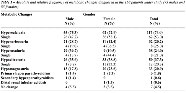
The most frequently found metabolic change
was hypercalciuria, present in 117 patients (74%), which was isolated
in 62 cases (53%) and was associated with other changes in 55 (47%). Hypocitraturia
was the second most frequent change, present in 59 (37,3%) patients. Single
presentation was found in 12 (20.3%). Slight hyperoxaluria (urinary oxalate
between 44 and 100 mg/24h) was found in 38 (24%) patients, with single
presentation in 8 (21%). Hyperoxaluria values higher than 100 mg/24h were
not seen. Hypomagnesuria was present in 33 (21%) patients. Hyperuricosuria
was diagnosed in 32 (20.2%) patients and, among these, only 8 (25%) had
single presentation. We found 4 cases of hypercalciuria associated with
hypercalcemia, with 3 cases of primary hyperparathyroidism and 1 case
secondary to chronic use of loop diuretic (furosemide). Distal renal tubular
acidosis was diagnosed in only one female patient. In 7 patients (4.5%)
it was impossible to establish a metabolic diagnosis.
COMMENTS
Urinary
lithiasis is a frequent disease that mainly affects young male individuals,
however recent publications showed less pronounced differences (6). In
the sample studied in this work, we found a slight predominance of female
(53.7%) over male gender (46.3%). When assessing the recurrence of lithiasis
(number stones/year) we observed it was significantly higher in males.
Studies show that the relative risk of stone formation is three times
higher in males (7).
As for fluid ingestion and urinary volume,
observations have shown that a low urinary volume is a real risk factor
for nephrolithiasis and that increased water ingestion should be the initial
therapy for preventing recurrent stones (8-10). When the patients’
urinary volume was assessed, we found only 8.2% of the cases presenting
diuresis lower than 1000 mL/24h. These findings could be explained by
the fact that these patients probably were instructed to increase their
fluid ingestion prior to the study. Therefore, in the present study, urinary
volume itself was not a significant risk factor for lithiasis, and was
significantly reduced in only 8.2% of patients.
Aspects related to the composition of water
consumed by the population and its relation to the incidence of renal
lithiasis are inconclusive and sometimes contradictory (11). Only calcium
and magnesium are found in significant concentrations in natural water,
so that water hardness is defined by the total concentration of magnesium
and calcium carbonate in the water. Smooth or soft waters contain approximately
50 to 75 mg/L of calcium carbonate (CaCO3), being suitable for public
consumption. Waters containing 75 to 150 mg/L of CaCO3 are considered
moderately hard with changes in taste and are considered as risk factor
for urinary lithiasis (11).
The water delivered for public consumption
in the city of Botucatu and 22 municipalities presents physical-chemical
characteristics that classify it as a soft water (10 to 67 mg/L of Mg
and CaCo3). Thus, values found concerning the hardness of water consumed
by patients with lithiasis do not characterize the water as a risk factor
for lithogenesis.
In Brazil, the experiences of several centers
for study on lithiasis showed that (93 to 97%) of patients with lithiasis
had metabolic diseases (12). In the present study, metabolic changes were
diagnosed in 95.5% of patients with lithiasis.
Of the 158 patients under study, 151 presented
at least one metabolic diagnosis; among these, 96 (63.6%) showed a single
metabolic disorder and 55 (36.4%) had mixed metabolic changes. This sample
was similar to a large Argentinean study with 2612 lithiasis patients,
where 61.5% of metabolic changes were single and 31.2% were mixed (13).
In the present study, hypercalciuria was
the most frequent change, being present in 117 (74%) cases. Hypocitraturia
was the second most frequent change, detected in 37.3% of patients. Studies
on hypocitraturia showed a highly variable prevalence in the single presentation,
reaching 62% when associated with other disorders (14). The citrate acts
on prevention of lithiasis due to its dissolving effects and by inhibiting
the crystallization of calcium and uric acid salts (15). Citraturia is
not directly influenced by the ingestion of alimentary citrate, but by
systemic acid-basic changes produced by a certain diet. Diets rich in
proteins and excessive physical exercise cause reduced citraturia (16).
Slight hyperoxaluria was present in 24.1%
of patients. The role of slight hyperoxaluria (< 100 mg/24h) in calcium
lithiasis is quite controversial. As for the association between hyperoxaluria
and hypercalciuria, no positive correlation was observed. Studies with
pure calcium oxalate stones concluded that they were more frequent in
patients with normal calcium excretion and that mixed stones made of calcium
phosphate and oxalate were more frequently found in patients with hypercalciuria
(17). Other authors observed that small increases in urinary oxalate would
be more critical than similar increases in calcium in the process of urinary
supersaturation by calcium oxalate (18). In our study, we found 43.6%
of hyperoxaluria associated with hypercalciuria in male patients and only
8% of this association in females.
Hypomagnesuria was diagnosed in 21% of patients
with lithiasis. The low urinary excretion of magnesium has been considered
as a potential risk for formation of calcium stones and its supplementation
has been shown to be effective in reducing or preventing the recurrence
of urinary calcium lithiasis (19).
Hyperuricosuria was observed in 20.2% of
our patients. National data published by MULTILIT (1995) (12) showed a
mean national frequency of 27.2% of excessive urinary excretion of uric
acid, however, when each Brazilian region was analyzed separately, large
different were detected in its frequency, ranging from 6.3% in Belo Horizonte
to 69% in Alagoas. Probably this could be due to different dietetic habits.
Primary hyperparathyroidism was diagnosed
in 1.8 % of patients with calcium lithiasis. Type I or distal renal tubular
necrosis with metabolic acidosis was diagnosed in only 1 patient (0.6%).
Recent randomized prospective studies, comparing
patients who received specific dietetic orientations, according to the
metabolic changes diagnosed and non-studied patients who followed general
orientations showed that after 3 years the development of new stones was
significantly lower in the group that had been given specific orientations,
when compared with patients receiving general orientations, showing the
importance of specific orientations derived from a metabolic diagnosis
(10).
REFERENCES
- Lancina Martin JA, Rodriguez-Rivera GJ, Novas Castro S, Rodriguez Gomez I, Fernandez Rosado E, Alvarez Castelo L, et al. [Metabolic risk factors in calcium urolithiasis according to gender and age of the patients] Actas Urol Esp. 2002; 26: 111-20 [Spanish].
- Joual A, Rais H, Rabii R, el Mrini M, Benjelloun S: Epidemiology of urinary lithiasis. Ann Urol. (Paris) 1997; 31: 80-3.
- Tiselius HG: Stone incidence and prevention. Clin Urol. 2000: 26: 452-62.
- Ekane S, Wildschutz T, Simon J, Schulman CC: Urinary lithiasis: epidemiology and physiopathology. Acta Urol Belg. 1997; 65: 1-8.
- Pak CY, Resnick MI, Preminger GM: Ethnic and geographic diversity of stone disease. Urology. 1997; 50: 504-7.
- Robertson WG, Peacock M, Baker M, Marshall DH, Pearlman B, Speed R, et al.: Studies on the prevalence and epidemiology of urinary stone disease in men in Leeds. Br J Urol. 1983; 55: 595-8.
- Shekarriz B, Stoller ML: Metabolic evaluation of stone disease. Int Braz J Urol. 2001; 27: 10-18.
- Curhan GC, Willett WC, Speizer FE, Stampfer MJ: Beverage use and risk for kidney stones in women. Ann Intern Med. 1998; 128: 534-40.
- Pak CY: Medical prevention of renal stone disease. Nephron. 1999; 81: 60-5.
- Pearle MS: Prevention of nephrolithiasis. Curr Opin Nephrol Hypertens. 2001; 10: 203-9.
- Agreste SA, Schor N, Heilerg IP: Papel da constituição físico química da água potável na litogênese renal. J Bras Nefrol. 2001; 23: 45-8.
- Laranja SMR, Heilberg IP, Coelho STSN, Novoa CG, Schor N: Estudo multicêntrico de litíase renal no Brasil (MULTILIT). In: Schor N, Heilberg IP (eds.), Calculose Renal, Fisiopatologia, Diagnóstico, Tratamento. São Paulo, Sarvier. 1995; pp. 295-338.
- Del Valle E, Spivacow R, Zanchetta JR: Metabolic changes in 2612 patients with nephrolithiasis. Medicina. (B Aires) 1999; 59: 417-22.
- Ayusso LL, Schor N: Avaliação de pacientes com litíase renal em região de clima quente. J Bras Nefrol. 2001; 23: 205-12.
- Hallson PC, Rose GA, Sulaiman S: Raising urinary citrate lowers calcium oxalate and calcium phosphate crystal formation in whole urine. Urol Int. 1983; 38: 179-81.
- Rebelo MAP. Hipocitratúria. In: Schor N, Heilberg IP (eds.), Calculose Renal, Fisiopatologia, Diagnóstico, Tratamento. São Paulo, Sarvier. 1995; p. 105.
- Grases F, Conte A, Coll R, Genestar C: The role of hyperoxaluria in the formation of calcium oxalate urinary calculi, and its association with other biochemical measurements. Scand J Urol Nephrol. 1990; 24: 211-3.
- Robertson WG, Hughes H: Importance of mild hyperoxaluria in the pathogenesis of urolithiasis- new evidence from studies in the Arabian peninsula. Scanning Microsc. 1993; 7: 391-402.
- Asplin JR, Murray JF, Coe FL: Nephrolithiasis. In: Brenner BM (ed.), The Kidney, Brenner & Rector’s. Philadelphia, WB Saunders. 2000; pp. 1774-819.
____________________
Received:
June 17, 2004
Accepted after revision: October 28, 2004
_______________________
Correspondence address:
Dr. Carmen R. Amaro
Av. Camilo Mazoni, 874, J. Paraíso
Botucatu, SP, 18610-460, Brazil
Fax: + 55 14 3882-5766
E-mail: btamaro@uol.com.br