LAPAROCOPIC
URETERAL REIMPLANTATION IN URETERAL STENOSIS AFTER GYNECOLOGIC LAPAROSCOPIC
SURGERY
(
Download pdf )
ANIBAL W. BRANCO, ALCIDES J. BRANCO FILHO, WILLIAM KONDO
Department of Urology and General Surgery, Vita Hospital and Cruz Vermelha Hospital, Curitiba, Paraná, Brazil
ABSTRACT
Pelvic surgery is the most common cause of iatrogenic ureteral injury, and traditionally repair of such injuries requires laparotomy. We report the case of a 48-year-old woman with an iatrogenic ureteral injury after laparoscopic ophorectomy which was laparoscopically reimplanted using the Lich-Gregoire technique. Total operating time was 150 minutes and estimated blood loss was 100 mL. Two months after surgery she is asymptomatic with normal renal function.
Key
words: ureter; iatrogenic disease; reconstructive surgical procedures;
laparoscopy
Int Braz J Urol. 2005; 31: 51-53
INTRODUCTION
The incidence of laparoscopic ureteral injuries in pelvic surgery range from less than 1% to 2% and laparoscopically assisted vaginal hysterectomy is the leading procedure in which injury occurs (1). Intraoperative injury to the ureter may result from ligation, angulation, transection, laceration, crush, ischemia, and resection. Most cases are only identified postoperatively and traditionally surgical repair is performed by laparotomy. We present a case of an iatrogenic ureteral injury managed laparoscopically by ureteral reimplantation.
CASE REPORT
A
48-year-old white woman underwent a laparoscopic oophorectomy for an ovarian
cyst and 4 months after the surgical procedure she came to our service
complaining about pain in her right flank, chills and fever.
Microscopic urinalysis revealed bacteriuria
and pyuria, and urine culture showed a growth of E. coli. Ultrasonography
showed a right pelviocaliceal dilatation and excretory urography demonstrated
a functional exclusion of the right kidney. Magnetic resonance imaging
urography identified right ureteral stenosis just after crossing the iliac
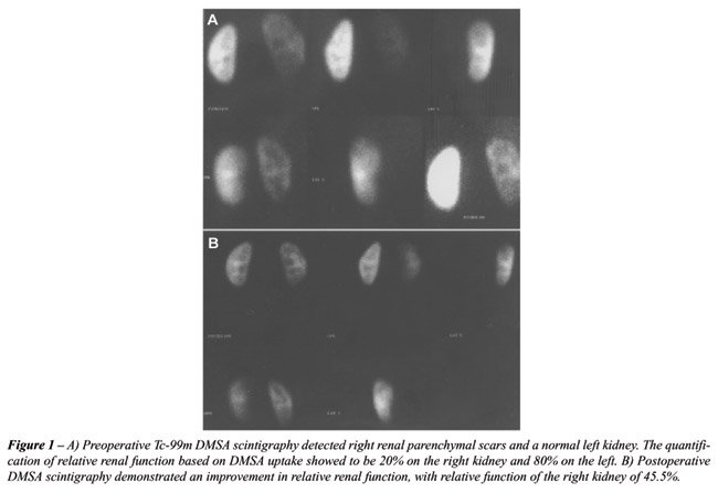
vessels. Technetium-99m dimercaptosuccinic acid (Tc-99m DMSA) scintigraphy
showed relative renal function of 20% in the right kidney (Figure-1).
The patient underwent laparoscopic ureteral
reimplatation according to Lich-Gregoir technique. The patient was placed
in a 45-degrees lateral decubitus position and a 10 mm trocar was placed
in the periumbilical area for the 30-degree laparoscope. Another 10 mm
trocar was placed in the anterior axillary line 4 cm below the umbilical
level and a 5 mm trocar was placed in the midline, approximately halfway
between the umbilicus and the pubis. The abdominal cavity was inspected
and intraperitoneal adhesions in the pelvis were identified. The right
colon was reflected and the dilated ureter was isolated. The ureteral
dissection in the inferior direction showed a fibrous area at the level
of the iliac vessels. The ureter was sectioned proximally to the obstruction
site and ureteral reimplatation was performed with the Lich-Gregoire technique.
The total operating time was 150 minutes and the estimated blood loss
was 100 mL. There were no intraoperative or postoperative complications
and the patient was discharged 36 hours after the surgical procedure with
the indwelling catheter being removed on day 5.
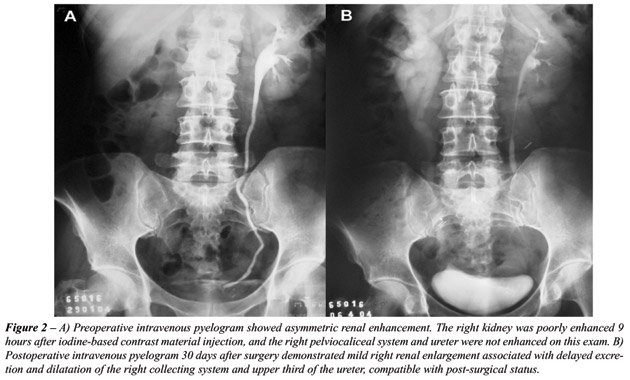
One month after surgery, the patient had
negative urine cultures and an unremarkable intravenous urogram, except
for a minimal delay in filling of the collecting system with contrast
material and residual ureteral dilatation (Figure-2).
In the second postoperative month the patient
was asymptomatic and was submitted to another DMSA scintigraphy which
showed a right relative function of 45.5% (Figure-1).
COMMENTS
The
concepts and techniques of the ureteroneocystostomy, most commonly performed
for the correction of vesicoureteral reflux in children, are also applicable
to ureteral reimplantation for the repair of ureteral injuries, including
stricture and intraoperative injury (2). A variety of techniques have
been described and we reported a case successfully managed using a laparoscopic
Lich-Gregoire procedure.
Most of the experimental studies report
a reduction of adhesion formation after laparoscopic surgery compared
to open surgery, and we did not find any difficulty in performing the
laparoscopic repair after the gynecologic laparoscopic surgery.
Although abdominal open surgeries cause
adhesions that may render subsequent laparoscopic access and dissection
problematic, we support the opinion that laparoscopy can be done even
after open surgeries. Parsons et al. (3) analyzed the effect of a previous
abdominal surgery on urological laparoscopy and they concluded that it
does not appear to adversely affect the performance of a subsequent urological
laparoscopy. So, laparoscopic ureteral repair can be done after open and
laparoscopic ureteral injuries.
In our literature review, it seems that
this is the first case of ureteral reimplantation by laparoscopic approach
after iatrogenic ureteral injury. Despite the limited experience, laparoscopic
repair of ureteral injuries seems to be feasible and safe.
REFERENCES
- Ostrzenski A, Radolinski B, Ostrzenska KM: A review of laparoscopic ureteral injury in pelvic surgery. Obstet Gynecol Surv. 2003; 58: 794-9.
- Koo HP, Bloom DA: Lower ureteral reconstruction. Urol Clin North Am. 1999; 26: 167-73.
- Parsons JK, Jarrett TJ, Chow GK, Kavoussi LR: The effect of previous abdominal surgery on urological laparoscopy. J Urol. 2002; 168: 2387-90.
_____________________
Received:
June 11, 2004
Accepted after revision: September 8, 2004
_______________________
Correspondence
address:
Dr. Anibal Wood Branco
Rua das Palmeiras, 170 / 201
Curitiba, PR, 80620-210, Brazil
E-mail: anibal@awbranco.com.br