TOTAL
LAPAROSCOPIC GASTROCYSTOPLASTY: EXPERIMENTAL TECHNIQUE IN A PORCINE MODEL
(
Download pdf )
FREDERICO R. ROMERO, CLAUDEMIR TRAPP, MICHAEL MUNTENER, FABIO A. BRITO, LOUIS R. KAVOUSSI, THOMAS W. JARRETT
The James Buchanan Brady Urological Institute, The Johns Hopkins Medical Institutions (FRR, CT, MM, FAB), Maryland, Baltimore, The Department of Urology, North Shore-LIJ Health System (LRK), Long Island, New York and Department of Urology, The George Washington University Medical Center (TWJ), Washington, DC, USA
ABSTRACT
Objective:
Describe a unique simplified experimental technique for total laparoscopic
gastrocystoplasty in a porcine model.
Material and methods: We performed laparoscopic
gastrocystoplasty on 10 animals. The gastroepiploic arch was identified
and carefully mobilized from its origin at the pylorus to the beginning
of the previously demarcated gastric wedge. The gastric segment was resected
with sharp dissection. Both gastric suturing and gastrovesical anastomosis
were performed with absorbable running sutures. The complete procedure
and stages of gastric dissection, gastric closure, and gastrovesical anastomosis
were separately timed for each laparoscopic gastrocystoplasty. The end-result
of the gastric suturing and the bladder augmentation were evaluated by
fluoroscopy or endoscopy.
Results: Mean total operative time was 5.2
(range 3.5 - 8) hours: 84.5 (range 62 - 110) minutes for the gastric dissection,
56 (range 28 - 80) minutes for the gastric suturing, and 170.6 (range
70 to 200) minutes for the gastrovesical anastomosis. A cystogram showed
a small leakage from the vesical anastomosis in the first two cases. No
extravasation from gastric closure was observed in the postoperative gastrogram.
Conclusions: Total laparoscopic gastrocystoplasty
is a feasible but complex procedure that currently has limited clinical
application. With the increasing use of laparoscopy in reconstructive
surgery of the lower urinary tract, gastrocystoplasty may become an attractive
option because of its potential advantages over techniques using small
and large bowel segments.
Key
words: laparoscopy; bladder; gastroplasty; experimental; pigs
Int Braz J Urol. 2007; 33: 94-9
INTRODUCTION
Introduced
by Sinaiko as an experimental study in 1956, (1) gastrocystoplasty was
later adapted for clinical practice by Leong and Ong (2,3).
Gastrocystoplasty was initially conceived
to avoid complications frequently present when using the small or large
bowel segments to augment the bladder, such as excessive mucus production,
hyperchloremic metabolic acidosis, and consequent bone rarefaction and
growth problems in the pediatric population (1-3).
The emergence of complications caused by
gastric secretion, including hematuria-dysuria syndrome and hypochloremic
metabolic alkalosis, as well as necessity for a large abdominal incision
to harvest the gastric wedge and anastomose it to the bladder, have restricted
the use of gastrocystoplasty (4-6).
Recently, many laparoscopic studies have
been performed, in an attempt to minimize the distress of urinary reconstruction,
avoiding large incisions and their destructive psychological and physical
consequences (7-11). These reports showed the feasibility of bladder augmentation
through laparoscopic approach, improving cosmesis and decreasing postoperative
morbidity. The majority of these publications have been done using intestinal
segments, (8-10) usually with a hand-assisted method (10,11).
To further increase the therapeutic options
and to reduce the morbidity of lower urinary tract reconstructive surgery,
we describe a unique simplified experimental technique for total laparoscopic
gastrocystoplasty in a porcine model.
MATERIAL AND METHODS
Ten
female Sus-scrofus domesticus piglets, with an average weight of 65 lb
were used in this study. The experiment protocol was approved by the Institutional
Animal Care and Use Committee. The animals received nothing per mouth
for 12 hours before the procedure. Each animal was premedicated with an
intramuscular injection of telazol, ketamine, and xylazine (TKX, 1 mL/50
lb). Once the animals were tranquilized, anesthesia was induced with intravenous
thiopental (10 mg/lb) and maintained with isofluorane inhalation (1.5%
- 2%).
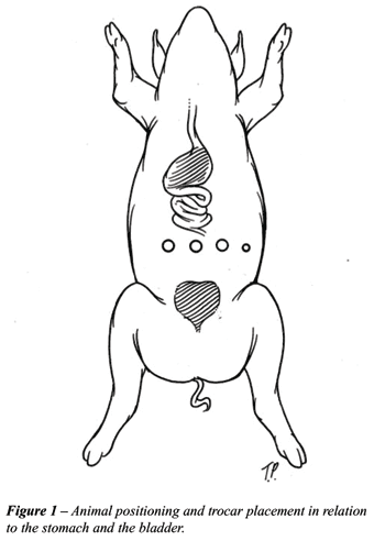
The animals were positioned supine. Pneumoperitoneum
(15 mm Hg) was achieved with a Veress needle at the level of the umbilicus,
followed by introduction of three 10 mm trocars under direct vision: in
the midline, four fingerbreadths to the right, and four fingerbreadths
to the left, at the level of the umbilicus. When necessary, a fourth 5
mm trocar was introduced laterally and in line with the other trocars
to assist with suction or traction (Figure-1).
The gastroepiploic arch was identified along
the greater gastric curvature. The branches of the right gastroepiploic
artery to the anterior and posterior wall of the antrum were carefully
mobilized, and transected between hemoclips. The use of electrocautery
and unnecessary grasping of the vessels was strictly avoided during this
dissection, to prevent injury to the pedicle. Dissection was carried from
the origin of the right gastroepiploic artery, at the level of the pylorus,
to the beginning of the segment of stomach which was gonna be used as
a graft. Adequate mobilization is important to allow enough length for
the pedicle to reach the bladder without tension.
After the pedicle was adequately freed,
a paper ruler was introduced into the abdomen and a segment of 4 to 6
cm of stomach was identified. The left gastroepiploic artery was transected
immediately after the distal end of the segment with the use of a linear
endoscopic stapler or titanium clips. The wedge-shaped segment of stomach
was delineated with electrocautery to facilitate the excision of the graft,
beginning at the posterior wall, around the pedicle, and at the anterior
wall of the stomach. The apex of the wedge was placed 2 cm away from the
lesser gastric curvature to avoid injury to branches of the vagus nerve
that control the gastric outlet. Initially, the resection of the gastric
wedge was performed by simultaneously cutting the seromuscular and the
mucosal layer of the stomach, duplicating the open technique. However,
the seromuscular layer retracted behind the mucosa, resulting in redundant
mucosal tissue that created difficulties with the visualization of the
gastric patch borders during the anastomosis to the bladder. This was
solved subsequently by incising the gastric wall in stages. The seromuscular
layer was opened first and was easily detached from the underlying mucosa.
The mucosal layer was then incised near to the border of the seromuscular
patch with the curve of the laparoscopic scissors pointing toward the
graft and using slight angulation of the scissors in the same direction,
to reduce the amount of mucosal tissue resected.
The native stomach was closed with one layer
of running sutures, taking care to invert the gastric mucosa. A stay suture
was positioned in the anterior angle of the gastrotomy and pulled outside
the abdomen with the assistance of a Carter-Thomason device (Inlet Medical,
Eden Prairie, Minnesota, USA), to help in the repair of the stomach and
facilitate the placement of the sutures.
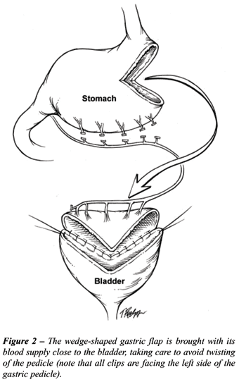
The gastric segment was positioned close
to the bladder. Care was taken to avoid twisting of the pedicle (Figure-2).
The bladder was opened in a sagittal plane in the midline from the bladder
neck anteriorly, through the dome, to the trigone posteriorly. Two stay
sutures were placed in each side of the bladder to assist in the anastomosis.
The first suture was placed in the left lateral aspect of the bladder
wall and the right corner of the gastric wedge. With another suture, the
right border of the bladder incision was sutured to the left corner of
the gastric wedge, and the wedge was approximated to the native bladder.
The posterior wall of the anastomosis was performed with absorbable running
sutures, with caution to include all the layers of the stomach. After
the posterior wall was concluded, the anterior wall of the anastomosis
was performed in the same way as with the posterior anastomosis.
At the end of the procedure, a gastrogram
was performed with 300 cc of contrast 50%, to confirm closure of the stomach.
A cystogram (300 cc of contrast 50%) or cystoscopy under intraabdominal
visualization was performed to confirm a watertight bladder reconstruction.
The complete procedure and the stages of
gastric dissection, gastric closure, and gastrovesical anastomosis were
separately timed for each laparoscopic gastrocystoplasty. The end-result
of the gastric suturing and the bladder augmentation were evaluated by
fluoroscopy or endoscopy, after which the animals were sacrificed. Postmortem
laparotomy was performed to inspect the final result of the gastrocystoplasty.
RESULTS
The mean total operative time was 5.2 hours (range 3.5 to 8 hours). The gastric dissection took an average of 84.5 minutes (range 62 to 110 minutes), the gastric suturing 56 minutes (range 28 to 80 minutes), and the gastrovesical anastomosis 170.6 minutes (range 70 to 220 minutes). Cystogram showed a small leakage from the gastrovesical anastomosis in the first two cases. No extravasation from the gastric closure was observed in the postoperative gastrogram. Laparotomy confirmed these results, showing a defect in the posterior anastomosis as the cause of the bladder leakage in the first two experiments.
COMMENTS
Docimo
et al. reported the first laparoscopic bladder augmentation in 1995 (7).
Since then, the laparoscopic approach has been increasingly used to perform
either augmentation or total replacement of the bladder (8-11). The objectives
have been to reduce the morbidity of these complex procedures and to expand
laparoscopic reconstructive surgery in both pediatric and adult urology
(8-13). Most techniques employ the small or large bowel and maintain the
same principles of conventional open surgery. Usually, the intestinal
segment is detubularized, refashioned, and sutured to the bladder using
either intracorporeal or, most frequently, extracorporeal suturing.
In the past decade, many authors restricted
the indications of open gastrocystoplasty because of the increasing appearance
of hematuria-dysuria (6,14), despite the small number of patients presenting
this complication in several series and the good response to clinical
management with proton pump inhibitors (14,15). Another complication reported,
the hypocloremic metabolic alkalosis (5), is a rarely seen entity that
can be prevented by proper electrolyte correction in the management of
acute diarrhea. Despite these particular complications, the stomach is
a useful alternative in selected patients with a poorly compliant or a
high-pressure bladder that need bladder augmentation. Short bowel syndrome,
renal insufficiency with metabolic acidosis, and previous pelvic irradiation
are clinical situations in which gastrocystoplasty would be the preferred
form of bladder augmentation (2,4,15,16). Other situations in which gastrocystoplasty
may be used include the necessity for a pouch with less mucous production
and in patients with recurrent urinary lithiasis (3,17).
With the increasing use of laparoscopy in
reconstructive surgery of the lower urinary tract, gastrocystoplasty may
be an attractive option because of its advantages over the techniques
using small and large bowel segments. It eliminates the need of detubularization
and refashioning of the bowel, reducing the amount of intracorporeal suturing.
Also, in contrast to other intestinal segments, the gastric flap is more
similar to the bladder wall in thickness, facilitating an even coaptation
during the anastomosis.
Pure laparoscopic gastrocystoplasty is a
feasible procedure (7). The porcine experimental model duplicates the
anatomy of the gastric and urinary tracts in humans relatively well, and
it is known to be a good training modality for advanced laparoscopic techniques.
Although this was not a survival study, this simplified surgical technique
may enable surgeons to practice both dissection and suturing skills required
in extirpative and reconstructive laparoscopic surgery. If this were a
survival procedure, some important technical aspects should be pointed.
Although intracorporeal gastric suturing is a well-established technique
for gastric closure and it is specially useful for surgical skills training,
laparoscopic stapling of the stomach avoids the risk of peritoneal contamination
and allows for a faster gastric wedge resection. The gastric patch pedicle
should be retroperitonealized by releasing the right colon medialy and
lying the right gastroepiploic pedicle in the retroperitoneum. The augmented
bladder should be drained by suprapubic cystostomy, and two abdominal
drains should also be placed through the ports to drain gastric and bladder
sutures.
Regardless of the gastrointestinal segment
chosen, several benefits are obtained with the laparoscopic approach.
Decreased perioperative morbidity, less need for postoperative analgesics,
faster recovery time, and improved cosmetic results are the main advantages
favoring laparoscopy over the open techniques (7,8,13). Furthermore, advancements
in tissue engineering technology may allow the use of demucosalized gastric
segments as a vehicle to transport in vitro expanded urothelial cells
during bladder reconstruction (18,19). The ease with which the gastric
mucosa can be detached from the seromuscular layer of the stomach may
allow gastrocystoplasty to be a handy transport matrix.
The development of the laparoscopic approach
and the recent advancements in tissue engineering may allow demucosalized
gastrocystoplasty to be applied more extensively in the near future, avoiding
the complications inherent to the gastric mucosa.
CONCLUSIONS
Total laparoscopic gastrocystoplasty is a feasible but complex procedure that currently has limited clinical application. With the increasing use of laparoscopy in reconstructive surgery of the lower urinary tract, gastrocystoplasty may become an attractive option because of its potential advantages over the techniques using bowel segments. Additional survival and clinical studies, specially with the use of the modified demucosalized technique with urothelial cells grafting, are necessary to evaluate the perspectives of laparoscopic gastrocystoplasty in the future.
ACKNOWLEDGEMENTS
The authors express appreciation to Carolyn Magee and Laurie Pipitone for assistance with animals and procedures, and to Timothy Phelps for the preparation of medical illustrations.
CONFLICT OF INTERERST
None declared.
REFERENCES
- Sinaiko E: Artificial bladder from segment of stomach and study of effect of urine on gastric secretion. Surg Gynecol Obstet. 1956; 102: 433-8.
- Leong CH, Ong GB: Gastrocystoplasty in dogs. Aust N Z J Surg. 1972; 41: 272-9.
- Leong CH, Ong GB. Proceedings: gastrocystoplasty. Br J Urol. 1975; 47: 236.
- Adams MC, Mitchell ME, Rink RC: Gastrocystoplasty: an alternative solution to the problem of urological reconstruction in the severely compromised patient. J Urol. 1988; 140: 1152-6.
- Gosalbez R Jr, Woodard JR, Broecker BH, Warshaw B: Metabolic complications of the use of stomach for urinary reconstruction. J Urol. 1993; 150: 710-2.
- Nguyen DH, Bain MA, Salmonson KL, Ganesan GS, Burns MW, Mitchell ME: The syndrome of dysuria and hematuria in pediatric urinary reconstruction with stomach. J Urol. 1993; 150: 707-9.
- Docimo SG, Moore RG, Adams J, Kavoussi LR: Laparoscopic bladder augmentation using stomach. Urology. 1995; 46: 565-9.
- Elliott SP, Meng MV, Anwar HP, Stoller ML: Complete laparoscopic ileal cystoplasty. Urology. 2002; 59: 939-43.
- Gill IS, Rackley RR, Meraney AM, Marcello PW, Sung GT: Laparoscopic enterocystoplasty. Urology. 2000; 55: 178-81.
- Hedican SP, Schulam PG, Docimo SG: Laparoscopic assisted reconstructive surgery. J Urol. 1999; 161: 267-70.
- Specht M, Pareek G, Lin DD, Bleustein C, Fahey TJ 3rd, Felsen D, et al.: Hand-assisted laparoscopic autoaugmentation gastrocystoplasty. Surg Endosc. 2002; 16: 1538-41.
- Anderson KR, Clayman RV: Laparoscopic lower urinary tract reconstruction. World J Urol. 2000; 18: 349-54.
- Chung SY, Meldrum K, Docimo SG: Laparoscopic assisted reconstructive surgery: a 7-year experience. J Urol. 2004; 171: 372-5.
- Chadwick Plaire J, Snodgrass WT, Grady RW, Mitchell ME: Long-term follow-up of the hematuria-dysuria syndrome. J Urol. 2000; 164: 921-3.
- DeFoor W, Minevich E, Reeves D, Tackett L, Wacksman J, Sheldon C: Gastrocystoplasty: long-term followup. J Urol. 2003; 170: 1647-9.
- Gearhart JP, Peppas DS, Jeffs RD: The application of continent urinary stomas to bladder augmentation or replacement in the failed exstrophy reconstruction. Br J Urol. 1995; 75: 87-90.
- Carr MC, Mitchell ME: Gastrocystoplasty. ScientificWorldJournal. 2004; 4 Suppl 1: 48-55.
- Shiroyanagi Y, Yamato M, Yamazaki Y, Toma H, Okano T: Urothelium regeneration using viable cultured urothelial cell sheets grafted on demucosalized gastric flaps. BJU Int. 2004; 93: 1069-75.
- Atala A, Koh C: Applications of tissue engineering in the genitourinary tract. Expert Rev Med Devices. 2005; 2: 119-26.
____________________
Accepted after revision:
August 25, 2006
_______________________
Correspondence address:
Dr. Frederico Ramalho Romero
The Johns Hopkins Medical Institutions
The James Buchanan Brady Urological Institute
600 North Wolfe Street, Marburg 1
Baltimore, Maryland, 21287, USA
Fax: + 1 410 502-7711
E-mail: frederico.romero@gmail.com
EDITORIAL COMMENT
Augmentation
cystoplasty is needed to treat some congenital pediatric and adult urological
diseases. Although several substitutes have been employed, the vascularized
intestinal segments are still the most commonly used tissue for this reconstruction,
despite the described long-term complications. The minimally invasive
approach for bladder augmentation has been described and may decrease
the perioperative morbidity of this procedure (1).
The
authors described a minimally invasive technique for gastrocystoplasty
in a non-survival porcine model and should be congratulate for their initiative.
Despite the authors’ large experience in advanced laparoscopic cases,
the mean operative time extended 5 h, most due to the gastrovesical anastomosis
(3 h), with posterior wall leakage occurring in the initial 2 cases of
this series. One alternative to this problem would be a laparoscopic-assisted
technique, employing the laparoscopic access to dissect and isolate an
adequate gastric segment, performing the anastomosis through a small abdominal
incision, in the same fashion as the reconstructive part of current technique
of laparoscopic radical cystectomy (2).
REFERENCES
- Gurocak S, De Gier RP, Feitz W: Bladder augmentation without integration of intact bowel segments: critical review and future perspectives. J Urol. 2007; 177: 839-44.
- Haber GP, Campbell SC, Colombo Jr JR, Fergany A, Gill IS: Perioperative outcomes with laparoscopic radical cystectomy: “pure laparoscopic” and “laparoscopic-assisted” approaches. Urology. 2007 (submitted).
Dr. Jose R. Colombo Jr.
Section of Laparoscopic and Robotic Surgery
Glickman Urological Institute, Cleveland Clinic
Cleveland, Ohio, USA
E-mail: columbj@ccf.org