CAN
INFUNDIBULAR HEIGHT PREDICT THE CLEARANCE OF LOWER POLE CALYCEAL STONE
AFTER EXTRACORPOREAL SHOCKWAVE LITHOTRIPSY?
(
Download pdf )
FM. ARZOZ-FABREGAS, L. IBARZ-SERVIO, F. J. BLASCO-CASARES, M. RAMON-DALMAU, F. J. RUIZ-MARCELLAN
USP, Instituto Universitario Dexeus, Barcelona, Spain
ABSTRACT
Purpose:
To evaluate the efficacy of extracorporeal shock wave lithotripsy (SWL)
on lower calyceal calculi in relation to the renal anatomical factors
and determine which of these factors can be used to select patients who
will benefit from SWL.
Materials and Methods: We analyzed retrospectively
78 patients with single radiopaque lower calyceal stones treated with
SWL. The patients were evaluated 3 months after lithotripsy with a simple
abdominal X-ray and a kidney ultrasound scan. The success of the treatment,
removal of all fragments, was correlated with renal anatomical factors
measured in the pre-treatment intravenous urography: infundibulopelvic
angle, lower infundibulum width, lower infundibulum length, ratio length/width,
infundibulum height, and number of minor calyces in the lower calyceal
group.
Results: Three months after SWL treatment,
39 patients were stone-free (NR group) and 39 had residual fragments (R
group). Both groups presented no differences in relation to infundibulopelvic
angle, width and length of the lower calyceal infundibulum, length/width
ratio of the lower infundibulum or number of lower calyces. Height of
the infundibulum, described as the distance between the line passing through
the lowest part of the calyx containing the calculus and the highest point
of the lower lip of renal pelvis, was the only parameter in which significant
differences (p = 0.002) were found between the NR and R groups.
Conclusions: Lower Infundibular height could
be a good measurement tool for deciding which patients with lower calyceal
lithiasis would benefit from SWL treatment. Height of less than 22 mm
suggests a good outcome from lithotripsy.
Key
words: kidney; kidney calculi; lithotripsy; anatomy; kidney calices
Int Braz J Urol. 2009; 35: 140-50
INTRODUCTION
The
objective of SWL is to obtain a fragmentation of the calculus into fragments
that can be expelled through the renal collecting system. However, the
success of SWL also depends on the size and composition of the calculus,
its location in the kidney, the anatomy of the urinary tract and some
personal factors such as body mass index or patient mobility (1-4). According
to Politis et al., although correct fragmentation is obtained in 98% of
cases after SWL, the fragments are eliminated in only 75% (4).
Calculi
in the lower calyceal group represent 24%-44% of all calculi requiring
treatment (1). In this location, there are some controversial aspects
as regards the efficacy of SWL, as the clearance rate is lower. It has
been suggested that this phenomenon could be explained by an antigravitational
position of the lower renal calyx (1,5), On the other hand, residual fragments
after SWL can cause complications such as chronic pain, obstruction, sepsis
and re-growth, which occasionally require an interventionist approach.
For these reasons, there is an obvious need for a method which helps us
to decide which treatment is the best option for each individual patient:
SWL, percutaneous surgery or flexible ureteroscopy (1,3).
Different
renal anatomic factors have been described since Sampaio et al. (1) first
described the anatomy of the renal collecting system using three dimensional
models and correlated the measurement of the infundibulopelvic angle with
the success of SWL, including infundibular width and length, the infundibular
width/length ratio, infundibular height, the number of minor calyces,
the volume of the renal collecting system and the pattern of dynamic urinary
transport (5-15). These measurements have been studied and correlated
with the success of SWL with different results.
The
objective of this study was to evaluate the outcome of SWL in patients
with single lithiasis of the lower renal pole and correlated it with the
aforementioned anatomical factors measured during the pre-treatment intravenous
urography (IVU), in order to determine which of them could be an effective
predictive factor to decide whether SWL could be successful.
MATERIALS AND METHODS
We
performed a retrospective analysis of 78 consecutive patients with single
radiopaque lithiasis of the lower calyceal group who were treated in only
one session with a Dornier Lithotripter S during a two-year period (from
June 2005 to June 2007).
Patients
with more than one calculus, residual fragments after prior lithotripsy,
urinary tract anomalies, prior surgical maneuvers, such as a double-J
catheter, or reduced mobility were excluded.
All
patients were treated by the same urologist under intravenous sedation.
The
results of the treatment were evaluated 3 months after lithotripsy. Stone
free status was defined as the absence of any residual fragments in a
simple abdominal X-ray film and kidney ultrasound scan. Depending on whether
there were remaining fragments after three months, the patients were divided
into two groups: group NR, (non-residual) composed of patients free from
calculi and group R (residual), composed of patients with residual fragments.
Personal
details as gender, age, body mass index (BMI) and affected kidney were
correlated for each patient with the existence or not of residual fragments
after the treatment.
The
following parameters, measured on the twenty minutes IVU pre treatment
film in a supine position, were correlated with the existence or not of
residual fragments three months after the treatment:
Calculus
Parameters
Estimated
surface area of the calculus (SA)(mm2): Measured at the pre-treatment
simple abdominal X-ray. Result of multiplying the length (L) and width
(W) diameters of the calculus by p and by 0.25 (16). SA= L x W x π
x 0.25
Number of shock waves applied: The number
of shockwaves required to completely fragment the calculus was recorded
in each case.
Calculus fragility index: Dividing the number
of shock waves by the surface of the calculus in mm2.
Anatomical
Parameters (measured at the pre-treatment IVU)
Infundibular width (mm): The narrowest point
on the axis of the lower infundibulum (Figure-1).

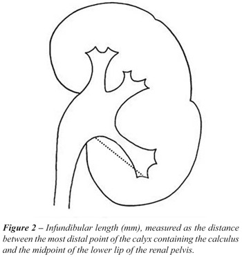
Infundibular length (mm): Distance between
the most distal point of the calyx containing the calculus and the midpoint
of the lower lip of the renal pelvis (Figure-2).
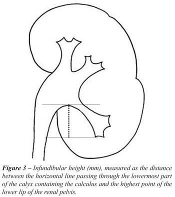
Infundibular height (mm): Distance between
the horizontal line passing through the lowest part of the calyx containing
the calculus and the highest point of the lower lip of the renal pelvis
(Figure-3).
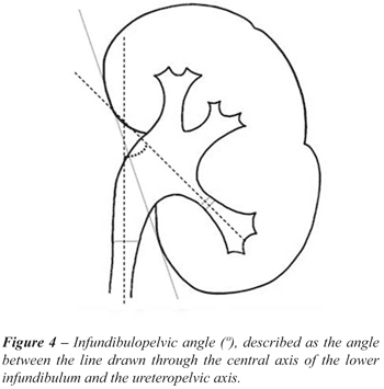
Infundibulopelvic angle (º): The angle
between the line drawn through the central axis of the lower infundibulum
and the ureteropelvic axis (Figure-4).
Infundibular length/width ratio.
Number of minor calyces.
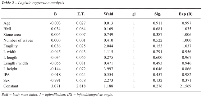
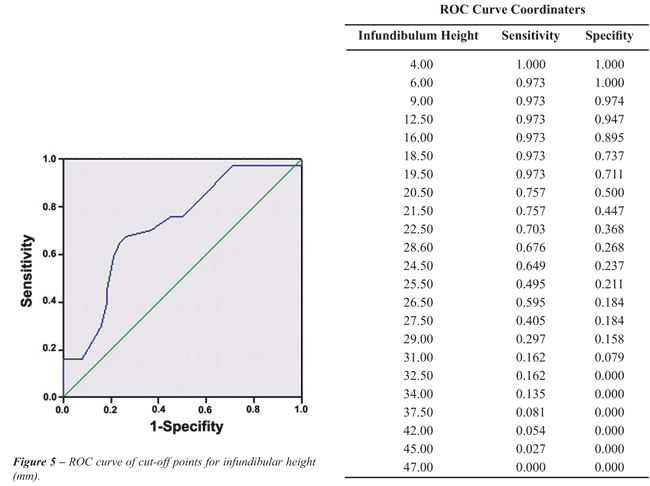
The statistical analysis was performed with the SPSS 13.0 Windows software program. We performed a descriptive analysis of all the aforementioned variables and compared them between the NR and R groups with Fisher’s exact test and the Mann-Whitney-Wilcoxon U-test for the qualitative and quantitative variables, respectively. A logistic regression analysis was also performed to study the correlation of the existence of residual fragments with all these parameters. Finally, a ROC curve was used to choose a cut-off point for the parameters showing significant differences in the logistic regression analysis.
RESULTS
Seventy-eight
patients were included in this study. Thirty-nine were classified in the
NR group and the remaining thirty-nine in the R group.
Fifty per cent of the studied population
was men and the other fifty per cent were women. The mean age of the patients
was 48 (SD 13.4) years, and the mean BMI was 25.1 Kg/m2 (SD 4.8). Thirty-seven
(47.4%) of the calculi were located in the right kidney and forty-one
(52.6%) in the left. There were no significant differences regarding gender
between NR and R groups (Table-1), but we found that women were more likely
than men to eliminate all the fragments after SWL in our population (p
= 0.023).
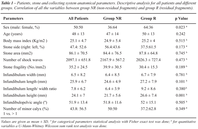
The median surface area of the calculi was
63 mm2 (9-450), the median number of shockwaves required to fragment them
was 2000 (1000-3300) and the median number of shock waves required to
fragment one surface area unit (calculus fragility index) was 31.7 waves/mm2
(111.1-7.3). Comparing the characteristics of the calculus between the
two groups, there were no statistically significant differences (Table-1.
Values are given as mean ± SD).
Concerning the anatomical measurements,
no significant differences were found between the two groups when comparing
infundibular length and width, infundibular length/width ratio, infundibulopelvic
angle (IPA) or number of minor calyces. On the other hand, significant
differences were found when comparing mean infundibular height in the
two groups (p = 0.002), with less infundibular height found in patients
who were stone-free after treatment (Table-1).
The logistic regression analysis for all
the factors studied (personal, pertaining to the calculus and anatomical
variables of the renal collecting system) show that only infundibular
height had a significant impact on the absence of residual fragments and
therefore, could be used as a predictive factor of the success of SWL
in calculi located in the lower calyx (Table-2). Furthermore, the ROC
curve shows that a height between 22 and 24 mm, and specifically 22.5
mm of height value could be the best cut-off point in our population for
predicting response to treatment with an approximate sensitivity and specificity
of 70% (Figure-5).
COMMENTS
Since
SWL appeared in the 1980s, most renal-ureteral calculi previously eligible
for open surgery or blind endoscopic maneuvers have been successfully
treated with few complications (3). However, with the development of new
therapeutic techniques such as percutaneous nephrolithotomy (PCNL) or
flexible ureteroscopy, the use of SWL in some situations, such as lithiasis
located in the lower calyceal group, is controversial.
The objective of this study was to evaluate
a population with single radiopaque lithiasis in the lower calyx, treated
by SWL and fragmented into expellable particles in a single session. Depending
on the response to treatment evaluated at three months with simple abdominal
X-ray and kidney ultrasound scan, we divided the patients into two groups
and compared them in relation to the factors which could be related to
fragment expulsion, with emphasis on anatomical variables, in order to
determine which of them would enable us to predict the success of SWL,
thus ruling out patients who would not benefit from this treatment and
who could be eligible for other therapeutic procedures such as ureteroscopy,
PCNL or control of evolution (17,18).
The purpose of SWL is to disintegrate the
stone into fragments of an expellable size (< 4 mm), in which success
represents the complete elimination of all fragments (3). However, this
often depends on factors affecting the particular patient, factors related
to the calculus and factors related to the anatomy of the renal collecting
system (1,3,4).
With reference to the size of lithiasis
for which PCNL should be used, instead of SWL in lithiasis of the lower
pole, continues to be subject to debate. Albala et al., in a multicenter
prospective study analyzing lithiasis located in the lower renal pole,
reached the conclusion that only calculi smaller than 1 cm are eliminated
in 50% of cases after lithotripsy, and they proposed that the cut-off
point for deciding between PCNL and SWL should be 1 cm (5). On the other
hand, with the development of new flexible ureteroscopes, remains debated
whether SWL should be the optimal choice of treatment for calculi in the
lower calyceal group measuring less than 1 cm. Pearl et al., in the second
phase of Lower Pole Study Group, conducted a prospective, randomized study
to compare treatment by SWL and ureteroscopy of lithiasis < 1 cm in
the lower pole, without finding statistically significant differences
(18). In our study, we analyzed patients with lithiasis with a median
surface area of 63 mm2 (9-450), equivalent to 8 mm diameter (3-21), which
were fragmented into expellable fragments in a single session, as the
objective was to evaluate the anatomical factors which could have an impact
on fragment expulsion, instead of studying the effect of the size of the
lithiasis on said expulsion. Moreover, we found no significant differences
between the NR and R groups in relation to the surface area of the calculus,
the number of shock waves required to fragment the stones or their fragility,
measured as the number of waves divided by the surface area of the calculus.
Concerning the location of the calculus,
there is some controversy concerning the efficacy of SWL, especially in
lithiasis of the lower calyceal group, where a large percentage of calculi
are not eliminated, regardless of their size or composition. This phenomenon
is believed to be due to an antigravitational problem, which could be
related to the anatomy of the calyx. The earliest studies of the anatomy
of the lower calyceal group were conducted by Sampaio et al., who used
polyester endocasts of cadaveric kidneys to study the length of the lower
infundibulum, the width of the calyx and the IPA. According to these authors.,
patients with an IPA of more than 90º are more likely to eliminate
the fragments after treatment with SWL (1,19). There have subsequently
been more studies, such as Elbahanasy et al., who performed a retrospective
analysis of the urograms examinations performed before SWL of lithiasis
smaller than 15 mm in the lower calyceal group, showing that patients
with a larger IPA, shorter infundibular length and greater infundibular
width are those who most often eliminate the fragments after the treatment
(9). Similar to Elbahanasy et al. studies we used urograms examinations
before SWL in order to measure the intrarenal geometry and to find if
there was any relationship with this anatomy and the stone-free status
after SWL and thus classify patients into favorable or unfavorable for
SWL.
Pace et al. (20) after analyzing the infundibular width on the 5, 10,
20 and compression films in supine position, on the prone film and a film
after voiding in erect position concluded that the compression film followed
by the 10 and 20 minute films are the most suitable to estimate the maximum
diameter of the infundibulum. In our study, we used the 20 minute film
in a supine position in all the patients in order to avoid different measurements
of each anatomic factor owing to the dynamic of the collecting system.
To avoid the interobserver variation of different measurement described
previously by Knoll et al. (2), all the parameters in our study where
evaluated by the same urologist.
Despite the fact that most of the studies
of the lower pole anatomy use urograms pre-treatment examinations to measure
anatomic factors, it has been discussed that some of these factors like
infundibular width or infundibular height should not be used because its
measurement can change with different urography phases, respiration and/or
postural movements or poor quality images (7,20,21). As we were more used
to evaluating the collecting system by urography in the period when the
study was done, we decided to perform this exploration on all the patients
included in the study. Although there have been some groups that evaluated
the possibility of using a three dimensional helical computed tomography
to measure the anatomy of the collecting system to avoid potential bias
as described above instead of using an urography some authors did not
find any statistical difference which concluded that IVU remains a good
method to analyze renal collecting system (20,22). IPA is the most widely
measured factor when evaluating the anatomy of the collecting system and
it has been measured using several methods. Sampaio et al. (1,19) calculated
the angle according to the location of the lithiasis, whereas Elbahanasy
et al. (9) calculated the IPA based on precise and reproducible anatomical
references, which seem more appropriate for defining the route to be followed
by stone fragments located in the inferior pole. We have therefore used
this method to measure the IPA in our study.
In our population, we found no statistically
significant differences between the two groups when comparing infundibular
length and width, the length/width ratio, the IPA or the number of minor
calyces (Table-1). Indeed, both in the univariate and logistic regression
analyses we found that only infundibular height (p = 0.002) had a significant
impact on calculus elimination and that it could be used to predict the
success of SWL in lithiasis of the inferior pole. These results are similar
to those of Tuckey et al. who, in order to simplify calculation of the
renal collecting system, analyzed the height of the infundibulum, found
that patients with a calyx height of < 15 mm eliminated the fragments
in 95% of the cases, whereas patients with a calyx height of > 15 mm
only do so in 52% of cases (15). Although this variable is easy to measure
compared with others such as the IPA and it is reproducible without requiring
a bevel protractor to measure it, some groups disregard it because they
believe that the measurement could vary in each urography according to
the patient’s respiratory movements and postural changes (7). Sorensen
et al. found a statistically significant difference in a wide range of
infundibular height (less than 15 mm or more than 30 mm) defined for stones
less than 10 mm (14). Poulakis et al. used an artificial neural network
in order to determine which anatomic measurements could predict the stone
free status after SWL. They found that infundibular height was one of
the most important variables to predict it with an excellent reproducibility
of this measurement (11). More recently, another study performed by Symes
et al. proved that infundibular height is useful to predict the success
rate after SWL when treating lower pole renal stones less than 20 mm (13).
Unlike Tuckey et al. (15), who used a 15
mm cut-off point to predict which patients were candidates for SWL, we
analyzed all the possible cut-off points with a ROC curve, finding that
the points with highest sensitivity and specificity in our population
were between 22 mm and 24 mm of infundibular height. The real cut-off
point in our ROC curve, with optimal sensibility and specificity, was
22.5 mm, but we reduced it to 22 mm because clinically it is very difficult
to measure 0.5 mm. Using the value of 22 mm as a cut-off, in our population
we found that 68.6% of patients with an infundibular height less than
22 mm were stone-free and only 35% of patients with an infundibular height
higher than 22 mm were free from fragments. In agreement with Tuckey and
Poulakis we suggest that this is one of the most easily and reproducible
anatomic factors to measure when evaluating the lower pole and should
be considered. Although our results are promising further prospective
studies comparing IVU and CT scans, with a larger number of patients are
warranted to confirm our data.
CONCLUSIONS
After analyzing all the aforementioned anatomical factors, our data suggests that the height of the calyx could be used in our population to predict which patients with lithiasis in the lower calyceal group would benefit from treatment with SWL. As the parameter is easy to calculate in outpatients without the need for specific instruments, it is certainly of great interest for consideration in future studies. Although we have found a possible range of cut-off points for distinguishing between these patients, further prospective studies with a larger number of patients are required to confirm our data.
CONFLICT OF INTEREST
None declared.
REFERENCES
- Sampaio FJ, D’Anunciação AL, Silva EC: Comparative follow-up of patients with acute and obtuse infundibulum-pelvic angle submitted to extracorporeal shockwave lithotripsy for lower caliceal stones: preliminary report and proposed study design. J Endourol. 1997; 11: 157-61.
- Knoll T, Musial A, Trojan L, Ptashnyk T, Michel MS, Alken P, et al.: Measurement of renal anatomy for prediction of lower-pole caliceal stone clearance: reproducibility of different parameters. J Endourol. 2003; 17: 447-51.
- Eisenberger F, Bub P, Schmidt A: The fate of residual fragments after extra-corporeal shock wave lithotripsy. J Endourol 1992; 6: 217-8.
- Politis G, Griffith DP: ESWL: stone-free efficacy based upon stone size and location. World J Urol 1987; 5: 255-8.
- Albala DM, Assimos DG, Clayman RV, Denstedt JD, Grasso M, Gutierrez-Aceves J, et al.: Lower pole I: a prospective randomized trial of extracorporeal shock wave lithotripsy and percutaneous nephrostolithotomy for lower pole nephrolithiasis-initial results. J Urol. 2001; 166: 2072-80. Erratum in: J Urol 2002; 167: 1805.
- Sumino Y, Mimata H, Tasaki Y, Ohno H, Hoshino T, Nomura T, et al.: Predictors of lower pole renal stone clearance after extracorporeal shock wave lithotripsy. J Urol. 2002; 168: 1344-7.
- Ghoneim IA, Ziada AM, Elkatib SE: Predictive factors of lower calyceal stone clearance after Extracorporeal Shockwave Lithotripsy (ESWL): a focus on the infundibulopelvic anatomy. Eur Urol. 2005; 48: 296-302; discussion 302.
- Danuser H, Müller R, Descoeudres B, Dobry E, Studer UE: Extracorporeal shock wave lithotripsy of lower calyx calculi: how much is treatment outcome influenced by the anatomy of the collecting system? Eur Urol. 2007; 52: 539-46.
- Elbahnasy AM, Shalhav AL, Hoenig DM, Elashry OM, Smith DS, McDougall EM, et al.: Lower caliceal stone clearance after shock wave lithotripsy or ureteroscopy: the impact of lower pole radiographic anatomy. J Urol. 1998; 159: 676-82.
- Keeley FX Jr, Moussa SA, Smith G, Tolley DA: Clearance of lower-pole stones following shock wave lithotripsy: effect of the infundibulopelvic angle. Eur Urol. 1999; 36: 371-5.
- Poulakis V, Dahm P, Witzsch U, de Vries R, Remplik J, Becht E: Prediction of lower pole stone clearance after shock wave lithotripsy using an artificial neural network. J Urol. 2003; 169: 1250-6.
- Desai MR, Raghunath SK, Manohar T, Prajay S: Lower-caliceal stone clearance index to predict clearance of stone after SWL. J Endourol. 2006; 20: 248-51.
- Symes A, Shaw G, Corry D, Choong S: Pelvi-calyceal height, a predictor of success when treating lower pole stones with extracorporeal shockwave lithotripsy. Urol Res. 2005; 33: 297-300.
- Sorensen CM, Chandhoke PS: Is lower pole caliceal anatomy predictive of extracorporeal shock wave lithotripsy success for primary lower pole kidney stones? J Urol. 2002; 168: 2377-82; discussion 2382.
- Tuckey J, Devasia A, Murthy L, Ramsden P, Thomas D: Is there a simpler method for predicting lower pole stone clearance after shockwave lithotripsy than measuring infundibulopelvic angle? J Endourol. 2000; 14: 475-8.
- Tiselius HG, Andersson A: Stone burden in an average Swedish population of stone formers requiring active stone removal: how can the stone size be estimated in the clinical routine? Eur Urol. 2003; 43: 275-81.
- Grasso M, Ficazzola M: Retrograde ureteropyeloscopy for lower pole caliceal calculi. J Urol. 1999; 162: 1904-8.
- Pearle MS, Lingeman JE, Leveillee R, Kuo R, Preminger GM, Nadler RB, et al.: Prospective, randomized trial comparing shock wave lithotripsy and ureteroscopy for lower pole caliceal calculi 1 cm or less. J Urol. 2005; 173: 2005-9.
- Sampaio FJ, Aragao AH: Inferior pole collecting system anatomy: its probable role in extracorporeal shock wave lithotripsy. J Urol. 1992; 147: 322-4.
- Gallagher HJ, Tolley DA: 2000 AD: still a role for the intravenous urogram in stone management? Curr Opin Urol. 2000; 10: 551-5.
- Pace KT, Weir MJ, Harju M, Tariq N, D’A Honey RJ: Individual patient variation and inter-rater reliability of lower calyceal infundibular width on routine intravenous pyelography. BJU Int. 2003; 92: 607-9.
- Filho DR, Favorito LA, Sampaio FJ: Inferior collecting system anatomy: comparative study between intravenous urogram and three dimensional helical computarizad tomography. J Urol. 2007; 177: (#431).
_____________________
Accepted
after revision:
December 10, 2008
_______________________
Correspondence
address:
Montserrat Arzoz Fàbregas
USP Instituto Universitario Dexeus
Calle Sabino de Arana, 5-19
Barcelona, 08028, Spain
Fax: + 34 93 227-4791
E-mail: 37031maf@comb.es
EDITORIAL COMMENT
The authors studied the influence of lower pole anatomy on the clearance of lower pole calyceal stone after extracorporeal shock wave lithotripsy (SWL). They found that only lower infundibular height of less than 22 mm was a favorable factor for a good outcome after SWL. In this study, the stone surface area was 86.1 mm2 in average. Previous studies showed that lower pole anatomy was important for the choice of treatment of lower pole stone sized 1-2 cm. I agree that the infundibular height is an easy method (cut off: 22 mm), nevertheless, several studies also demonstrated the importance of other factors such as infundibular width, infundibular length and mainly infundibulopelvic angle. Sampaio et al. (1) and Lojanapiwat et al. (2) demonstrated the effect of infundibulopelvic angle in the outcome of SWL treatment for lower pole calyceal stone sized between 1 to 2 cm (1,2). I would suggest that the combination of these factors is still important for understanding this clinical problem.
REFERENCES
- Sampaio FJ, D’ Anunciacao AL, Silva EC: Comparative follow-up of patients with acute and obtuse infundibulum-pelvic angle submitted to extracorporeal shockwave lithotripsy for lower caliceal stones: preliminary report and proposed study design. J Endourol. 1997; 11: 157-61.
- Lojanapiwat B, Soonthornpun S, Wudhikarn S: Lower pole caliceal stone clearance after ESWL: Effect of infundibulo-pelvic angle. J Med Assoc Thai. 1999; 82: 891-4.
Dr. Bannakij Lojanapiwat
Division of Urology, School of Medicine
Chiangmai University
Chiang Mai, Thailand
E-mail: blojanap@mail.med.cmu.ac.th
EDITORIAL COMMENT
The
purpose of this study was the prediction of stone clearance after shock
wave lithotripsy (SWL) of small lower pole stones (LPS). In the time before
modern endourology with flexible ureterorenoscopy and minimally invasive
percutaneous nephrolitotripsy, SWL was with no doubt the treatment of
choice in cases of LPS. Following the prospective randomized trials of
Albala et al. (1) and Pearle et al. (2), despite their low statistical
power, which were both cited in this contribution, stone free rates of
SWL seem to be inferior to modern endourological approaches. Therefore,
pre-procedure predictive factors are needed to increase predictive stone
clearance after SWL and to customize the therapy for each patient, either
SWL or an endourological procedure.
There
are two principals of pre-procedure prediction, anatomical factors like
skin to stone distance, calyx geometries or stone characterization like
density (Hounsfield units) or dual source computed tomography.
Until
now, several attempts for prediction of stone clearance have been published;
however, Knoll et al. (3) showed their insufficient reproducibility by
different investigators. However, infundibular height, which was previously
published by Tuckey et al. (4) seems to be easily reproducible and could
be one of the missing prediction factors for decision of treatment, either
effective SWL or an endourological procedure. Studies comparing the infundibular
height in intravenous urograms and CT scans would be needed to further
see the potential of this method.
REFERENCES
- Albala DM, Assimos DG, Clayman RV, Denstedt JD, Grasso M, Gutierrez-Aceves J, et al.: Lower pole I: a prospective randomized trial of extracorporeal shock wave lithotripsy and percutaneous nephrostolithotomy for lower pole nephrolithiasis-initial results. J Urol. 2001; 166: 2072-80. Erratum in: J Urol 2002; 167: 1805.
- Pearle MS, Lingeman JE, Leveillee R, Kuo R, Preminger GM, Nadler RB, et al.: Prospective, randomized trial comparing shock wave lithotripsy and ureteroscopy for lower pole caliceal calculi 1 cm or less. J Urol. 2005; 173: 2005-9.
- Knoll T, Musial A, Trojan L, Ptashnyk T, Michel MS, Alken P, et al.: Measurement of renal anatomy for prediction of lower-pole caliceal stone clearance: reproducibility of different parameters. J Endourol. 2003; 17: 447-51.
- Tuckey J, Devasia A, Murthy L, Ramsden P, Thomas D: Is there a simpler method for predicting lower pole stone clearance after shockwave lithotripsy than measuring infundibulopelvic angle? J Endourol. 2000; 14: 475-8.
Dr. Udo Nagele
Klinik für Urologie
Universitätsklinikum Tübingen
Tübingen, Germany
E-mail: udo.nagele@med.uni-tuebingen.de
EDITORIAL COMMENT
The
present study focused on the role of measuring infundibular height (IH)
as a predictor of success in the treatment of lower pole calyceal stones
after SWL and suggests that a cutoff value of 22 mm should be used as
a reference. IH is determined on intravenous pyelography. As such, contemporary
imaging for the diagnosis of urolithiasis relies primarily on non-contrast
CT scan imaging; therefore, the information needed to calculate infundibular
height may not be available prior to shockwave lithotripsy.
Stone
free condition was determined by X-ray and ultrasonography; methods which
have faded into historical significance when it comes to imaging to define
outcomes in clinical research protocols.
As
gender was demonstrated to impact stone-free results, a multivariate analysis
controlling for this would be needed to confirm that IH remains an independent
predictor.
Finally,
when analyzing infundibular length (IL) and height as components of a
right triangle (see figure) the IL line is approximately parallel to AA,
which means that angle B approximately equals the infundibular pelvic
angle (IPA).
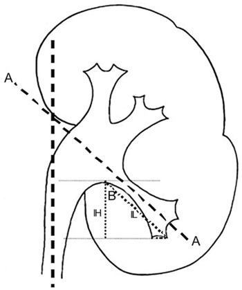
By
extending the line at the base of IH from the lowest part of the calyx
to the most distal point of the calyx, we have a right triangle with IL
as the hypotenuse.
Assuming
that angle B equals the IPA then the formula that relates IPA, IH and
IL is: cos(IPA) = IH/IL or IH = IL * cos(IPA)
Therefore,
since the three measures are dependent, given any 2, one should be able
to find the third. One would therefore expect that if IH is a predictor
of stone clearance, the relationship between IPA and IL would also be
a predictor of stone clearance.
Dr.
Ricardo Miyaoka,
Dr. W. K. Durfee & Dr. Manoj Monga
University of Minnesota
Edina, Minnesota, USA
E-mail: endourol@yahoo.com