OPTIMAL MINIMALLY
INVASIVE TREATMENT OF
URETEROLITHIASIS
M. HAMMAD ATHER
Section of Urology, Department of Surgery, The Aga Khan University, Karachi, Pakistan
ABSTRACT
Objective:
To determine the relative safety and efficacy of lithotripsy (intra-and
extra-corporeal) in the management of ureterolithiasis at various anatomical
subdivisions by auditing our experience between 1996 -1998.
Material and Methods: Medical records and
radiographic studies of patient with primary ureteric calculi treated
at this hospital between January 1996 and December 1998 were reviewed
for demographic data, site and size of calculi, treatment sessions, complications
and treatment outcome. Patients were divided into extra-corporeal lithotripsy
(ESWL) group and were treated on Dornier MPL 9000ä echo-guided lithotriptor
and intra-corporeal lithotripsy (ISWL) group treated with Swiss lithoclastä
(EMS, Switzerland).
Results: During this period 364 patients
with primary ureteric calculi were treated, of these 150 patients had
ESWL and 214 patients were treated by ISWL. Proximal abdominal ureteric
calculi were successfully treated in 92% by ESWL compared to 75% with
ISWL. Iliac stone were only treated by ISWL with a stone free (SF) rate
of 97%. For pelvic calculi SF was 95% for both groups.
Conclusions: In-situ ESWL is an ideal treatment
for ureteric calculi. URS + ISWL is an equally effective and safe modality.
ESWL is ideally suited for upper abdominal ureteric calculi. URS + ISWL
has a high SF for iliac ureterolithiasis. For pelvic ureteric calculi
results of the two treatment modalities are comparable with relatively
better safety profile for ESWL.
Key words:
ureter; ureteral calculi; lithotripsy; ureteroscopy
Braz J Urol, 27: 128-132, 2001
INTRODUCTION
Although
present day urologist’s armamentarium, is so replete with tools to
treat ureterolithiasis, management options are by no means less controversial
today to what it was nearly a decade back (1). Each individual stone,
presents the physician and the patient with a dilemma, in the era in which
myriad of management options are available.
Evolution of technology in the last two
decades has revolutionized the treatment of ureteric calculi. At one time,
open ureterolithotomy and stone basket manipulation were the mainstay
of treatment. But with the advent of lithotripsy, management of urolithiasis
has taken a quantum leap. Now with high safety and comparable efficacy
profile lithotripsy has superseded (2) all other treatment modalities
for ureteric calculi.
Presently the two most frequently used options
for ureteric calculi that require intervention, are ESWL and ISWL or contact
lithotripsy applied by attaining endoscopic access to the calculi. In
the present study we are presenting data from our institution on the usefulness
of these two modalities in the last three years in over 350 consecutive
patients with primary ureterolithiasis. Efficiency quotient (E.Q.) (3)
is used to determine the efficacy, whereas the safety profile is determined
by reviewing peri-operative morbidity. The relative efficacy of either
forms of lithotripsy in various anatomical subdivisions of ureter is also
determined to establish indications at different locations.
MATERIAL AND METHODS
We
retrospectively reviewed medical records and radiographic studies of consecutive
patients treated for primary ureteric calculi by either ESWL or URS +
ISWL between January 1996 and December 1998. Patients with multiple calculi,
or secondary ureteric calculi were excluded. Charts reviewed for demographic
data, site and size of calculus and complications during and after treatment
were noted and statistically analyzed. Size of the calculus is measured
in two dimensions on a plain x-ray (perpendicular and parallel to the
long axis of ureter). For the ease of description, the ureter is divided
into three parts. Abdominal ureter from ureteropelvic junction to upper
border of sacro-iliac (SI) joint; Iliac ureter lying parallel to SI joint
and pelvic ureter from lower border of SI joint to uretero-vesical junction.
Pre-procedural intravenous urogram was done
in all patients, unless contraindicated. A post treatment plain x-ray
and/or ultrasound confirmed complete stone clearance. Additional information
for patients who underwent ESWL included number of shock waves and average
energy setting. All patients in the ESWL group were treated on Dornier
MPL 9000ä echo-guided lithotriptor. In the URS + ISWL group, rigid
ureteroscope with a straight channel to accommodate the lithoclast probe
was used. Stone fragments were retrieved either with forceps or stone
basket.
Ureteroscopy was performed with a rigid
tapered (7 - 8.5 and 8 - 9.8F) scope after initial dilatation of ureteric
orifice and intra- mural ureter with either a tapered metal dilator or
balloon. Stone fragmentation performed by using pneumatic lithotripsy
with the Swiss lithoclastä (EMS, Switzerland). Post treatment JJ
stents (multi-length 4.7 and 6.0F) was left in place, whenever, the surgeon
felt that stone burden was large or ureteroscopy was prolonged or traumatic.
In all other patients a 5F open-ended ureteric catheter was placed, attached
externally to a 16F Foley’s catheter (4). The attending physician
responsible in the consulting clinics did post procedure evaluation. Treatment
outcome was defined as radiographic evidence of fragmentation and stone
clearance. Efficacy was subjectively assessed by efficiency quotient (EQ)
(3). EQ was determined by using the following formula:
![]()
All patients with abdominal (except for stones in the proximal 1-2 cm), iliac and proximal pelvic ureteric calculi were offered URS + ISWL as a primary therapeutic option. Patients with calculus in distal pelvic ureter were given an option to choose between the two options once they were explained the pros and cons of both modalities. Stones in the proximal 1 - 2 cm of abdominal ureter were primarily treated by ESWL unless there is difficulty in localization. Pushed back calculi were not considered in this study.
RESULTS
Overall
364 patients of primary ureteric calculi were treated in the 3-year period.
They are divided into ESWL and ISWL groups.
In the ESWL group, there were a total of
150 patients treated during the period. There was a clear male preponderance
with a male to female ratio of 2.5: 1 (108 males and 42 females). Age
ranged from 11 to 75 years (± 19.2) with a mean of 38 years. ESWL
patients are further considered in three groups; based upon the number
of treatment required to attain stone free status.
The ESWL patients who achieved complete
stone clearance in a single sitting included a total of 134 (89%) patients
with a male to female distribution of 2.9: 1 (100 males and 34 females).
There were 76 left sided and 58 right-sided calculi. Stone size determined
in two dimensions varied from 4 - 24 mm (7.2 mm) and 2 – 12 mm (10.2
mm) respectively. Majority (77%) had stones in the proximal and distal
ends of the ureter. Sixty-nine (51%) patients had stones in abdominal
ureter, 12 (9%) in proximal pelvic ureter, and 53 (40%) in distal pelvic
ureter. Average shock waves used were 1995.7. Energy setting was between
14 - 20 with a mean of 18.8 kV.
Thirteen (8.7%) patients in ESWL group required
two sittings for stone clearance. Size of the stone varied from 7 - 18
mm (mean 10.8) and 5 - 13 mm (mean 6.3). Average number of shock waves
was 3735 at an energy setting of 18.2 kV. There were 9 calculi in abdominal
ureter and 4 in the pelvic.
Three patients required three sittings of
ESWL for complete stone clearance. Mean stone size was 12 and 22 mm. Two
patients had calculi in proximal abdominal and one in proximal pelvic
ureter. They received a mean of 5553.3 shock waves at an average energy
setting of 18 kV.
Complications, requiring active intervention
or extra hospital stay were noted in 19 patients (13%). This included
pain requiring in-patient stay with injectable analgesics in 8 patients
(4.2%), and sepsis requiring injectable antibiotics in 8 (4.2%). Eight
patients developed stein-strasse; of these five had spontaneous passage;
1 patient, however, required ESWL to the leading fragment and 2 had to
have endoscopic procedure to clear the obstruction. These two patients
had residual fragments at a site not amenable to ESWL thereby requiring
ureteroscopy.
Of the 10 patients who failed to achieve
stone clearance with ESWL alone, 5 had primary treatment failure while
5 others had fragmentation without complete clearance. Of these 4 were
later treated endoscopically, using ISWL. One patient, however, required
open ureterolithotomy from impacted stone.
In the URS + ISWL group during the same
period, there were 214 patients. This comprises of 176 males and 38 females.
Age ranged from 18 - 70 (±17.2) years with a mean of 32 years.
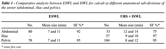
Mean stone size was 11 and 08 mm. Fifty-three stones were in abdominal
ureter, 57 in iliac and 104 in pelvic. Eighty-seven (41%) patients had
double J stent placed following ureteroscopy when URS is prolonged, there
are residual calculi, or there is mucosal edema or injury. Overall success
rate was 90% with URS + ISWL, whereas, 21 (10%) had some ancillary procedure
performed to attain stone free status. Differential success rate at various
sections of the ureter is detailed in (Table-1). High failure rate for
upper ureteric calculi was due to inadvertent push back into the lower
pole calyx or proximal sinuous ureter (in both conditions beyond the reach
of semi-rigid ureteroscopes), ureteric injury and mucosal edema rendering
further treatment difficult.
Of the 21 failed cases 16 had ESWL with
or without push back, whereas 5 patients had open surgery. Analysis of
the failed cases showed that there was technical difficulty in 6, inadvertent
pushback in 5 and large impacted calculi in the other ten warranting an
added procedure.
Complication rate was 10%; this included
prolonged pain requiring extra hospital stay for parenteral analgesics,
sepsis requiring parenteral antibiotics, and damage to ureteric wall requiring
placement of stent and percutaneous nephrostomy tube. Stone clearance
was clearly better in pelvic and Iliac ureter (92 - 97%) than in the abdominal
ureter, where it ranged from 60 - 81% (Table-2).
DISCUSSION
Technological
advancements in the last decade have made access to symptomatic ureteric
calculi possible from all directions. Antegrade approach for complex upper
ureteral calculi (5), retrograde approach with contact lithotripsy and
extracorporeal lithotripsy are all well established. A small subgroup
of patients can, however, only be managed by ureterolithotomy using either
conventional open approach (6) or laparoscopy (7). Though for routine
ureteral calculi general consensus is to go for either ISWL or ESWL (1,8).
Introduction of ureteroscopes in late 1970’s
opened a rare insight into the live anatomy of ureter. Initially, however,
because of the size constraint and rigidity of the instrument use of endoscopes
was confined to the distal ureter. Refinement of technology in the 1990’s
with the use of fibreoptic instrument has totally replaced large (11 and
13F) rigid ureteroscopes with 7F semi rigid and flexible ureterorenoscopes.
Simultaneously, development in the field of intra-corporeal lithotripsy
modalities has made possible to use finer less traumatic instruments through
the fibreoptic ureterorenoscopes. The transformation from the era of stone
baskets, forceps and electro-hydraulic lithotripsy to laser and lithoclast
has changed the way ureteric calculi are treated. All these have made
URS + ISWL a safe and effective means of treatment, even in the age dominated
by newer generation extra corporal shock wave lithotriptors.
ESWL was introduced in 1981 and rapidly
transformed the management of ureteric calculi. Although it initially
was only used for upper ureteral calculi, modification in the first generation
lithotriptors and development of 2nd generation machines with dual localization
have made possible to treat vast majority of ureteral calculi. Initially
most ureteral stones treated by ESWL were stented but reports in the last
five years have proved the efficacy of in situ ESWL (9). Besides the use
of stents increases the cost and morbidity of the procedure as well. It
is only indicated in the presence of significant obstruction (4,9).
We are reporting our early experience with
ISWL, which was performed by several urologists with varying degrees of
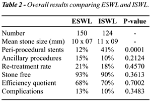
expertise. Complication rate was comparable to ESWL (13% for ESWL versus
10% for ISWL). Stone free rate for ISWL was 90%, marginally lower than
ESWL (93%). Efficiency quotient for the groups was also comparable (68
and 70%) for ESWL and ISWL groups respectively. Since only Ureteroscopy
was used for iliac ureterolithiasis, due to echo-guided nature of the
lithotripsy devise, it is obviously not possible to make comparison between
the two modalities for this anatomical site.
For abdominal ureteral calculi controversy
exists concerning in situ treatment and ESWL following push back. We had
comparable SF rate in the various sections of the ureter (92 - 95%) with
ESWL (Table-1). In this particular study we have not looked into the results
of calculi that were pushed back and as such our results may be biased.
Still the facts remain that stone that were subjected to in situ ESWL
had 92% success rate. Mueller et al. (10) reported a significant difference
in success rate (62 - 97%) between in situ treated upper ureteral calculi
and those that were pushed back. Graff et al. (11) concluded that obstructing
proximal ureteral calculi have poorer clearance (70%) compared to in situ
treated non obstructing (83%) and displaced calculi (95%). Liong et al.
(12), however, felt that results of by passed calculi are better than
pushed back (87% versus 81%). However, consensus, in the contemporary
urological literature (1) is that stone manipulation for second-generation
lithotriptors is not required. In our series for SF rate for abdominal
ureterolithiasis is 75% with URS + ISWL. This was in comparison to 95%
with ESWL. In our opinion, ESWL should be considered as a first option,
whenever the stone could be adequately focused.
Mid-ureteral stones (in section 4) are not
generally considered ideal for ESWL (13). We did not treat any iliac ureteral
calculi as our lithotriptor only has ultrasound localization. Although
both anterior and posterior approaches have been employed to treat section
4 calculi. Either approach is marred by significantly high re-treatment
rate (13) as shock waves are absorbed by bowel gas in the former and by
the dense pelvic bone in the later. In our study with URS + ISWL SF rate
is 97%.
Success of ESWL for pelvic ureterolithiasis
is dependent only upon adequate localization. Many investigators have
shown that in situ treatment is an ideal option for this location. All
of our patients were treated with in situ ESWL in pelvic ureter with SF
rate of 95%. With URS + ISWL similarly the clearance was in the range
of 95%. Although ISWL and ESWL have comparable stone clearance but safety
profile of ESWL is better and it should offered as a primary option.
Ideal treatment for ureterolithiasis would
render the patient stone free without anesthesia, has low morbidity and
cost. Such a modality is not currently available, however, significant
advances in the management of symptomatic ureterolithiasis have been made
recently most noticeable the in situ ESWL.
REFERENCES
- Netto Junior NR, Claro JA, Esteves SC, Andrade EF: Ureteroscopic stone removal in the distal ureter. Why change. J Urol, 157: 2081-2083, 1997.
- Singal RK, Denstedt JD: Contemporary management of ureteral stones. Urol Clin North Am, 24: 59-70, 1997.
- Clayman R, McClennan B, Garvin T: Lithostar: An electromagnetic acoustic unit for extracorporeal lithotripsy. J Endourol, 3: 307, 1989.
- Memon A, Ather MH, Sulaiman MN: Three points of technique to make rigid ureteroscopy simpler, safer and cost effective. Tech Urol, 2001 (unpublished).
- Alken P: Percutaneous ultrasonic destruction of renal calculi. Urol Clin North Am, 9 145-151, 1982.
- Paik ML, Wainstein MA, Spirnak JP, Hampel N, Resnick MI: Current indications for open stone surgery in the treatment of renal and ureteral calculi. J Urol, 159: 374-378; discussion 378-379, 1998.
- Raboy A, Ferzli GS, Ioffreda R, Albert PS: Laparoscopic ureterolithotomy. Urology, 39: 223-225, 1992.
- Turk TM, Jenkins AD: A comparison of ureteroscopy to in situ extracorporeal shock wave lithotripsy for the treatment of distal ureteral calculi. J Urol, 161: 45-46; discussion 46-47, 1999.
- Mobley TB, Myers DA, Jenkins JM, Grine WB, Jordan WR: Effects of stents on lithotripsy of ureteral calculi: treatment results with 18,825 calculi using the Lithostar lithotriptor. Urol, 152: 53-56, 1994.
- Mueller S, Wilbert D, Thueroff J, Alken P: Extracorporeal shock wave lithotripsy of ureteral stones: Clinical experience and experimental findings. J Urol, 135: 831-834, 1986.
- Graff J, Berding C, Pastor J: Extracorporeal shock wave lithotripsy for ureteral stones: A retrospective analysis of 417 cases. J Urol, 139: 513-533, 1988.
- Liong ML, Clayman RV, Gittes RF, Lingeman JE, Huffman JL, Lyon ES: Treatment options for proximal ureteral urolithiasis: review and recommendations (see comments). J Urol, 141: 504-509, 1989.
- Deliveliotis C, Kostakopoulos A, Stavropoulos NJ, Koutsokalis G, Dimopoulos C: Extracorporeal shock wave lithotripsy of middle ureteral calculi: ventral shock wave application. Urol Int, 56: 21-22, 1996.
__________________________
Received: September 22, 2000
Accepted after revision: April 16, 2001
_______________________
Correspondence address:
Dr. M Hammad Ather
Section of Urology, Department of Surgery
The Aga Khan University
Stadium Road, PO Box 3500, Karachi, Pakistan
Fax. ++ (92) (21) 493-4294
E-mail: hammad.ather@aku.edu