INTRAMURAL
LEIOMYOMAS OF THE BLADDER IN ASYMPTOMATIC MEN
(
Download pdf )
ROBERTO I. LOPES, ROBERTO N. LOPES, MIGUEL SROUGI
Women’s Beneficent Society, Syrian and Libyan Hospital, São Paulo, SP, Brazil
ABSTRACT
Bladder
leiomyomas are rare benign mesenchymal tumors, which account for less
than 0.43% of all bladder tumors with approximately 200 cases described
in the literature. These tumors may be classified into 3 different locations:
endovesical, intramural and extravesical. Endovesical is the most common
form, accounting for 63-86% of the cases, while intramural occurs in 3-7%
and extravesical in 11-30%.
The intramural form, especially small tumors,
may not produce symptoms hardening detection. We report two cases of intramural
bladder leiomyomas in asymptomatic men observed incidentally by transabdominal
ultrasonography during the follow-up of benign prostatic hyperplasia.
We discuss the diagnosis and management
of these lesions.
Key
words: leiomyoma; bladder; benign neoplasm
Int Braz J Urol. 2003; 29: 245-7
INTRODUCTION
Bladder leiomyomas are rare benign mesenchymal tumors that account for less than 0.43% of all bladder tumors (1). Approximately 200 cases have been described in the literature (1).
CASE REPORTS
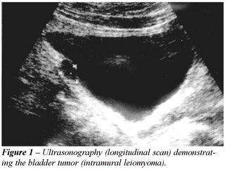
Case
1 - 59 year-old man with a 3-year history of benign prostatic hyperplasia
without clinical manifestations. During follow-up, a pelvic ultrasonography
demonstrated a well-circumscribed hypoechoic mass at the postero-superior
bladder wall measuring 1.74 x 1 cm (Figure-1). Cystoscopy demonstrated
a lesion covered with normal bladder mucosa. A transurethral resection
was performed and the pathologic examination revealed a leiomyoma. No
recurrence was observed after 10 months.
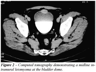
Case 2 - A 59 year-old asymptomatic man
had been accompanied for benign prostatic hyperplasia for 9 years. Transabdominal
ultrasonography revealed a 2.8 x 2.2 x 1.8 cm well-circumscribed hypoechoic
mass at the antero-superior bladder wall thought to be an urachal cyst,
due to its midline location. Computed tomography scan showed bilateral
renal cysts and a lesion at the bladder apex (Figure-2). Open segmental
resection was performed for the latter and pathologic examination revealed
leiomyoma. There has been no evidence of recurrence after 10 months.
COMMENTS
Bladder
leiomyomas have been reported to occur equally in both men and women (2).
However, more recently a review demonstrated predominance in women (3),
which may be attributed to the increased use of pelvic ultrasonography
in female patients (1). In our 2 cases, pelvic ultrasonography performed
during follow-up of benign prostatic hyperplasia led to incidental diagnosis
of bladder leiomyomas, suggesting that the reported predominance of these
tumors in women is questionable.
These tumors may be classified into 3 different
locations: endovesical, intramural and extravesical. Endovesical is the
most common form, corresponding to 63-86% of the cases, while intramural
occurs in 3-7%, and extravesical in 11-30% (2,3). Based on cystoscopic
findings, an intramural leiomyoma can be distinguished from an endovesical
tumor. Endovesical tumors are usually pedunculated or polypoid, while
intramural myomas are usually well encapsulated and surrounded by bladder
wall muscle.
The endovesical form usually causes irritative
or obstructive symptoms and gross hematuria (2) that result in detection
(1). Intramural form, especially small tumors, may not produce symptoms.
Radiologically, leiomyomas appear as well-circumscribed
hypoechoic masses at ultrasonography and as in case 2, these tumors may
be misinterpreted as other bladder lesions such as an urachal cyst when
observed in a bladder midline position. To rule out other benign lesions
and, especially, bladder cancer, the tumor should be biopsed.
Intramural tumors may be managed according
to their size and location. Small easily accessible tumors may be treated
with transurethral resection, while unfavorable positioning and recognition
difficulties may require segmental resection as in case 2. Management
of unfavorable lesions comprises open segmental resection or laparoscopic
partial cystectomy.
Histopathologically, leiomyoma of the bladder
is composed of fascicles of smooth muscle fibers separated by connective
tissue. The etiology of these tumors remains unknown. It is proposed that
bladder leiomyomas may arise from chromosome abnormalities (1), hormonal
influences, bladder musculature infection, perivascular inflammation or
dysontogenesis (3).
REFERENCES
- Cornella JL, Larson TR, Lee RA, Magrina JF, Kammerer-Doak D: Leiomyoma of the female urethra and bladder: report of twenty-three patients and review of the literature. Am J Obstet Gynecol. 1997; 176:1278-85.
- Knoll LD, Segura JW, Scheithauer BW: Leiomyoma of the bladder. J Urol. 1986; 136:906-8.
- Goluboff ET, O’Toole K, Sawczuck IS: Leiomyoma of bladder: report of case and review of literature. Urology 1994; 43:238-42.
_______________________
Received: January 10, 2003
Accepted after revision: April 14, 2003
_______________________
Correspondence address:
Dr. Roberto Iglesias Lopes
Rua Baronesa de Itu, 721 / 121
São Paulo, SP, 01231-001, Brasil
Fax: + 55 11 3666-8266
E-mail: robertoiglesias@
terra.com.br
EDITORIAL COMMENT
Leiomyomas
of the bladder is distinctly unusual as the author’s report. They
provide a concise case report of 2 men who were discovered to have this
unusual lesion of the bladder.
They provide a wide range of incidences
for the 3 locations for a leiomyoma of the bladder. It would seem that
given the paucity of this tumor, that it would be difficult to indicate
other than the most common location, is what they term endovesicle. I
am not even certain what they mean by endovesicle and how they differentiate
this from intramural with any precision.
It would seem that an important part of
this manuscript, which is overlooked, is whether one can make the diagnosis
based upon radiographic configuration and avoid any surgery. The authors
do not provide this as an option and simply state that there are several
ways of removing these tumors. Since this is a benign neoplasm and if
there are no signs or symptoms, one would wonder why it would be necessary
to remove the lesion. For instance, if a percutaneous biopsy was performed
and the diagnosis was a benign leiomyoma, would it be necessary to proceed
with any further surgery, such as removal?
Dr.
Mark S. Soloway
Professor and Chairman of Urology
University of Miami School of Medicine
Miami, Florida, USA
REPLY BY THE AUTHORS
The
term endovesical refers to the submucosal growth of leiomyoma, first described
by Campbell & Gislason (1). The endovesical (submucosal) tumors are
usually pedunculated or polypoid, while intramural leiomyomas are surrounded
by the musculature of the bladder wall (as in these 2 cases reported)
and are usually well encapsulated. Distinction between these 2 types is
based on cystoscopic findings.
To rule out other benign lesions, and, especially,
bladder cancer that may have the same radiologic appearance of an intramural
leiomyoma, the tumor should be biopsed. Since bladder leiomyomas are rare
tumors, there is no trial comparing tumor observation and surgery for
the management of these lesions.
The Authors