RENAL
LIPOSARCOMA
(
Download pdf )
DIOGO A.L. BADER, LUIS A.B. PERES, SÉRGIO L. BADER
West Paraná State University, UNIOESTE, Cascavel, Paraná, Brazil
ABSTRACT
Introduction:
Liposarcoma is a malignant mesenchymal tumor frequently located in retroperitoneum,
and rarely presenting an isolated lesion in kidney.
Case Report: Female, Caucasian, 49-year
old patient, with family history of renal polycystic disease, was selected
for organ donation. During preoperative examinations a renal pleomorphic
liposarcoma was detected. She was treated with radical nephrectomy and
remains asymptomatic, without evidences of recurrence in control ecographic
examinations after a 4-year follow-up.
Comments: Renal liposarcoma is a rare tumor.
We report one case incidentally diagnosed during a routine pre-transplantation
assessment in renal donor.
Key
words: kidney; kidney neoplasms; liposarcoma
Int Braz J Urol. 2004; 30: 214-5
INTRODUCTION
Liposarcoma is a malignant mesenchymal tumor frequently located in the retroperitoneum (1). Isolated lesion in kidney has rarely been described (2). We present a case of renal liposarcoma incidentally diagnosed during the assessment of a candidate to renal donation for transplantation.
CASE REPORT
Female,
Caucasian, 49-year old patient, with family history of renal polycystic
disease, was selected for organ donation. During preoperative examination
a rounded, heterogeneous, well-defined mass with solid aspect was detected
by renal ultrasonography, adjacent to the lower pole of the left kidney.
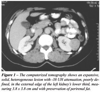
A computerized tomography was performed, showing an expansive, solid,
heterogeneous lesion with -38 UH attenuation, poorly defined in the lower
third’s external edge, measuring 3.8 x 3.8 cm and with preservation
of perirenal fat. There was no contrast medium impregnation in the tumoral
lesion during the late phase (Figure-1). The angiography showed a hypovascularized
and hypodense mass. The intravenous urography was normal. Radical nephrectomy
was performed following the intraoperative freezing diagnosis of malignant
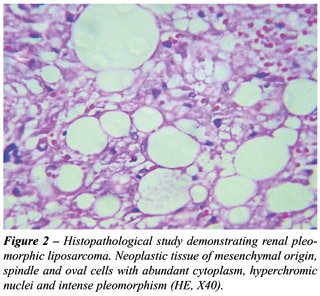
lesion. The pathological examination revealed a brownish nodular structure
with 4.8 cm in diameter, and the microscopy detected neoplastic tissue
of mesenchymal origin, spindle and oval cells with abundant cytoplasm,
hyperchromic nuclei and intense pleomorphism (Figure-2), characteristic
of a renal pleomorphic liposarcoma. The patient has been followed up for
4 years and remains asymptomatic, without evidence of recurrence on control
ecographic examinations.
COMMENTS
Renal
liposarcoma is a rare tumor. There are few well-documented reports in
the literature, many of those are associated with tuberous sclerosis and
probably correspond to angiomyolipomas. The majority of published cases
refer to well-differentiated tumors, with dimensions greater than 5 x
5 x 4 cm and presenting symptoms such as pain, hematuria, abdominal mass
or loss of weight. The liposarcoma is classified according to the histological
type, in well-differentiated, myxoid and pleomorphic. The myxoid type
occurs in 60%, the well-differentiated in 25% and the pleomorphic in 10%
of the cases. The pleomorphic type is highly aggressive with high rates
of metastases (2). We describe an incidentaloma of the pleomorphic type
with 4.8 cm in diameter.
Perirenal localization is often observed
in such tumors, which can mimic renal cystic tumor. The differential diagnosis
must include renal cell carcinoma or atypical angiomyolipoma. Some features
in the computerized tomography, such as linear vascularization, aneurismal
dilatation, hematoma and presence of tissue with fat attenuation speak
for angiomyolipoma. Frequently the definitive diagnosis is achieved only
through the pathologic examination (3).
The prognosis of liposarcomas depends on
the degree of differentiation, size, histological type and tumor staging.
The total surgical resection with free margins offers a good probability
of cure (2). The standard treatment has been radical nephrectomy, associated
or not with radiotherapy. Clinical follow-up is important to monitor tumor
recurrence. There is a report of recurrence 13 years after the initial
surgery (2). The case we described here was treated with radical nephrectomy,
presenting a 4-year follow-up, without evidence of recurrence to this
moment.
Dr. José R. L. Ferreira and Dr. Alexandre Galvão
Bueno assisted in preparing the images and
the histological material.
REFERENCES
- Lopes RI, Lopes RN, Filho B: Giant retroperitoneal liposarcoma. Int Braz J Urol. 2002; 28: 227-9.
- Mayes DC, Fechner RE, Gillenwater JY: Liposarcoma renal. Amer J Surg Pathol. 1990; 14: 268-73.
- Wang LJ, Wong YC, Chen CJ, See LC: Computerized tomography characteristics that differentiate angiomyolipomas from liposarcomas in the perinephric space. J Urol. 2002; 167: 490-3.
__________________________
Received: September 29, 2003
Accepted after revision: January 21, 2004
________________________
Correspondence address:
Dr. Diogo Alberto Lopes Bader
Praça Getúlio Vargas, 55 / 10
Cascavel, PR, 85801-220, Brazil
E-mail: diogobader@hotmail.com