MANAGEMENT
OF COMPLETE URETERAL OBSTRUCTIONS WITH A TRANSLUMINAL PUNCTURE TECHNIQUE
(
Download pdf )
TULGA EGILMEZ, SEZGIN GUVEL, FERHAT KILINC, OZGUR YAYCIOGLU, HAKAN OZKARDES
Department of Urology (TME, SG, FK, OY), Adana Clinic and Research Center, Baskent University School of Medicine, Adana, and Department of Urology (HO), Baskent University School of Medicine, Ankara, Turkey
ABSTRACT
Introduction:
The traditional delayed treatment of iatrogenic complete ureteral obstruction
is open surgery. An easy endourological technique, transluminal re-canalization
of the ureter by guide-wire puncture under fluoro-endoscopic control,
which has been performed on 4 patients, is described.
Surgical technique: With the guidance of
C-arm fluoroscopy, by moving the C-arm to different planes, the tip of
the ureteroscope is directed to the correct plane to meet the obliterated
proximal end of the ureter and under direct vision, transluminal puncture
is performed using the stiff end of a 0.035-inch guide wire. Once the
stiff end of the guide-wire is in the lumen of the proximal ureter, an
ureteral catheter is introduced over the guide wire, the guide wire is
then removed and reinserted through the ureteral catheter with its soft
end leading and a double J catheter is inserted. Ureteral stricture, if
later encountered, is treated with balloon dilatation.
Results: Continuity of the ureter was restored
in all 4 patients. The double J stents were removed 6 weeks later and
a retrograde pyelography revealed resolution of the hydronephrosis without
extravasation of urine.
Conclusion: Although a very satisfactory
result was achieved in our cases, more cases are needed to show if it
can be an alternative to conventional surgical repair. However, we believe
that this minimally invasive technique can be used for short obliterated
ureteral segments and neither delays nor does it preclude further management
using open surgery.
Key
words: ureteral obstruction; urinary fistula; ureteroscopy; surgical
procedures, minimally invasive
Int Braz J Urol. 2005; 31: 264-8
INTRODUCTION
Ureteral
injury during gynecologic surgery is an infrequent but serious complication,
with an estimated incidence from 0.5 to 4 percent of all procedures (1).
The traditional delayed treatment for complete ureteral injuries is open
surgery preceded by several weeks to months of nephrostomy drainage.
We report an easy technique, transluminal
re-canalization of the ureter by guide-wire puncture under fluoro-endoscopic
control, used to treat 4 patients with postoperative ureteral damage leading
to complete ureteral obliterations and an ureterovaginal fistula.
MATERIALS AND METHODS
Total ureteral obstruction was revealed by intravenous pyelography (IVP) in 3 patients, 35 to 45 years old, who had admitted to the outpatient clinic with flank pain and also in another 41 years old patient with concomitant ureterovaginal fistula who presented with urinary incontinence. A proximal ureteral obstruction in one patient and distal ureteral obstructions in 3 patients were due to pyelolithotomy and total abdominal hysterectomy, respectively, which they had underwent 20 days to 3 months ago. The patient with total ureteral obstruction and also an ureterovaginal fistula noted urine leakage from her vagina with a need of approximately 30 pads per day to remain dry. Methylene blue instilled into the bladder showed no dye in the vagina and ureterovaginal fistula was confirmed by spiral-computed tomography. IVP revealed grade 3 hydronephrosis and non-visualization of the distal ureteral segment in all of the patients with distal ureteral obstruction and non-visualization of the ureter below the ureteropelvic junction in the patient with the proximal ureteral obstruction. All of the patients were initially treated with percutaneous nephrostomy catheter drainage followed by re-canalization of the ureter by guide-wire puncture under fluoro-endoscopic control. Appropriate antibiotics according to the patients’ urine cultures were initiated preoperatively to all of them and were continued for 5 days after the operation.
Surgical Technique
1. The patient
is positioned in the lithotomy position. Retrograde ureterography and
an antegrade pyelography are performed simultaneously revealing the distance
between the 2 ureteral ends (Figures-1 and 2).

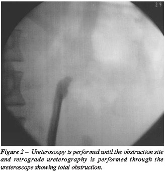
2. Ureteroscopy is performed until the site of obstruction. With the guidance
of C-arm fluoroscopy, by moving the C-arm to different planes, the tip
of the ureteroscope is directed to the correct plane to meet the obliterated
proximal end of the ureter (Figure-1) and under direct vision, transluminal
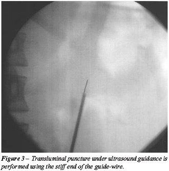
puncture is performed using the stiff end of a 0.035-inch guide wire (Figure-3).
3. Once the stiff end of the guide-wire is in the lumen of the proximal
ureter, an ureteral catheter is introduced over the guide wire, the guide
wire is then removed and reinserted through the ureteral catheter with
its soft end leading.
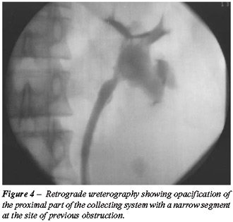
4. Leaving the guide wire in place, retrograde pyelography is carried
out through a double lumen catheter or through the side port of the ureteroscope
(Figure-4).
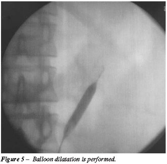
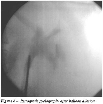
5. If necessary balloon dilation is performed (Figures-5 and 6).
6. A double J catheter is inserted.
In
the patient with a concomitant ureterovaginal fistula, retrograde pyelography
showed complete obliteration of the ureter approximately 4 cm from the
ureteral orifice and an antegrade pyelography performed simultaneously
revealed an approximately 5 mm distance between the two ureteral ends
together with an ureterovaginal fistula tract originating from the distal
dilated ureter. In the other 2 patients with total distal ureteral obstructions,
the distance between the 2 ureteral ends was approximately 4 mm and they
underwent the same procedure.
To the patient with a total proximal ureteral
obstruction just below the ureteropelvic junction, the procedure was performed
with ultrasound guidance instead of antegrade pyelograpy due to spontaneous
dislocation of the percutaneous nephrostomy catheter prior to surgery.
The retrograde pyelography carried out after the procedure, revealed the
ureteropelvic junction to be slightly narrow and balloon dilation was
performed to the ureteropelvic junction.
RESULTS
Continuity
of the ureter was restored in all 4 patients. In the patient with a concomitant
ureterovaginal fistula, the vaginal leakage ceased on the day of the operation
allowing removal of the percutaneous nephrostomy catheter. The patients
tolerated their stents reasonably well preventing early removal. The double
J stents were removed six weeks later and a retrograde pyelography revealed
resolution of the hydronephrosis without urine extravasation. Follow-up
urine cultures were sterile in all of the patients. In 2 patients (50%)
with distal ureteral obstruction, IVP performed in the 3rd postoperative
month showed mild stenosis at the previous obstruction site. Although
Mag-3 diuretic renal scintigraphy showed no urinary obstruction, these
patients were managed with balloon dilatation due to intervals of flank
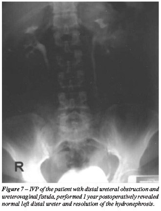
pain. Follow-up IVP 1 year postoperatively was normal in all of the patients
(Figure-7).
COMMENTS
Management
of an ureteral injury is usually complicated by a delay in diagnosis.
After the diagnosis is confirmed, immediate corrective surgery, temporary
nephrostomy catheter insertion with delayed corrective surgery, or observation
and delayed repair for persistent fistula are the conventional treatment
options. The technical difficulties associated with reoperation in an
anatomically distorted field has led search for endourological techniques.
Percutaneous nephrostomy and/or antegrade ureteral stent insertion have
previously been reported to be effective. Persky and associates reported
iatrogenic ureteral injuries in which percutaneous nephrostomy was utilized
to demonstrate the site of injury, to relieve the symptoms or to control
wetness by urinary diversion (2). Lang and associates have reported 5
successfully treated ureteral injuries managed with percutaneous ureteral
catheterization alone in which 4 were accompanied with fistulas (3). Retrograde
passage of ureteral stents may not always be successful due to angulation,
stenosis or complete obstruction. Ureteroscopy has been used to overcome
angulation and stenosis allowing visual assessment of the injured area
and passage of a guide-wire or catheter if the ureteral lumen is identified
(4). However, ureteroscopy alone fails when complete obliteration of the
ureteral lumen exists. We have used a technique similar to a needle puncture
procedure previously utilized in the management of urethral obliterations
in order to discard the need for open surgery (5).
Although a very satisfactory result was
achieved in our cases, more cases are needed to show if it can be an alternative
to conventional surgical repair. However, we believe that this minimally
invasive technique can be used for short obliterated distal ureteral segments
and neither delays nor does it preclude further management using open
surgery.
REFERENCES
- Bunkin IA: Prevention of ureteral injury in gynecologic surgery. Clin Obstet Gynecol. 1965; 17: 383-98.
- Persky L, Hampel N, Kedia K: Percutaneous nephrostomy and ureteral injury. J Urol. 1981; 125: 298-300.
- Lang EK, Lanasa JA, Garrett J, Stripling J, Palomar J: The management of urinary fistulas and strictures with percutaneous ureteral stent catheters. J Urol. 1979; 122: 736-40.
- Bagley DH: Indications for Ureteropyeloscopy. In: Huffman J, Lyon E, Bagley D (eds.). Ureteroscopy. Philadelphia, WB Saunders. 1988; pp. 17-30.
- Takeuchi T, Ishihara S, Nagatani Y, Koide T, Sakai S, Tamaki M et al.: Endourological re-establishment by transluminal puncture for a complete obliteration or traumatic urethral disruption. Nippon Hinyokika Gakkai Zasshi. 1991; 82: 750-7.
_________________________
Received: September 29, 2004
Accepted after revision: March 24, 2005
_______________________
Correspondence address:
Dr. Tulga M. Egilmez
Department of Urology, Baskent University
Dadaloglu Mah. 39 Sok. No: 6
01250, Yuregir, Adana, Turkey
Fax: + 90 322 327-1273
E-mail: tulgaegilmez@yahoo.com