MALE
PERINEAL SLING WITH AUTOLOGOUS APONEUROSIS AND BONE FIXATION – DESCRIPTION
OF A TECHNICAL MODIFICATION
(
Download pdf )
LUIS A.S. RIOS, RODRIGO T. TONIN, RENATO PANHOCA, OSMAR E.R. DE SOUZA, LIMÍRIO L. DA FONSECA FILHO, DEMERVAL MATTOS JR.
Department of Urology, State Civil Servant’s Hospital, São Paulo, SP, Brazil
ABSTRACT
Post-prostatectomy
urinary incontinence is an uncommon complication of adenomectomies, occurring
in approximately 1% of cases and being more frequent following radical
prostatectomies. There is a significant implication in the quality of
life for these patients. The surgical techniques employed for its treatment
are the implantation of an artificial sphincter, peri-urethral injections
and suburethral slings.
Considering the low efficacy of peri-urethral
injections and the high cost of artificial sphincters, we present in this
work a technical modification of the suburethral sling, whose preliminary
results are satisfactory.
The fundamental modification in this technique
is due to the replacement of the synthetic material usually employed for
making the sling for autologous tissue, constituted by an aponeurotic
strip taken from the rectus muscle of abdomen.
This modification aims to minimize risks
of urethral erosion that, despite it was not described in this population
due to the use of synthetic materials, is a possibility when facing the
tension that is used over the bulbar urethra. In addition to such aspects
the autologous aponeurosis does not have a cost except for a short prolongation
of the surgical act.
Key
words: urinary incontinence; male; perineum; urinary sphincter,
artificial; transplantation, autologous
Int Braz J Urol. 2003; 29: 524-527
INTRODUCTION
Post-prostatectomy
urinary incontinence (PPUI) is an uncommon complication of adenomectomies,
whether they are performed by transurethral or transvesical route. It
is estimated that approximately 1% of patients who undergo these surgeries
develop urinary incontinence.
After the advent of the routine use of PSA,
however, the number of radical surgeries for treatment of prostate adenocarcinoma
has significantly increased and brought along an equally important increase
in the diagnoses of incontinence following radical prostatectomies. It
is estimated that the increase in the number of diagnosis of prostate
cancer has been around 467% in the last 12 years, leading to a real increase
of 404% in the number of prostate radical surgeries in the same period.
The actual incidence of PPUI is hard to
assess, since the definition used for diagnosing incontinence varies substantially
between authors and expressions such as “socially continent”,
“minimal urinary leaks” contribute to make the objective interpretation
of results expressed in the literature even more complicate. It is possible
to find incidence reports from 2 to 87% when analyzing specific publications
(1). Recently published, an analysis of PPUI indexes based on data from
the North-American public health system (Medicare), revealed that 50%
of patients presented some degree of urinary incontinence. Among the incontinent
patients 32% used penile clamps continuously and 6% required surgical
treatment for the problem. A study on incidence conducted at a University
Hospital revealed that 8.2% of patients who underwent radical prostatectomy
(RP) evolved with PRPUI (2).
The surgical alternatives used in the treatment
of PPUI include the use of peri-urethral injectables, artificial sphincter
or suburethral slings.
Peri-urethral injections are technically
simple and minimally invasive procedures, which can be performed at an
outpatient setting. Among the substances most often employed, bovine collagen,
autologous fat, texturized silicone and more recently pyrolitic carbon
stand out. Despite the simplicity of these procedures, there are serious
issues concerning the method’s costs and efficacy. In our setting,
injectables are marketed at high costs, making their routine use unfeasible,
especially when one considers the frequent need of repeat injections for
obtaining and maintaining the results. The analysis of results from peri-urethral
injections, in turn, shows that the indexes of cure and significant improvement
in urinary leaks lies around 30-60%, with a follow-up longer than 24 months,
being variable according to the substance employed (3). The artificial
sphincter is considered as the standard therapy for treating PPUI today.
Introduced in 1973, by Scott and colleagues, the device underwent some
modifications and technical refinements that made it highly reliable in
terms of biocompatibility and function, as well as in terms of efficacy
in controlling urinary incontinence. The currently available design, AMS
800, provides positive results in 60 to 95% of treated patients (4).
Even though the artificial sphincter is
currently a routine and low-risk procedure, its use is not free from complications
and technical problems. Among the possible complications, the most common
are infection, urethral erosion and poor mechanical function. All of those
complications require a mandatory surgical treatment and, in the case
of infection, it involves the early removal of the sphincter and its connections.
Urethral erosion is due to bulbar ischemia and forces the removal of the
urethral cuff until the wound is completely healed. Bulbar atrophy, which
can occur as a result of chronic bulbar compression, manifests through
recidivation or worsening of the urinary incontinence, and can be treated
by replacing the cuff or placing a second cuff distal to the first one.
The cost of the AMS 800 sphincter, associated
with the fact that the device is not available in the public health service
prevents its application in the vast majority of potential candidates
in our country.
Recently, Comiter (5) described the placement
of a suburethral sling in men with PPUI using the perineal approach and
the synthetic strip fixed by means of screws placed in the ischiopubic
rami. The procedure, technically simple, and with low indexes of complications
in the first series reported, showed quite satisfactory results in short
and medium-term follow-up.
Even though there was no erosion in the
first series of male slings that were presented, we know from previous
experience with female population that the use of synthetic material under
the urethra bears a risk of erosion of adjacent tissues. Such complication,
quite frequent with the use of some substances that were employed in the
past for treating female SUI, has turned the autologous fascia into the
standard material for treatment of SUI in incontinent women.
The use of orthopedic anchors placed in
the ischiopubic rami allows the proper fixation of the fascial flap to
the bony tissue in a safe and technically simple way. The option for the
use of anchors is based in the easy handling, low cost, safety and comfort
since the suture thread is already present in the material. The cost of
these anchors is less than 5% of the artificial sphincter’s value,
making the procedure widely advantageous even when using 4 units for proper
fixation.
SURGICAL TECHNIQUE
The
patient is placed in lithotomy position, under general or regional anesthesia;
the lower abdomen, the genitals, perineum and the thighs are prepared.
Through a transverse incision in the lower
abdomen, a strip is removed from the aponeurosis of the rectus muscle
of abdomen, measuring approximately 6.0 x 2.0 cm. This graft is kept in
warm saline solution with antibiotics (gentamicin 160 mg in 500 mL of
saline) until the moment of its use.
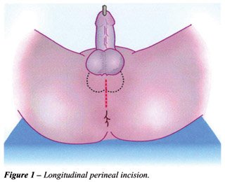
A 5-cm longitudinal perineal incision is
made (Figure-1), the bulbocavernous muscle is identified and then incised.
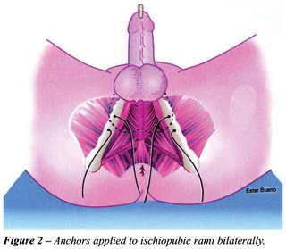
The ischiopubic ramus is dissected bilaterally and the periosteum is identified
through a 3-cm incision over the ischiocavernous muscle. Two special ortopedic
screws are placed (anchor FASTIN Mitekâ) in each ischiopubic ramus
2 cm apart. These screws have a polyester wire measuring approximately
25 cm in length, coupled to its plane surface (Figure-2).
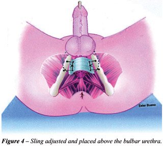
Once the screws have been placed, the aponeurotic
fascia (Figure-3) is fixed with wires to the bones, over the bulbo-urethral
muscle, compressing it with a controlled pressure (Figure-4). The pressure
with which the sling compresses the urethra is controlled using retrograde
perfusion sphincterometry. An 8F urethral catheter is inserted in the
distal urethra, occluding the glans around the catheter so that there
is no leakage of fluid. This catheter is connected to the liquid column
placed 60 cm above the patient’s pubic symphysis. When the fluid
is open in order to run backwards through the urethra, the bulbo-urethral
muscle is compressed with the sling until the dripping stops. In this
way we impose a resistance of 60 cm of water to the passing of fluids
through the urethra, a pressure that is similar to the one used in the
artificial sphincter.

After fixing the sling, the surgical wound
is copiously irrigated with antibiotic solution and the incision is drained
and closed in 2 planes.
COMMENTS
The use of this procedure for correcting PPUI has shown to be an alternative to the employment of synthetic material, minimizing, like those slings used for correction of female UI, the risk of urethral erosion. It is a technical modification that is easy to reproduce and has an acceptable cost when compared with the use of artificial sphincters.
REFERENCES
- Rudy DC, Woodside JR, Crawford ED: Urodynamic evaluation of incontinence in patients undergoing modified Campbell radical retropubic prostatectomy: A prospective study. J Urol. 1984; 132: 708-11.
- Goluboff ET, Said JA, Mazer S, Bagiella E, Heitjan DF, Benson MC, et al.: Urinary continence after radical prostatectomy: The Columbia experience. J Urol. 1998; 159: 1276-9.
- Rios LAS, Panhoca R Borrelli IM: Injectables in the Treatment of Urinary Incontinence. In: Rubinstein I (ed.), Brazilian Clinics of Urology. 2001; vol 1: 265-74 [in Portuguese].
- Gundian JC, Barret DM, Parulkar BG: Mayo Clinic experience with use of the AMS 800 artificial urinary sphincter for urinary incontinence following radical prostatectomy. J Urol. 1989; 142: 1459-967.
- Comiter C V: The male sling for stress urinary incontince: A prospective study. J Urol. 2002; 167: 597-601.
____________________
Received: May 16, 2003
Accepted after revision: October 6, 2003
_______________________
Correspondence address:
Dr. Luis Augusto Seabra Rios
Rua Conselheiro Brotero, 1539 / 2o andar
São Paulo, SP, 01232-011, Brazil
Fax: + 55 11 3826-2871
E-mail: seabrarios@uol.com.br