STRUCTURAL
ALTERATIONS OF THE BLADDER INDUCED BY DETRUSOR INSTABILITY. EXPERIMENTAL
STUDY IN RABBITS
(
Download pdf )
JOAO L. AMARO, KARINA T. BALASTEGHIN, CARLOS R. PADOVANI, RENATA MONTENEGRO
Department of Urology (JLA, KTB, RM) and Department of Statistics (CRP), School of Medicine, State of Sao Paulo University, UNESP, Botucatu, SP, Brazil
ABSTRACT
Objectives:
The aim of this study was to evaluate the histopathological and immunohistochemical
alterations induced by detrusor instability in the bladder of rabbits
submitted to partial bladder outlet obstruction.
Materials and Methods: Thirty male Norfolk
rabbits were divided into 2 groups, a clinical control and a group with
detrusor instability. Urine culture, cystometric study, histopathological
and immunohistochemical analysis were performed in all animals prior to
surgery (M1) and 4 weeks after-surgery (M2).
Results: Partial obstruction (G2) resulted
in a 2.5 fold increment (p < 0.05) in bladder weight when compared
to control (G1). Four weeks after surgery, 93% of animals in G2 developed
cystitis. Partial obstruction resulted in detrusor instability at M2 and
bladder capacity was significantly increased (p < 0.05) from M1 to
M2. The incidence of mild to moderate mucosal and adventitious fibrosis
at M2 was higher in G2 (p < 0.05) when compared to G1. Inflammatory
reaction at M2 was statistically higher (p < 0.05) in G2. There was
no difference in muscular hypertrophy between M1 and M2 in G1. However,
67% of G2 bladders showed a moderate to intense muscular hypertrophy at
M2. Hyperplasia of the epithelium was also increased in G2 when M1 and
M2 were compared (p < 0.05).
Conclusion: Detrusor instability induced
by partial bladder outlet obstruction caused significant histopathological
and immunohistochemical alterations in the bladder of rabbits.
Key
words: bladder; rabbits; bladder outlet obstruction; histopathology
Int Braz J Urol. 2005; 31: 579-86
INTRODUCTION
Detrusor
instability is a common phenomenon in urological practice that may be
seen in men with bladder outlet obstruction (BOO) (1) and in women with
urge incontinence, causing psychosocial and sexual problems (2).
Experimental model of partial bladder outlet
obstruction in pigs (3) and rats (4) showed structural and functional
detrusor changes quite similar to those caused by voiding dysfunction
in human. In rabbits, morphologic changes often associated with BOO include
progressive denervation and hypertrophy of the bladder wall (5,6).
Physiological changes that occur in the
bladder in response to obstruction, which may lead to disorders in function
including detrusor instability, are poorly understood. Thus, there is
a need for a suitable animal model that mimics the response of the bladder
detrusor instability, for further understanding of BOO.
Our objective was to evaluate the histopathological
and immunohistochemical changes in bladder using an experimental model
of detrusor instability in rabbits with partial bladder outlet obstruction
(PBOO).
MATERIALS AND METHODS
Thirty-five
male Norfolk rabbits weighing between 1.700g and 2.820g (average 2.140
± 200) were randomly divided into 2 groups. Group 1 (G1, n = 15)
served as clinical control with no surgical intervention. The remaining
20 animals were submitted to laparotomy, and an adjustable polyethylene
bracelet was placed around the bladder neck without compression of the
urethra, previously catheterized with a 10F catheter, as previously described
(7). From this group, only 15 rabbits developed detrusor instability verified
by cystometric studies one week after surgical intervention. Since the
objective of this trial was to investigate vesical alterations in animals
with detrusor instability due to partial bladder obstruction, the 5 rabbits
that did not show involuntary contraction of the bladder were not included
in the trial. Therefore, group 2 (G2) consisted of the 15 animals with
detrusor instability.
Urine culture, serum creatinine and cystometric
evaluation were performed in all animals at different moments: one week
after surgical procedure (M1) and 4 weeks after surgery (M2). Animals
with positive urine culture at M1 were treated with 1 mg/Kg/day of intramuscular
trimethropim.
The cystometric evaluation was performed
using the UrobyteTM 5000 computed urodynamics system. To measure intravesical
pressure a 10F Foley catheter was used and to measure intraabdominal pressure,
a catheter with a rectal balloon was used (8). During this exam, the vigil
animals were placed in wooden cages, with posterior extremities held by
cotton strings. After antisepsis of the penile area with topic povidone
and lubrication with 2% xylocaineTM jelly, a 10F double-way Foley catheter
was introduced into the bladder with vesical emptying, and urine was drawn
in a sterile tube for urine culture.
After lubricating the rectal balloon with
2% XylocaineTM jelly, it was introduced 2 cm beyond the anal margin and
connected to a 2-way tap, injecting 0.4 mL of sterile water in one of
the ways in order to the balloon wall and the rectal mucosa came into
contact. The other way connected to the pressure transmitter. The vesical
catheter was connected to a 2-way tap, and one of the ways was connected
to the pressure transmitter and the other to a continuous infusion pump
(2 mL/min). To verify the good placement of the catheters a slight compression
of the abdomen was made in order to obtain an abdominal pressure curve,
which was transmitted to the bladder, and, consequently obtaining the
vesical curve.
The cystometry was initiated proceeding
to the simultaneous measurements of the abdominal and vesical pressures.
When urinary leakage around the vesical catheter was observed, the exam
was discontinued. At that moment, we verified the vesical pressure (VP)
and the abdominal pressure, also measured in the beginning of the urinary
leakage (VP-AbdP) expressing maximal detrusor pressure (DeiP). Maximal
bladder capacity was considered the one, which attained a filling volume
enough to yield bladder contractions. Vesical compliance was obtained
by the formula: Fv-Iv/Fp-Ip where Fv = Final volume, Iv = Initial volume,
Fp = Final pressure and Ip = Initial pressure. Uninhibited contractions
were considered those detrusor involuntary contraction with low vesical
volume, yielding or not simultaneous urinary leakage.
At M2, after the cystometric evaluation,
blood and urine collection all animals were euthanised and their bladders
collected and weighed using a laboratory micro scale. Samples from the
bladder were fixed in formaldehyde for 24 hours. Then they followed the
protocol of the automated inclusion processor Leica TP-1020, where they
were dehydrated in 100% ethanol, followed by clarification in xylol. The
samples then were embedded in liquid paraffin using the automated inclusion
system EG: 1160 Leica. The blocks were sectioned (4 microns) and stained
Hematoxylin and Eosin for histopathological studies. To investigate the
occurrence of hyperplasia, immunohistochemistry was carried out using
KI 67 (primary antibody diluted in PBS 1:100, Dako, Carpinteria, CA, USA),
BA2000 and Pk6100 (secondary antibodies diluted in PBS 1:200, Dako, Carpinteria,
CA, USA).
For the different groups, the Mac Neman
test was used in the study of the combination of urine culture and uninhibited
contractions in the beginning and the end of the evaluation. Comparison
of the groups’ mean profile along both moments of evaluation was
performed through the analysis of repeated measures, considering both
groups independently. For the histological and immunohistochemical analysis,
the Goodman test was used.
RESULTS
Four
weeks after surgery, bladder weight in G2 was 2.5 times heavier than G1
(p < 0.05). Partial outlet obstruction was also responsible for an
increase in bladder infection. Urine samples collected at M2 showed that
93% of G2 had positive urine cultures compared to 13% in the G1 group
(p < 0.001).
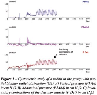
The cystometric studies showed that detrusor
instability was observed in all G2 animals (Figure-1). In G2, maximum
cystometric capacity (Volmax) was higher (p < 0.05) at M2 if compared
to M1 (Table-1). Maximum detrusor pressure and bladder compliance showed
no statistical difference between M1 and M2 in both groups (Table-1).
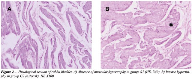
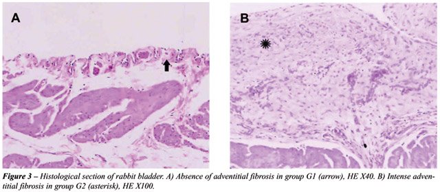
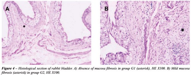
The histopathological analysis showed that
animals in G1 had normal epithelium at the end of the trial. However,
animals in G2 underwent histological changes (Table-2), including muscle
hypertrophy (94%) (Figure-2), Inflammatory reaction (87%), mild to moderate
fibrosis of the adventitia (80%) (Figure-3) and mild to moderate fibrosis
of the mucosa (53%), Figure-4.
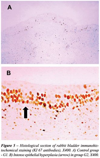
The immunohistochemical analysis showed
significantly higher hyperplasia of the epithelium and muscle layer in
G2 (Figure-5).
COMMENTS
Increase
in bladder weight is predominant in vesical obstruction models (9,10).
In this experiment, after 4 weeks of PBOO, bladder weight in G2 animals
was 2.5 times that of controls. The increase in bladder weight is explained
in part by the hypertrophy of the detrusor muscle, found in 98% of the
G2 animals. This is in line with previous studies using a rabbit model
of BOO (11). Indeed, the hypertrophy of smooth muscle (SM) cells in the
bladder of obstructed rabbits, was followed by changes in the expression
of cytoskeleton and cytocontractile proteins such as SM alpha-actin and
SM myosin (12). Recent studies suggest that the mechanical stretch due
to obstruction is responsible for stimulating the expression of growth
factors and other specific proteins through the activation of stretch-activated
ion channels (SACs) and protein kinase (PKC) sarcolemal proteins (13).
The immunohistochemistry analysis, using
the cell proliferation marker KI 67, showed that hyperplasia of the epithelium
and muscle cells was also responsible for the G2 group increase in bladder
weight. This confirms earlier studies where hyperplasia of the extramural
conjunctive tissue (14), epithelium and muscle cells (15,16) was observed
in animals with bladder obstruction.
A high incidence of positive urine cultures
in G2 (93%) after partial obstruction is most likely a combination of
overdistesion of the bladder and animal manipulation. In G2 87% of the
bladder epithelium had severe inflammatory reaction and 13% presented
ulcerations. At M2, the urine culture of G2 revealed a predominance of
Escherichia coli (data not shown). This corroborates with earlier data
which demonstrated that the integrity of the urothelium is compromised
after the bladder is stretched (17,18), thus making it easier for bacterial
colonization.
In the cystometric study, no significant
difference was observed in maximum vesical volume (Volmax) at the different
moments for G1. However, G2 had significantly (p < 0.05) increased
Volmax at M2 when compared to M1. In other partial obstruction models,
using silk ligature (19,20) and silicon sleeve (21) vesical capacity was
also significantly increased.
There was no statistical difference in maximum
detrusor pressure between groups at M1 and M2. These findings disagree
with (16,22), where an increase in detrusor pressure was observed, following
BOO. These discrepancies in the results are expected due to the different
methodology used to cause BOO, the timing of the cystometric study (6)
and individual differences (4). Recent experiments showed that despite
detrusor hypertrophy, some bladders with BOO work normally while others
are unable to empty properly (23). This was associated with over expression
of non-muscular caldesmon (l-CaD), a protein capable of inhibiting actin-activated
myosin ATPase, compromising the detrusor contractions. The quantification
of l-CaD in further BOO studies could be useful for separating the different
degrees of detrusor dysfunction.
Lost of vesical compliance is associated
with lost of elasticity due to increase in conjunctive tissue deposition
in different layers of the bladder. Cystometric studies showed no difference
in vesical compliance between groups. This is supported by histological
data where mild to moderate fibrosis of the mucosa and adventitial layers
were predominant and interstitial muscle fibrosis was absent in 80% of
the G2 group; thus, not yet compromising vesical compliance. However,
other studies showed higher incidence of mucosal (16), adventitial (15)
and interstitial muscle (24) fibrosis, suggesting that the G2 group could
develop loss in vesical compliance if PBOO had persisted for longer.
Detrusor instability was persistent in G2
at M2 while there was an absence of contraction in the control group.
This demonstrates that the experimental model employed is adequate for
studying vesical instability. This is extremely important for it will
enable further studies on the role of muscarine receptors subtypes (25,26),
adrenoreceptors subtypes (27,28), and myosin isoform (29) expression,
which have been described as being important in the control of contractility
of the detrusor muscle.
CONCLUSION
The described experimental PBOO model in rabbits induced detrusor instability and histological alteration, similar to changes caused by obstructive pathologies, and therefore is a useful tool for further physiological and pharmacological research.
ACKNOWLEDGEMENT
This work was supported by the São Paulo Foundation for Research Support - FAPESP.
CONFLICT OF INTEREST
None declared.
REFERENCES
- Andersson KE: Current concepts in the treatment of disorders of micturition. Drugs. 1988; 35: 477-94.
- Abrams P, Wein AJ: Introduction: Overactive bladder and its treatments. Urology. 2000; 55 (Suppl. 5A): 1-2.
- Sibley GN: An experimental model of detrusor instability in the obstructed pig. Br J Urol. 1985; 57: 292-8.
- Lluel P, Duquenne C, Martin D: Experimental bladder instability following bladder outlet obstruction in the female rat. J Urol. 1998; 160: 2253-7.
- Schroder A, Chichester P, Kogan BA, Longhurst PA, Lieb J, Das AK, et al.: Effect of chronic bladder outlet obstruction on blood flow of the rabbit bladder. J Urol. 2001; 165: 640-6.
- Gosling JA, Kung LS, Dixon JS, Horan P, Whitbeck C, Levin RM: Correlation between the structure and function of the rabbit urinary bladder following partial outlet obstruction. J Urol. 2000; 163: 1349-56.
- Balasteghin KT, Nardo AM, Amaro JL, Padovani CR: Experimental model of bladder instability in rabbits. Int Braz J Urol. 2003; 29: 62-7.
- Amaro JL, Cury PR, Fabris VH, Trindade JC: Ampliação vesical utilizando dura-máter e pericárdio bovino. Estudo comparativo em coelhos. J Bras Urol. 1997; 23: 88-92.
- Levin RM, High J, Wein AJ: The effect of short-term obstruction on urinary bladder function in the rabbit. J Urol. 1984; 132: 789-91.
- Nigro DA, Haugaard N, Wein AJ, Levin RM: Metabolic basis for contractile dysfunction following chronic parcial bladder outlet obstruction in rabbits. Mol Biol Biochem. 1999; 200: 1-6.
- Kato K, Monson FC, Longhurst PA, Wein AJ, Haugaard N, Levin RM: The functional effects of long-term outlet obstruction on the rabbit urinary bladder. J Urol. 1990; 143: 600-6.
- Roelofs M, Wein AJ, Monson FC, Passerini-Glazel G, Koteliansky VE, Sartore S, et al.: Contractility and phenotype transitions in serosal thickening of obstructed rabbit bladder. J Appl Physiol. 1995; 78: 1432-41.
- Yamaguchi O: Response of bladder smooth muscle cells to obstruction: signal transduction and the role of mechanosensors. Urology. 2004; 63 (Suppl 1): 11-6.
- Levin RM, Wein AJ, Buttyan R, Monson FC, Longhurst PA: Update on bladder smooth-muscle physiology. World J Urol. 1994; 12: 226-32.
- Ghoniem GM, Regnier CH, Biancani P, Johnson L, Susset JG: Effect of vesical outlet obstruction on detrusor contractility and passive properties in rabbits. J Urol. 1986; 135: 1284-9.
- Kuo HC: Effects of mild bladder outlet obstruction on rabbit bladder structure and function. J Formos Med Assoc. 1995; 94: 555-61.
- Leppilahti M, Kallioinen M, Tammela TL: Duration of increased mucosal permeability of the urinary bladder after acute overdistension: an experimental study in rats. Urol Res. 1999; 27: 272-6.
- Buttyan R, Chen MW, Levin RM: Animal models of bladder outlet obstruction and molecular insight into the basis for the development of bladder dysfunction. Eur Urol. 1997; 32: 32-9.
- Malmgren A, Sjogren C, Uvelius B, Mattiasson A, Andersson KE, Andersson PO: Cystometrical evaluation of bladder instability in rats with infravesical outflow obstruction. J Urol. 1987; 137: 1291-4.
- Steers WD, De Groat WC: Effect of bladder outlet obstruction on micturition reflex pathways in the rat. J Urol. 1988; 140: 864-71.
- Kato K, Wein AJ, Kitada S, Haugaard N, Levin RM: The functional effect of mild outlet obstruction on the rabbit urinary bladder. J Urol. 1988; 140: 880-4.
- Gray M: Progressive changes in detrusor function with bladder outlet obstruction. J Urol. 1997; 158: 631-5.
- Zhang EY, Stein R, Chang S, Zheng Y, Zderic SA, Wein AJ, et al.: Smooth muscle hypertrophy following partial bladder outlet obstruction is associated with overexpression of non-muscle caldesmon. Am J Pathol. 2004; 164: 601-12.
- German K, Bedwani J, Davies J, Brading AF, Stephenson TP: Physiological and morphometric studies into the pathophysiology of detrusor hyperreflexia in neuropathic patients. J Urol. 1995; 153: 1678-83.
- Hegde SS, Eglen RM: Muscarinic receptor subtypes modulating smooth muscle contractility in the urinary bladder. Life Sci. 1999; 64: 419-28.
- Chapple CR, Yamanishi T, Chess-Williams R: Muscarinic receptor subtypes and management of the overactive bladder. Urology. 2002; 60 (Suppl. 1): 82-8; discussion 88-9.
- Wanajo I, Tomiyama Y, Yamazaki Y, Kojima M, Shibata N: Pharmacological characterization of beta-adrenoceptor subtypes mediating relaxation in porcine isolated ureteral smooth muscle. J Urol. 2004; 172: 1155-9.
- Andersson KE: Storage and voiding symptoms: pathophysiologic aspects. Urology. 2003; 62 (Suppl 2): 3-10.
- Austin JC, Chacko SK, DiSanto M, Canning DA, Zderic SA: A male murine model of partial bladder outlet obstruction reveals changes in detrusor morphology, contractility and Myosin isoform expression. J Urol. 2004; 172: 1524-8.
____________________
Received: April 26, 2005
Accepted after revision: August 08, 2005
_______________________
Correspondence
address:
Dr. João Luiz Amaro
Faculdade de Medicina de Botucatu
Dept de Urologia, Distrito de Rubião Júnior, s/n
18618-970, Botucatu, SP, Brazil
Telephone / Fax: + 55 14 3811-6271
E-mail: jamaro@fmb.unesp.br
EDITORIAL COMMENT
The
authors present an interesting experimental model as well as a careful
experimental design conferring credibility to the results obtained.
The authors have pointed out fibrosis of
the adventitia and “mild to moderate fibrosis of the adventitia
and mild to moderate fibrosis of the mucosa”. Immunohistochemical
analysis showed a high hyperplasia of the epithelium and muscle layer.
However, all observations are expressed in subjective assessments. In
a work in which methodological care with different parameters involved
in the experiment are clear, we would expect the use of morphometric methods
(stereologic) which application has showed to be efficient and the results
much more reliable than the method where crosses or personal assessments
are used. The results expressed after the use of this methodology can
be easily and correctly interpreted and, thus, open the way to researches
that shall certainly result in efficient therapeutic conducts as foreseen
by the authors.
Dr.
Waldemar Silva Costa
Urogenital Research Unit
State University of Rio de Janeiro
Rio de Janeiro, Brazil
E-mail: wscosta@gmail.com