PATHOLOGICAL
STAGING OF MUSCLE INVASIVE BLADDER CANCER. IS SUBSTAGING OF PT2 TUMORS
REALLY NECESSARY?
(
Download pdf )
HUSNU TOKGOZ, KADIR TURKOLMEZ, BERKAN RESORLU, KENAN KOSE, OZDEN TULUNAY, YASAR BEDUK
Departments of Urology, Biostatistics and Pathology, Ankara University School of Medicine, Ankara, Turkey
ABSTRACT
Objective:
Compare clinical outcomes in patients having urothelial tumors invading
less than one half of the depth of bladder muscle and greater than one
half of bladder muscle and, to determine various clinical variables as
predictive factors for survival.
Materials and Methods: According to our
inclusion criteria, 57 patients among cases with T2 bladder tumor were
selected. Thirty-five patients (61.4 %) had pT2a (Group-1) and 22 patients
(38.6%) had pT2b (Group-2) muscle invasive tumors. Mean follow up time
was 7.3 years for Group-1, and 6.1 years for Group-2. Multivariate analysis
was performed in order to identify possible correlation of clinical variables
like age, gender, grade of primary tumor, appearance of local and/ or
distant metastasis with patient outcome.
Results: Five year recurrence-free and overall
survival rates were 69.1% and 44.3% for patients with pT2a tumor, whereas
these ratios were 66.1% and 43%, respectively for patients with pT2b tumor
(p = 0.896; p = 0.975). Mean overall and progression-free survival times
were 87.7 ± 13.8 and 116 ± 13.12 months for Group-1, while
they were 73.8 ± 13.7 and 88.85 ± 12.55 months for Group-2,
respectively. On both univariate and multivariate analysis, age was noticed
as an independent predictive factor for survival.
Conclusions: The depth of muscle invasion
in bladder tumors has no prognostic significance. Recurrence of the disease
either locally or at distant sites dramatically shortens patients’
life. Being older than 60 years old during the time of radical surgery,
is also a bad prognostic factor for overall and progression-free survival.
Key
words: bladder neoplasms; TNM staging; neoplasm staging; survival
Int Braz J Urol. 2007; 33: 777-84
INTRODUCTION
Bladder
carcinoma is the second most frequent urogenital tumor disease, more than
90% of which are transitional cell carcinoma (TCC). Nearly 30% of these
tumors already are or will progress into muscle invasive tumor during
their follow up (1).
After classification of bladder tumors in
1992, the AJCC (American Joint Committee on Cancer) and UICC (Union Internationale
Contre le Cancer) revised this TNM (tumor, lymph node and metastasis)
bladder cancer staging system (2). They divided T2 tumors (muscle invasive
tumor) into superficial (T2a, tumor invasion in less than one half of
the depth of the bladder muscle) and deep (T2b, tumor invasion in more
than one half of the depth of the bladder muscle). This need for sub grouping
of T2 tumors arose after Jewett had reported a significant prognostic
difference in bladder tumor patients with superficial and deep muscle
invasion. Including 18 patients with muscle invasive bladder tumor, overall
survival rate was 80% for T2a tumors, whereas it was only 8% for T2b tumors
(3). Presently, since the 2002 version did not include any changes for
the staging of bladder cancer, the use of the 1997 version is still encouraged
in most guidelines.
After this study, several studies were conducted
to compare clinical outcomes in terms of overall and recurrence-free survival
rates (4-9). In contrast with the findings of Jewett in 1952, most of
these studies were unable to find a prognostic difference between these
subgroups. Last study published on this subject was by Yu et al. (10).
They found significant difference when 10 year recurrence free survival
rate in pT2a and pT2b cases were compared (79% vs. 64%). However, when
they re-compared after sub grouping as lymph node negative and positive,
the difference disappeared.
The paper by Cheng et al., revealed somewhat
more interesting findings. They evaluated the depth of invasion by micrometer
measurement and its relation to clinical outcome (11). They found that
tumor size, rather than the degree of depth of muscle invasion, had much
more prognostic value in terms of cancer specific and distant metastasis
free survival rates.
Similarly, the aim of our study was to evaluate
the practicality of subdividing the muscle invasion into pT2a and PT2b
in routine pathological examination and to assess if substaging of T2
bladder tumors is predictive for overall and recurrence free survival.
MATERIALS AND METHODS
We
performed retrospective evaluation of 225 patients who had undergone radical
cystectomy and bilateral pelvic lymphadenectomy for bladder urothelial
carcinoma in our department between January 1990 and December 2005. The
1997 TNM system was used for staging. From records, we detected that all
specimens had been assessed by the same pathology team. Multiple sections
were obtained from the tumor, bladder wall and adjacent mucosa. In addition,
sections from distant mucosa, regional lymph nodes and ureters were evaluated.
In men, tissue blocks from the seminal vesicles and prostate were examined.
In women, tissue blocks obtained from ovaries, uterus and vagina were
examined. Palliative cystectomy cases; cases with upper urinary tract
tumors and non-transitional cell carcinomas, were excluded. Also, cases
with histological features of squamous differentiation were excluded.
We included cases with T2 urothelial carcinoma
of the bladder which did not have lymph node metastasis on pathological
evaluation of cystectomy specimens, and which were not given preoperative
or early postoperative radio and/ or chemotherapy.
Finally, 57 patients were included in the
study. Of these, 35 patients (61.4 %) had pT2a and 22 patients (38.6 %)
had pT2b muscle invasive tumors. As mentioned previously, none of those
patients had evidence of pelvic lymph node invasion.
Histological grading was performed according
to the World Health Organization / International Society of Urologic Pathology
Classification (12).
The patients were initially seen 2 months
after surgery and then every 3 months for 2 years and every 6 months until
disease progression or death. Each examination consisted of full physical
examination, abdominal tomography, thorax X-rays and laboratory investigations
like complete blood count, serum biochemical analysis.
The correlation of clinical and pathological
variables with survival was investigated by the Cox proportional hazards
test. Two, 3 and 5 year recurrence free and overall survival rates were
compared between patients with pT2a (Group-1) and pT2b (Group-2) tumor.
The Kaplan-Meier method was used to estimate progression free and cumulative
survival probabilities. Differences between groups on the basis of progression
free and overall survival rates were assessed with log-rank test. Multivariate
analysis was performed using the Cox regression survival method. A two-
tailed p value of < 0.05 was accepted as statistically significant.
RESULTS
The
mean patient age at surgery was 62.2 years for Group-1 and the mean follow
up time was 7.3 years, while the mean age at surgery was 59.3 years in
Group-2. Mean follow up time was 6.1 years for this group (p = 0.657;
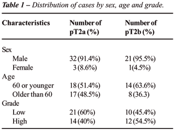
p = 0.798). Demographic distributions of patients were given in Table-1.
During follow-up, 19 patients died because of primary tumor progression.
No patient had died from unrelated causes during follow up.
On both univariate and multivariate analysis,
age was noticed as a predictive factor for survival. Patients younger
than 60 years of age, had 2.5 times higher advantage for survival on univariate
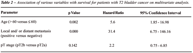
analysis (p = 0.05; hazard ratio = 2.45). On multivariate analysis, age
(p = 0.02; Hazard ratio: 5.6), appearance of local and / or distant metastasis
during follow up, were also statistically significant effective factors
on survival (Table-2).
Initial surgical management consisted of
radical cystectomy, bilateral pelvic lymph node dissection and urinary
diversion including neobladder replacement in 16 patients, continent urinary
reservoir in 12 patients, ileal conduit in 28 patients and ureterocutaneostomy
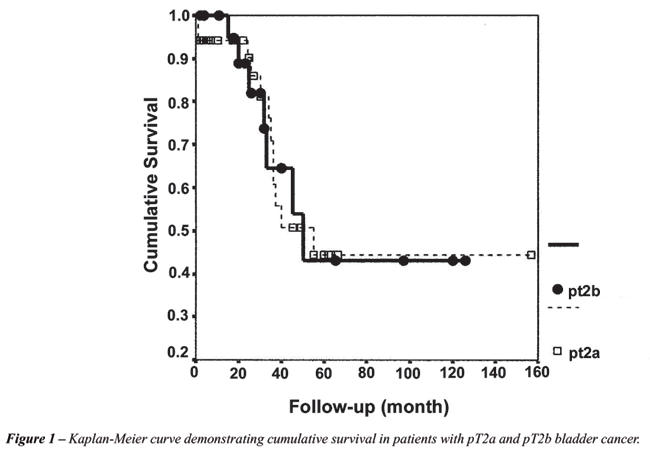
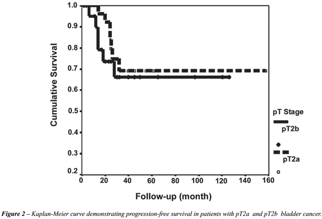
in 1 patient. Two, 3 and 5 year recurrence free survival rates were 83.7%;
69.1% and 69.1%, whereas 2,3,5 year overall survival rates were 90.1%;
71% and 44.3% for patients with pT2a tumor. Two, 3 and 5 year recurrence
free survival rates for patients with pT2b tumor, were 73.5%; 66.1% and
66.1%, respectively. Overall survival rates for this group were 88.9%;
64.6% and 43%. Although, 2,3 and 5 year recurrence free and overall survival
rates were lower in pT2b patients, both recurrence free survival and overall
survival rates were statistically insignificant between Group-1 and 2
(p = 0.896; p = 0.975) (Figure-1 and 2). On multivariate analysis, pT
substaging (pT2b versus pT2a) had a hazard ratio greater than 1 (2.2),
but the p value was again insignificant (p = 0.142) (Table-2).
Mean survival time was 87.7 ± 13.8
months for Group-1, and 73.8 ± 13.7 months for Group-2 (p = 0.97).
Mean progression-free survival time was 116 ± 13.12 months for
Group-1, while it was 88.85 ± 12.55 months for Group-2 (p = 0.53).
When we divided each group into two as patients
with low grade and high grade tumors; mean survival times for patients
with low and high grade tumors in Group-1, were 105.18 ± 16.54
and 37.8 ± 5.57 months, respectively (p = 0.10). In Group-2, these
were 53.33 ± 17.06 months for patients with low grade tumors and
84.4 ± 16.4 months for patients with high grade tumors (p = 0.32).
Although the differences seem great especially in Group-1, no statistical
significance was present.
Local pelvic recurrence or distant metastasis
developed in 13 of 57 patients (22.8%) with pT2 disease. Seven (20%) out
of 13 patients who showed progression of the primary tumor during follow
up, were belong to Group-1 (distant metastasis in 6; both local recurrence
and distant metastasis in 1 case), whereas 6 patients (27.2%) in Group-2
showed progression of primary tumor (local pelvic recurrence in 1; distant
metastasis in 5 cases). There was no statistically significant difference
when groups were compared (p = 0.496).
None of the variables, including gender
and primary tumor grade were significantly associated with patient outcome.
Thus, none of them were believed to be predictive of patient survival.
COMMENTS
Presently,
TNM staging system revised in 1997 seems to provide the most valuable
prognostic information in patients with transitional cell carcinoma of
the bladder. Superficial (T1), muscle invasive (T2) and perivesical invasive
(> T2) urothelial tumors have significantly different 5 year recurrence
free and survival rates. Nicely, T1 tumors form the major and best prognostic
group. Only 25% of these cases do not survive 5 years (13). For T2 tumors,
5 year survival rates were given as approximately 60% (40-70%) (4-9).
Nevertheless, Skinner and Herr, reported that urothelial cancers invading
perivesical fat tissue had much worse prognosis than those not invading
the perivesical fat tissue (14-17).
For high grade, non metastatic, muscle invasive
urothelial tumors, radical cystectomy is accepted as the gold standard
treatment. Although it is a major surgical procedure including reconstructive
techniques for neobladder replacement, this operation provides high 5
year survival rates. In addition, cystectomy with pelvic lymph node dissection
allows the clinician to know accurate pathological staging of the primary
tumor, and aids in further treatment planning, including adjuvant radio
and / or chemotherapy.
For organ confined lymph node negative T2
tumors, May et al., reported a 74% progression free 5 year survival rate,
which was very close to the ratios given for T1 tumors (18). In a study
by Wishnow and colleagues, 5 year mean survival and disease free survival
rates after radical cystectomy, were 82 and 77%, respectively for patients
with Stage B disease (19). In 2001, Stein et al. presented their long
term surgical experience and patient outcomes in 1054 cases treated with
radical cystectomy for invasive transitional cell carcinoma of the bladder
(1). Their 5 and 10 year recurrence free survival rates for organ-confined
lymph node negative tumors were, 85% and 82%, respectively. However, it
was not the case for lymph node positive cases. Five year recurrence free
and overall survival rates were, 35% and 31%, respectively for those patients.
In last years, major criticism on TNM system,
was focused on if substaging of T2 tumors was really necessary. The findings
of the studies evaluating the correlation of depth of muscle invasion
with patient survival, indicate that this parameter is of no prognostic
value (4-9). For this reason, some of the uro-oncologists offer the removal
of substaging of T2 bladder cancers from TNM systems.
Although our study was retrospective, parameters
like age, gender, and mean follow up times were similar in each group.
Tumor progression rates for Group-1 and 2 were 20% and 27.2% respectively,
which were also close (p > 0.05). Grade of the primary tumor was not
correlated with survival of patients in both groups. Statistically significant
difference was only observed when mean survival times of patients who
developed progression of the disease were compared with survival times
of the cases who were recurrence or metastasis free during their follow
up.
Instead of substaging, Cheng et al., mentioned
the prognostic value of primary tumor size (11). Actually, we had problems
in accurate evaluation of primary tumor size, since they were not always
mentioned in pathology reports. So, we were not able to include this parameter
for survival analysis. Cheng and associates found lower results than our
study, revealing 62% 5 year survival rate for patients with pT2a tumors
and 56% for patients with pT2b tumors (11). But, they included node positive
cases. After they divided study population according to their lymph node
status, they reported 10 year recurrence free survival as 74% for lymph
node negative cases.
We were interested in age as an independent
predictive factor for survival, and we observed statistically significant
difference in the 6th decade. This means that, for bladder tumor patient,
being older than 60 years old at the time of radical surgery is a bad
prognostic factor for both progression-free and overall survival.
One should reasonably think that the appearance
of local and / or distant metastasis during follow-up, is a progression
of the disease, not a real predictive factor. So, high correlation of
this variable with survival might be expected. But, it is not the case
for age. As Cheng et al. insisted on the importance of primary tumor size,
we aimed to take the attention of uro-oncologists on the predictive value
of age (11). Most prominent difference was observed in the 6th decade
in our study. Thus, we have taken the age 60 as a cut-off point.
Regarding that all cases in our study population
were lymph node negative, the rates Yu and colleagues reported, were higher
than our results (69.1% vs. 66.1%). They reported 10 year recurrence free
survival rate for node negative pT2a disease as 84% and for pT2b disease
as 74% (10).
But, we cannot ignore the effect of substaging
on survival. In our study, although the p value was insignificant; odds
ratio of higher than 1 (2.2) on multivariate analysis, suggest possible
effect of pT substaging on cancer specific survival of these patients.
Similarly, in the study by Cheng, 10 year survival rates were lower in
cases with pT2b disease compared with pT2a disease (51% vs. 48%). But,
the p value was again insignificant (11). When they re-compared 10 year
cancer specific survival rates in lymph node negative cases between the
2 groups, they found a risk ratio of 2 (pT2b versus pT2a); a value which
was so close to ours (2.2).
Limitations of the study should be discussed.
First of all, our sample size is relatively small. But, in order to form
homogenous study groups, we excluded cases; with histological features
of squamous differentiation, having nodal involvements on pathological
evaluation of cystectomy specimens, and who were given preoperative or
early postoperative radio and / or chemotherapy. We hope in the near future,
prospective studies with larger series, will be published and, will give
more valuable data.
CONCLUSIONS
We think that the depth of muscle invasion in bladder tumors has no prognostic significance. But, we cannot fully exclude its role on cancer specific survival. In addition, the results underline the importance of patient age at the time of surgery, as age was found an independent prognostic factor for both overall and progression-free survival.
CONFLICT OF INTEREST
None declared.
REFERENCES
- Stein JP, Lieskovsky G, Cote R, Groshen S, Feng AC, Boyd S, et al.: Radical cystectomy in the treatment of invasive bladder cancer: long-term results in 1,054 patients. J Clin Oncol. 2001; 19: 666-75.
- Fleming ID, Cooper JS, Henson DE: American Joint Committee on Cancer, Cancer Staging Manual. (5th ed,). Philadelphia, Lippincott-Raven. 1997; pp. 241-3.
- Jewett HJ: Carcinoma of the bladder: influence of depth of infiltration on the 5-year results following complete extirpation of the primary growth. J Urol. 1952; 67: 672-80.
- Ghoneim MA, el-Mekresh MM, el-Baz MA, el-Attar IA, Ashamallah A: Radical cystectomy for carcinoma of the bladder: critical evaluation of the results in 1,026 cases. J Urol. 1997; 158: 393-9.
- Pollack A, Zagars GK, Cole CJ, Dinney CP, Swanson DA, Grossman HB: The relationship of local control to distant metastasis in muscle invasive bladder cancer. J Urol. 1995; 154: 2059-63; discussion 2063-4.
- Roehrborn CG, Sagalowsky AI, Peters PC: Long-term patient survival after cystectomy for regional metastatic transitional cell carcinoma of the bladder. J Urol. 1991; 146: 36-9.
- Sorensen BL, Soeborg Ohlsen A, Barlebo H: Carcinoma of the urinary bladder. Clinical staging and histologic grading in relation to survival. Scand J Urol Nephrol. 1969; 3: 189-92.
- Cuesta JA, Chapado MS, Cid MG, Corral NF, Pontes EJ, Grignon DJ: Survival of patients with stage T2-3a bladder cancer treated by radical cystectomy. Arch Esp Urol. 1997; 50: 17-25.
- Pagano F, Bassi P, Galetti TP, Meneghini A, Milani C, Artibani W, et al.: Results of contemporary radical cystectomy for invasive bladder cancer: a clinicopathological study with an emphasis on the inadequacy of the tumor, nodes and metastases classification. J Urol. 1991; 145: 45-50.
- Yu RJ, Stein JP, Cai J, Miranda G, Groshen S, Skinner DG: Superficial (pT2a) and deep (pT2b) muscle invasion in pathological staging of bladder cancer following radical cystectomy. J Urol. 2006; 176: 493-8; discussion 498-9.
- Cheng L, Neumann RM, Scherer BG, Weaver AL, Leibovich BC, Nehra A, et al.: Tumor size predicts the survival of patients with pathologic stage T2 bladder carcinoma: a critical evaluation of the depth of muscle invasion. Cancer. 1999; 85: 2638-47.
- Epstein JI, Amin MB, Reuter VR, Mostofi FK: The World Health Organization/International Society of Urological Pathology consensus classification of urothelial (transitional cell) neoplasms of the urinary bladder. Bladder Consensus Conference Committee. Am J Surg Pathol. 1998; 22: 1435-48.
- Angulo JC, Lopez JI, Grignon DJ, Sanchez-Chapado M: Muscularis mucosa differentiates two populations with different prognosis in stage T1 bladder cancer. Urology. 1995; 45: 47-53.
- Herr HW: Staging invasive bladder tumors. J Surg Oncol. 1992; 51: 217-20.
- Skinner DG: Current perspectives in the management of high-grade invasive bladder cancer. Cancer. 1980; 45: 1866-74.
- Herr HW: A proposed simplified staging system of invasive bladder tumors. Urol Int. 1993; 50: 17-20.
- Skinner DG: Current state of classification and staging of bladder cancer. Cancer Res. 1977; 37: 2838-42.
- May M, Helke C, Nitzke T, Vogler H, Hoschke B: Survival rates after radical cystectomy according to tumor stage of bladder carcinoma at first presentation. Urol Int. 2004; 72: 103-11.
- Wishnow KI, Ayala AJ, Levinson AK, Logothetis CJ, Johnson DE, Swanson DA, et al.: Stage B (P2/ 3A/ N0) transitional cell carcinoma of bladder highly curable by radical cystectomy. Urology 1992; 39: 12-6.
____________________
Accepted after revision:
August 8, 2007
_______________________
Correspondence
address:
Dr. Hüsnü Tokgöz
Çukuranbar Mah. 41. Cad.
No: 2 / 35 06520
Balgat, Ankara, Turkey
Tel: + 90 312-2847285
E-mail: h_tokgoz@hotmail.com
EDITORIAL COMMENT
In
1952, Jewitt proposed the subclassification of T2 (prior stage B) tumors
based on their depth of involvement of the muscular wall - T2a (B1) involving
the inner half and T2b (B2) infiltrating the outer half based on survival
differences observed in just 18 patients. Over the ensuing decades, increasing
worldwide experience suggested that perhaps such substaging may not be
in fact predictive leading Jewitt to revise his comments stating in 1978
that “the arbitrary dividing line drawn 30 years ago at the halfway
level to separate B1 and B2 was too superficial.” His remarks seem
to also acknowledge the greater relevance of deeper invasion - i.e. involvement
of the perivesical soft tissue - as having worse disease-associated outcomes.
The
present study supports this contention by failing to demonstrate any survival
differences in bladder cancer patients when stratified by depth of muscle-invasion
- T2a versus T2b. These results support the recent findings Cheng et al.
from the Mayo Clinic. Furthermore, although Yu et al. recently demonstrated
decreased survival in T2b vs. T2a patients, these differences disappeared
when patients were re-analyzed according to lymph node status.
The
ongoing study to help identify which clinical and pathological parameters
have a relevant impact on a patient’s prognosis remains an important
and worthy research endeavor. However, the answers may ultimately lie
beyond the clinical and histological findings alone, and are likely to
be found in the molecular and genetic signatures of tumors occurring at
the cellular level. Tumor suppressor genes (e.g. p53 and Rb), proliferative
indices (Ki-67), urinary growth factors (e.g. epidermal growth factor,
basic fibroblast growth factor, and CD44), matrix metalloproteinses (e.g.
MMP-9), and urinary plasminogen activators are just some of the indicators
that one day may prove to be valuable adjuncts in the management of urothelial
cancers.
These
include blood group antigens (e.g., LewisX), tumor suppressor genes (e.g.
p53 and Rb), proliferative indices (Ki-67), urinary growth factors (e.g.
epidermal growth factor, basic fibroblast growth factor, and CD44), matrix
metalloproteinses (e.g. MMP-9), and urinary plasminogen activators. One
day such indicators may prove to be valuable adjuncts to stage and grade
in the management of noninvasive urothelial cancers.
Dr. Raj
S. Pruthi
Associate Professor of Surgery/Urology
Director of Urologic Oncology
The University of North Carolina at Chapel Hill
Chapel Hill, North Carolina, USA
E-mail: rpruthi@med.unc.edu