SCHISTOSOMAL
EPIDIDYMITIS
(
Download pdf )
LEONARDO S. ALVES, BERNADO P. S. ASSIS, MÁRCIA M. B. REZENDE
Procriar Instituto de Urologia, Belo Horizonte, Minas Gerais, Brazil
ABSTRACT
Epididymitis
is a frequent inflammatory process. It is related to sexually transmitted
diseases, urinary tract infections by E. coli, or scrotal trauma. We describe
the case of a Caucasian 32-year old man, who presented scrotal pain for
3 months, with difficult management with medication. Testis was normal;
however, the left epididymis was extremely painful and hardened. Following
the unsuccessful use of analgesic and anti-inflammatory medication, a
left epididymectomy was performed, with resolution of the pain.
The pathological examination showed the
presence of chronic inflammatory process associated with eggs of the parasite
Schistosoma mansoni in the resected epididymis. Patient evolved without
pain in the post-operative period and was medicated with a single dose
of oxamniquine after etiologic confirmation.
Key
words: epididymis; epididymitis; Schistosoma mansoni
Int Braz J Urol. 2004; 30: 413-5
INTRODUCTION
Epididymitis is an inflammatory process that affects the epididymis. It occurs around the testis and can appear at any age. Clinical presentations of epididymitis are most frequently related to sexually transmitted diseases, trauma and contamination by surgical instruments. The present work reports the case of a patient with chronic epididymal pain and hardening due to infestation by the parasite Schistosoma mansoni.
CASE REPORT
Patient
W.A.O., 32 years old, born in Belo Horizonte, Minas Gerais, Brazil, developed
a painful scrotal process, with difficult management, 3 months earlier.
He denied trauma, recent surgical procedures (vasectomy 2 years earlier)
or suspected sexual activity.
On physical examination, a hardened and
painful left epididymis was observed. Ipsilateral testis and spermatic
cord were normal. Scrotal ultrasound demonstrated only an increase in
echogenicity, characteristic of chronic inflammatory process. After the
unsuccessful use of analgesic and anti-inflammatory drugs, we chose to
perform unilateral epididymectomy, with the patient’s consent. The
procedure had no intercurrences, and during the surgery, we observed hardening
of the entire epididymis, without testicular involvement. The patient’s
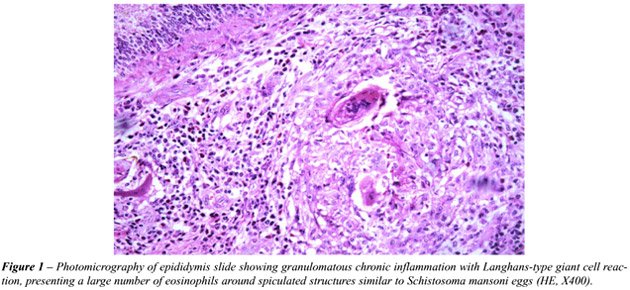
pain resolved shortly after surgery. Histopathological study detected
granulomatous chronic inflammatory process due to the presence of Schistosoma
mansoni eggs (Figure-1). After the diagnosis, the patient’s contact
with a lagoon infested by the parasite was confirmed. Patient received
a single dose of the antiparasitic drug oxamniquine.
COMMENTS
Epididymitis
is a common inflammatory process frequently involving the ipsilateral
testis as well, a condition known as orchiepididymitis (1). The acute
presentation constitutes the most common cause of acute scrotum in adults.
Isolated epididymitis can occur at all ages, and is related to bacterial
infection (E. coli, N. gonorrhea, H. influenza, Chlamydia trachomatis,
Thichomonas vaginalis). Other etiologic causes are contamination by endoscopic
instruments, testicular torsion, trauma, vasectomy, orchiopexy, etc.
Etiologic agents such as M. tuberculosis,
Brucella, fungi (coccidiodomycosis and blastomycosis) and parasites such
as Schistosoma mansoni, are less frequently diagnosed due to technical
difficulties (1,2). In these cases, the confirmation is achieved only
by histopathological study. Most frequently, the contamination occurs
by the canalicular route, through the deferens vas, where germs from the
urogenital tract ascend to the epididymis (1).
The diagnosis of epididymitis can be made
from the anamnesis and physical examination. Through correlation with
the patient’s age range, it is possible to deduce the etiologic
agent. Viral and bacterial infections are common until sexual initiation.
From this period up to 40 years old, gonococcus and Chlamydia are the
major responsible pathogens, while after this age, E. coli is the most
frequently found agent. The physical examination can determine if the
process is acute (enlargement and pain) or chronic (hardening). Imaging
methods, as well as blood, urine and feces tests, can help to determine
the etiology of the symptoms.
Schistosomiasis mansoni is an endemic parasitosis
in Brazil caused by a trematode, the Schistosoma mansoni. It usually parasitizes
the venous system presenting tropism to the hepatic portal system. In
this system, the inferior mesenteric vein, liver and spleen are the most
frequently affected sites (3). Contamination by Schistosoma mansoni occurs
by hematogenic route according to the parasite’s life cycle (3).
When a person comes in contact with water that is infested by schistosomes,
the parasites in the form of cercariae penetrate the skin and reach blood
or lymph circulation. At this moment, they can be destroyed by the immunological
system or reach the peripheral venous circulation. The parasite couple
goes towards the hemorrhoidal plexus at the time of oviposition, and can
go to the seminal plexus as well, which would explain the present case
(3). When affected by schistosoma, the epididymis increases in size due
to the inflammatory process, similarly to other affected organs. A granulomatous
inflammatory pattern with Langhans-type giant cell reaction, presenting
a large number of eosinophilic cells around spiculated structures (eggs)
is characteristic in this process (Figure-1).
The difficulty for etiologic diagnostic
in this case was because the only sign of disease was a hardened epididymis
and pain. The pathological result, until then unexpected, draw our attention
to the clinical fault in the anamnesis, which failed to investigate the
contact of the patient with “still waters”, since schistosomiasis
mansoni is an endemic disease in Brazil.
Prof. Lúcia Porto Fonseca Castro performed
the
pathological analysis.
REFERENCES
- Costa, M: Orquiepididimites In: Guia Prático de Urologia, SBU, São Paulo, BG Cultural Editora. 1999; pp. 139-46.
- Kaufman, JJ: Current Urologic Therapy. Philadelphia, WB Saunders Co. 1980; pp. 357-60.
- Pessoa SB: Parasitologia Médica. Rio de Janeiro, Guanabara Koogan. 1958; p. 471-558.
_____________________
Received: April 04, 2004
Accepted after revision: July 29, 2004
_______________________
Correspondence address:
Dr. Leonardo de Souza Alves
Rua Gonçalves Dias, 142, Funcionários
Belo Horizonte, MG, 30140-090, Brazil
Tel.: + 55 31 3225-0907
E-mail: procriar@bol.com.br