RETROPERITONEOSCOPIC
SURGERY WITH EXTRACORPOREAL URETERO-URETERAL ANASTOMOSIS FOR TREATING
RETROCAVAL URETER
(
Download pdf )
M. TOBIAS-MACHADO, MARCO T. LASMAR, ERIC R. WROCLAWSKI
Department of Urology, ABC Medical School, Santo André, Sao Paulo, Brazil
ABSTRACT
We
present a case of retrocaval ureter featuring laparoscopic technique treatment
using extraperitoneal access and extracorporeal suture of the ureteral
stumps. Surgical time was 130 minutes, and the anastomosis was performed
in 40 minutes. There were no intra- or postoperative complications, and
the patient was discharged from hospital on the second postoperative day.
The medium-term outcome featured similar results to pure laparoscopic
technique.
We conclude that this technical variation
for treatment of retrocaval ureter makes the procedure easier and provides
a drastic reduction in surgical time, without compromising the minimally
invasive aspect of this kind of approach.
Key
words: ureter; urogenital abnormalities; vena cava, inferior;
laparoscopy
Int Braz J Urol. 2005; 31: 147-50
INTRODUCTION
Retrocaval
ureter is a rare congenital entity that ultimately results in a developmental
abnormality of the vena cava inferior, located postero-laterally to the
ureter.
Though it is a congenital pathology, symptoms
usually arise during the 3rd or 4th decade of life and result from the
compressive effect exerted by the vena cava on the ureter with consequent
proximal uretero-hydronephrosis. Diagnosis can be suggested through excretory
urography and confirmed through helical computerized tomography.
The treatment is surgical and is always
suggested in symptomatic cases and/or when signs of renal obstruction
are verified. Currently, laparoscopic surgery has been employed as the
minimally invasive therapeutic option. Our objective was to present a
new technical approach that uses laparoscopic surgery with extraperitoneal
suture as a strategy for reducing surgical time.
CASE REPORT – SURGICAL TECHNIQUE
A
24-year old woman complained on recurring lumbar colic pain associated
with repeated episodes of acute pyelonephritis on the same side. Laboratory
tests showed preserved renal function.
The ultrasound revealed moderate dilation
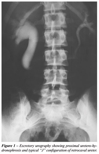
of the pyelocaliceal system in the right kidney. The excretory urography
showed a J-shaped proximal ureter, suggesting retrocaval ureter (Figure-1).
Helical tomography confirmed this diagnosis.
Due to the clinical picture and findings
of complimentary tests, we decided for surgical treatment. We used the
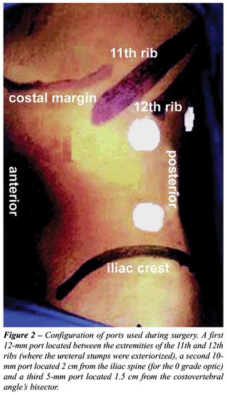
standard extraperitoneal laparoscopic access through 3 ports (Figure-2).
We dissected the entire proximal ureter
and released the lower renal pole, and then dissected the distal ureter
up to the point of maximum visualization in the interaortocaval position,
while preserving the periureteral fat. The ureter was identified and brought
to the extremity of the trocar positioned at the extremity of the 12th
rib, while observing which would be the point with the least tension for
exteriorization. The ureter was sectioned at this site. The ureteral stumps
were exteriorized through the incision of the 12-mm port after enlarging
the skin incision to 20 mm. The proximal and distal ureteral margins were
resected and a good vascularization was observed. We performed a continuous
spatulate suture on the posterior aspect of the ureter using monocryl
4-0. A double-J catheter was then inserted. The anterior ureter was sutured
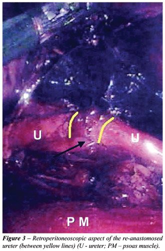
like the posterior aspect. The ureter was repositioned at its usual location
(Figure-3) and a Penrose was inserted for drainage. Total surgical time
was 130 minutes, 40 minutes of which were employed for the anastomosis.
Estimated bleeding was 50 mL and there were no intraoperative complications.
The patient was discharged from hospital on the 2nd postoperative day
with no intercurrence. The double-J catheter was maintained for 6 weeks,
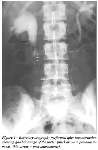
and the patient remained asymptomatic following removal. A control urography
performed 3 months after surgery showed good drainage of the ureter with
correction of the anatomic abnormality (Figure-4).
COMMENTS
The
classical treatment for retrocaval ureter consists of separating the ureter,
re-anastomosing its stumps and replacing the ureter in its usual position
while maintaining its irrigation.
The transperitoneal laparoscopic surgical
repair for retrocaval ureter was first described by Baba et al. in 1994
(1). Comparisons between previous work using a laparoscopic approach and
historical reports on open surgery for treatment of retrocaval have shown
the advantages of minimally invasive access, specifically a shorter hospital
stay, reduced postoperative pain, early return to daily activities and
a remarkably superior esthetic effect while maintaining efficacy (2,3).
The main limiting factor for laparoscopic
treatment of this pathology has been the intracorporeal anastomosis of
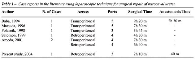
the ureter, which considerably increases surgical time (1-3), Table-1.
The majority of reports have used a transperitoneal
access. In 1999, Salomon et al. described the feasibility of the surgical
repair of retrocaval ureter through laparoscopic access using the retroperitoneal
approach (3).
We are presenting here a technique that
offers a feasible, efficacious and fast alternative for cases where it
is possible to exteriorize the stumps that will be anastomosed. Difficulties
resulting from the intracorporeal suture, which is more significant when
the extraperitoneal access is preferred, are thus eliminated. Antegrade
insertion of the double-J catheter prevents the endoscopic approach from
being performed at the beginning. The patient’s body habitus is
important in order to safely accomplish this approach. The proper planning
of the site where the ureter should be sectioned, the perfect maintenance
of its irrigation properties and a proper assessment of the ureteral segment’s
feasibility are fundamental for success using this technical variation.
The long-term postoperative result shows an effectiveness that is similar
to the pure laparoscopic technique. Surgical time was the lowest posted
in the literature so far.
The
authors acknowledge Dr. Pedro Forseto Jr. and
Dr. Marcelo M. Gava for patient assistance and
cooperation in the conduction of this project.
REFERENCES
- Baba S, Oya M, Miyahara M, Deguchi N, Tazaki H: Laparoscopic surgical correction of circumcaval ureter. Urology. 1994; 44: 122-6.
- Matsuda T, Yasumoto R, Tsujino T: Laparoscopic treatment of a retrocaval ureter. Eur Urol. 1996; 29: 115-8.
- Salomon L, Hoznek A, Balian C, Gasman D, Chopin DK, Abbou CC: Retroperitoneal laparoscopy of a retrocaval ureter. BJU Int. 1999; 84: 181-2.
__________________________
Received: September 17, 2004
Accepted after revision: February 4, 2005
_______________________
Correspondence address:
Dr. Marcos Tobias-Machado
Rua Graúna, 104 / 131
São Paulo, 04514-000, SP, Brazil
Phone: + 55 11 288-1003
E-mail: tobias-machado@uol.com.br