TEXTILOMA
NINE YEARS AFTER NEPHRECTOMY
(
Download pdf )
MIGUEL ZERATI FILHO, PAULO S. FURTADO. LEONARDO A. P. DE ANDRADE
Institute of Urology and Nephrology, São José do Rio Preto, São Paulo, Brazil
ABSTRACT
Introduction:
Foreign bodies after surgical procedures are not very reported in the
literature. It is estimated to have 1 case for 1,300 operations, although
in practice the frequency might probably be higher.
Case Report: A woman, 38 years, submitted
to left nephroureterectomy for renal transplantation in 1993. During 9
years, she was asymptomatic, and then she presented intermittent left
flank pain. Radiographic workup demonstrated a textiloma.
Discussion: In a review of the literature
since 1950, solely 8 cases of textiloma in renal surgeries were reported,
probably due to legal implications.
Key words:
nephrectomy; postoperative complications; foreign bodies
Int Braz J Urol. 2002; 28: 537-8
INTRODUCTION
Foreign bodies (gauzes, dressing) left after surgical procedures are not frequently described occurrences, probably due to legal implications, and are related in 1/1,300 laparotomies (1), despite all protocols applied. Some patients may present asymptomatically for several years (20%), and others develop early persistent infected discharge through the surgical wound, digestive fistulae, and even septic conditions.
CASE REPORT
The
patient was a 38 years old woman submitted to a left donor nephroureterectomy
for renal transplantation in August, 1993. She was asymptomatic in the
pos-operative period. She had an annual follow-up. In November 2001, after
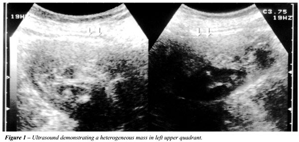
a mild intermittent left flank pain, she had an ultrasound study that
evidenced a nodular formation of approximately 280 mL, heterogeneous,
in the left renal bed (Figure-1). The abdominal CT scan showed a round
image of regular contours with soft tissue density, measuring about 7.0x6.3x5.0
in the topography of the left renal bed, with calcifications within it
(Figure-2). During the operation, it was identified as a solid mass with
well defined limits, mildly adhered to adjacent structures, and poor vascularization.

The opening of the mass after its removal
was performed, confirming the initial diagnosis of surgical foreign body
(bandage).
DISCUSSION
Literature review shows few reports of foreign bodies in surgery. Ballesteros et al. (2) described in a recent review 8 cases of textiloma in renal surgeries, one after a nephrectomy performed 24 years before textiloma diagnosis. Risk factors implied are primarily related to the type of surgery (urgent or elective) and to the preventive routine executed. There are several forms of clinical presentation, making diagnosis very difficult sometimes. Radiographic methods are useful and, occasionally, conclusive (3). Many centers use radiopaque labeled material. Occasionally only surgery define the diagnosis.
REFERENCES
- Fernandez LR, Marin LFJ, Fradejas LJM, Dias GLM, Camarero ME, Moreno AM: Postoperative textilomas: review of 14 cases. Int Surg. 1998; 83:63-6.
- Ballesteros SJJ, Alameda QF, Pares PME: 3 rare cases of textilomas after renal surgery: review of the literature. Arch Esp Urol. 2002; 55:25-9.
- Kopka L, Fischer U, Grooss AJ, Funke M, Oestermann JW, Grable E: CT of retained surgical sponges (textilomas): pitfalls in detection and evaluation. J Comput Assist Tomogr. 1996; 20:919-23.
_________________________
Received: September 6, 2002
Accepted after revision: October 30, 2002
_______________________
Correspondence address:
Dr. Miguel Zerati Filho
Instituto de Urologia e Nefrologia
Rua Voluntários de São Paulo, 3826
São José do Rio Preto, SP, 15015-200, Brazil
Fax: + 55 17 3214-7147
E-mail: iunsjrp@terra.com.br