TRANSPERITONEAL
LAPAROSCOPIC NEPHRECTOMY IN CHILDREN: SURGICAL TECHNIQUE WITH 3 TROCARS
(
Download pdf )
ROBERTO K. B. MIURA, CARLOS E. R. JUNQUEIRA, LEOLINDO TAVARES, ROBERTO R. MAROCLO, ROGÉRIO DE M. MATTOS, RONALDO DAMIÃO
Section of Urology, State University of Rio de Janeiro (UERJ), and Cardoso Fontes Municipal Hospital, Rio de Janeiro, RJ, Brazil
ABSTRACT
Introduction:
The first videolaparoscopic nephrectomy in children was performed in 1992,
and since then, little experience, and small series of pediatric patients
have been reported. The technique, described by Clayman and accepted worldwide,
requires the insertion of 4 or 5 trocars. Introduction of trocars is an
important cause of complication in videolaparoscopic surgery. The authors
report laparoscopic transperitoneal nephrectomy in children using only
3 trocars, to minimize risk of vascular injury or visceral perforation.
Surgical Technique: The patient is placed
in supine position with flank rotated at approximately 45°. After
pneumoperitoneum is established, the first trocar is introduced in umbilicus
for the laparoscope. Under direct vision, the second trocar is placed
at ipsilateral midclavicular line, and the third and last trocar in the
epigastric region. Laparoscopic transperitoneal nephrectomy was performed
in 3 children aged 7, 8, and 14 years old. Right nephrectomy was performed
in 2 cases, and left nephrectomy in one. Mean operative time was 163 min
(100 to 230 min), and no transfusion was necessary. Patients were discharged
from hospital on day 2 to 4 after the procedure. There were no conversions
to open surgery, and no intra or postoperative complications.
Comments: Every trocar and instrument introduction
into the abdominal cavity presents an important risk of vascular injury
or visceral perforation. The risk per patient is naturally increased with
the number of trocars utilized. Injuries during laparoscopic procedures
can theoretically damage every intra- or retroperitoneal organ. The majority
of these lesions will need immediate or delayed open surgery, due to hematoma
formation, postoperative bleeding, abscess, or peritonitis. Transperitoneal
videolaparoscopic nephrectomy in children can be performed using only
3 trocars. The technique allows a better cosmetic result, and reduces
the risk of trocar introduction injuries, like vascular and visceral lesions.
Key words:
kidney; nephrectomy; laparoscopy; children
Int Braz J Urol. 2002; 28: 346-8
INTRODUCTION
The
introduction of videolaparoscopic surgery brought a new dimension to surgical
practice. In urologic scope, laparoscopic procedures are performed only
in few centers, especially in children, for which this technique was described
to the diagnosis of cryptorchidism, intersexual disorders, and gonadectomy
(1).
Even though the first laparoscopic nephrectomy
in children has been performed in 1992 (1), at present little experience
and small series have been published (2,3), and the most diffused technique
needs the introduction of 4 or 5 trocars (2,3).
Injuries due to trocar insertion are major
and potential complications of the laparoscopic technique, comprehending
from visceral injuries to serious vascular trauma (4,5). The authors describe
the transperitoneal laparoscopic nephrectomy technique in children performed
with only 3 trocars.
SURGICAL TECHNIQUE
Patients
were admitted on the day before surgery and were not submitted to a specific
bowel preparation, only an 8 hour fasting. Antimicrobial prophylaxis with
1st generation cephalosporin was administered before the procedure. After
general anesthesia, nasogastric and bladder tubes were inserted. Patients
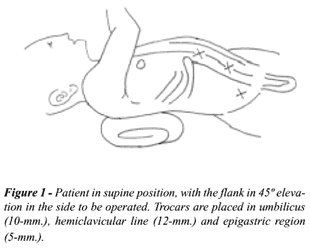
were placed in supine position, with the flank in 45º elevation in
the side to be operated.
A small circular incision in the inferior
umbilicus edge was made, the rectus abdominalis aponeurosis was fixed,
and the Veress needle was introduced. The pneumoperitoneum was established
with 12-mm. Hg and the first 10-mm. trocar was introduced for the 30º
optical insertion. The second 12-mm. trocar was placed under direct vision
at the midclavicular line in the ipsilateral flank, and the third and
last 5-mm. trocar in the epigastric region (Figure-1).
Nephrectomy itself was performed by incision
of the paracolic gutter, with medial mobilization of the ascendant colon
for right nephrectomy, and mobilization of the descendent colon for left
nephrectomy; anterior renal fascia opening and identification of the renal
parenchyma and ureter during lower pole dissection. Ureter was clipped,
divided, and fixed to facilitate approaching renal hilus, without being
necessary introducing a fourth trocar for kidney traction and major vascular
exposure.
Renal pedicle was approached with dissection,
clipping and division of renal artery and vein individually. Both arterial
and vein duplications may exist, and if a large vein is found, endoscopic
vascular staplers, as Endo-GIA, can be used for a safe ligature. Then
proceed to kidney upper pole dissection and removal of surgical specimen.
The cavity was revised with special care to renal bed hemostasis. Surgical
specimens were removed through 12-mm. incision, after been placed in an
endo-bag.
Three children aged 7, 8 and 14 years old
underwent a transperitoneal videolaparoscopic nephrectomy using this technique.
Two of them were male and one was female. All had urinary tract infection
and the radiologic exams (ultrasonography, urography, and cintigraphy)
showed lost of renal unity.
None was submitted to previous renal or
ureteral surgery, and in all cases, adrenal glands were carefully preserved.
Right nephrectomy was performed in 2 cases, and left nephrectomy in one,
and no surgical field draining was performed in any of them.
Operating time ranged from 100 to 230 minutes
(mean 163 minutes). Patients were discharged between postoperative days
2 and 4. There were no intra- or postoperative complications, and no patient
required a blood transfusion. Patients are in medical follow up and returned
to their normal activity on day 10 after the surgery.
COMMENTS
Trocar
and other instruments insertion in abdominal cavity present an important
risk of vascular injury or visceral perforation. The risk per patient
is naturally increased with number of trocars utilized. Based on videolaparoscopic
splenectomy with 3 trocars experience, this technique has been recently
performed for nephrectomy aiming to achieve a lower risk (4).
Desgrandchamps et al. (4) reported videolaparoscopic
nephrectomy results in 20 patients using only 3 trocars. In this study,
operative time was similar for 3 and 5 trocars use, indicating that reducing
the number of instruments did not make performing the same laparoscopic
procedures more difficult (4).
Injuries during laparoscopic procedures
can theoretically affect any intra- or retroperitoneal organ. Most of
these injuries will need immediate or delayed open surgery conversion,
due to hematoma formation, postoperative bleeding, abscess, or peritonitis.
Bowel, gastric and colonic injuries, when not identified, lead to major
complications as ileus, peritonitis and abdominal sepsis (5). Safety device
trocars were developed to prevent risks of visceral and vascular perforation,
even though these are not 100% safe (5).
When detecting a visceral injury, the surgeon
may decide if it can be laparoscopically restored or if an immediate conversion
to a laparotomy is required. The injury will be limited when an appropriate
and immediate treatment is established.
Transperitoneal videolaparoscopic nephrectomy
in children can be performed using only 3 trocars. The technique provides
better cosmetic results, lesser surgical trauma and reduces the risk of
injuries related to trocar insertion, as vascular and visceral lesions.
REFERENCES
- Ehrlich RM, Gershman A, Mee S, Fuchs G: Laparoscopic nephrectomy in a child: expanding horizons for laparoscopy in pediatric urology. J Endourol. 1992; 6:463-5.
- Gillick J, Mohla DJ, Nicholas JL, Fitzgerald RJ: Pediatric laparoscopic nephrectomy: review of 5 years experience at three centers. Pediatr Endosurg Innov Techn. 2000; 4:237-41.
- Borer JG, Atala A: Endoscopic retroperitonel nephrectomy. Pediatr Endosurg Innov Techn. 2000; 4:229-36.
- Desgrandchamps F, Gossot D, Jabbour ME, Meria P, Teillac P, Le Duc A: A 3 trocar technique for transperitoneal laparoscopic nephrectomy. J Urol. 1999; 161:1530-2.
- Fahlenkamp D, Rassweiler J, Fornara P, Frede T, Loening SA: Complications of laparoscopic procedures in urology: experience with 2,407 procedures at 4 German centers. J Urol. 1999; 162:765-71.
______________________
Received: October 2, 2001
Accepted after revision: May 5, 2002
_______________________
Correspondence address:
Dr. Roberto Kazumi Baldas Miura
Av. das Américas, 5001 / 226
Rio de Janeiro, RJ, 22631-004, Brazil
Tel.: + 55 21 2432-7828